What do songbirds tell us about ES cells?
Posted by Raj Ladher, on 8 January 2016
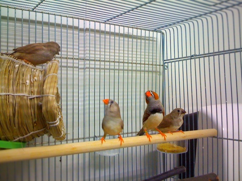
A running joke amongst avian developmental biologists is that the chicken (Gallus gallus) is the tastiest of the model organisms. A typical response from some of my mouse, frog or fish friends, would be that that is where the advantages end – the lack of the ability to do genetics in birds present limitations in the types of molecular analyses that could be done. Despite being so tasty, the chick is not the most cost effective lab animal to keep: It is big, and requires a large amount of space for housing, and it has a slow generation time of almost a year. These points make the development of genetic techniques in chick prohibitive to most labs. About 5 years ago, we decided to see what could be done with the zebra finch. Zebra finches have been widely used in studying behaviour and neural circuitry. They have the advantage of being small and have a generation time of about 3-4 months, the same as that for mouse. With support from the RIKEN CDB Director’s fund (Masatoshi Takeichi at that time) and from the Animal Facility (particularly Shinichi Aizawa, its then head), establishing our finch colony in the RIKEN CDB was straightforward. We bought the founders from pet shops in the Kansai region of Japan and housed them in our newly appointed finch room at the CDB. At this point we realised that many of the published protocols for finch husbandry were not optimal for egg production and so we decided to invest time optimizing these protocols.
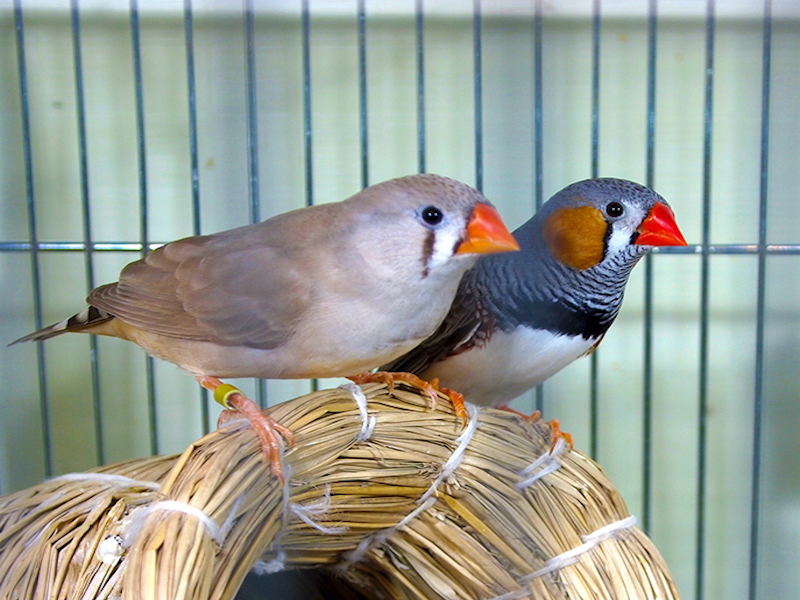
Siu-Shan Mak (Suzanne) the lead post-doc who really drove the study, and Anna Wrabel-Suzuki, the animal technician in charge of looking after the birds, spent the first year studying the behaviour of the birds, and checking the quality of the eggs, tweaking the diet, lighting, and cage conditions so that we could house them in the best conditions for optimal fertile egg production. It was during these checks of egg quality that Suzanne noticed that the finch embryo was at a younger stage than the chick at laying. A small note here; birds undergo some development in utero, before the egg is laid. Chicks and quails lay their eggs when the embryos are just about to gastrulate (Eyal-Giladi and Kochav 1976). We found that finches lay fairly early, with the embryo at a late blastula stage. We thought that this difference would allow us some additional insights into early avian development.
Around this time, the groups of Jennifer Nichols and Austin Smith published a series of papers describing the stage in mouse at which embryonic stem cells (ES cells) that showed naïve pluripotency could be derived (Boroviak et al. 2014). In this state, naïve ES cells can give rise to all cell types found in the embryo. It contrasts with primed pluripotency, where the cells (also called epiblast-derived stem cells or EpiSCs) have a more restricted potential (Nichols and Smith 2009). Another key difference of these cells is their sensitivity to the inhibition of an important signalling pathway intermediate MAP kinase: naïve ES cells were resistant to its inhibition whereas EpiSCs were not (Nichols and Smith 2012). The group showed that mice blastocysts at around E4 were able to generate naïve ES cells. At this stage the epiblast has been specified (as shown by molecular markers) but is yet to epithelialise. We found that the finch blastoderm at laying is similar: The epiblast, and indeed the hypoblast, are both specified but there is little morphological segregation, reminiscent of the pre-epithelialised epiblast. When we took cells from newly laid finch embryos, and cultured in the presence of a MAP kinase inhibitor, they retained the expression of pluripotent markers, and markers of naïve ES cells. Cells taken from later staged finches or from newly laid chicken embryos did not express pluripotent markers when MAP kinase was inhibited, behaving more like primed pluripotent cells. These results were presented in our recent paper in eLife (Mak et al. 2015).
We think that at the very least, the finch embryo could provide some comparative insights into the restriction of pluripotency in amniotes, and possibly would enable the generation of testable hypotheses when thinking about the evolution of pluripotency in mammals. Our hope that by combining new genome engineering tools with long-term finch ES cell cultivation and subsequent chimera generation, this work sets the stage for a tractable, genetic avian model system. These are some of the ideas we will be thinking about in our new labs in Kumamoto (Guojun) and in Bangalore (Raj).
Boroviak T, Loos R, Bertone P, Smith A, Nichols J. 2014. The ability of inner-cell-mass cells to self-renew as embryonic stem cells is acquired following epiblast specification. Nat Cell Biol 16: 516-528.
Eyal-Giladi H, Kochav S. 1976. From cleavage to primitive streak formation: a complementary normal table and a new look at the first stages of the development of the chick. I. General morphology. Dev Biol 49: 321-337.
Mak S-S, Alev C, Nagai H, Wrabel A, Matsuoka Y, Honda A, Sheng G, Ladher RK. 2015. Characterization of the finch embryo supports evolutionary conservation of the naive stage of development in amniotes. Elife. doi:10.7554/eLife.07178
Nichols J, Smith A. 2009. Naive and primed pluripotent states. Cell stem cell 4: 487-492.
Nichols J, Smith A. 2012. Pluripotency in the embryo and in culture. Cold Spring Harbor perspectives in biology 4: a008128.
 (10 votes)
Get involved
Create an account or log in to post your story on the Node.
Sign up for emails
Subscribe to our mailing lists.