Tough decisions for the developing brain
Posted by Misato Iwashita, on 3 October 2014
To form complex organs, somatic stem cells proliferate and then differentiate during development. In this process, intrinsic factors, i.e. the sequential expression of transcriptional genes, and extrinsic factors, i.e. extracellular microenvironment, are intimately involved. Recent in vitro studies have revealed that the physical properties of the extracellular niche, possibly tissue stiffness, may play an important role in cellular behavior, growth and differentiation. This is referred to as “mechanotransduction”. However, the procedure by which physical cues are sensed and translated into gene expression, and their physiological significance in vivo,are essentially unknown.
To understand the role of mechanotransduction in cellular behavior and fate specification during development, one of the critical points is to determine whether there are any spatiotemporal shifts in stiffness in a given developing tissue. Atomic force microscopy (AFM) is a strong tool for this purpose. Using this system, the stiffness in certain postnatal tissues including the brain has been previously examined. In our recent paper in Development, we combined this system with a structural support to measure stiffness in the embryonic mouse brain, one of the softest tissues in our body. We further combined this technique with immunostaining, to increase the spatiotemporal resolution of the measured tissue based on anatomical information (Figure 1A).
As a result, we obtained hitherto unknown results about the shift in stiffness in the developing brain tissue (Iwashita et al, 2014; Figure 1). First, stiffness in the proliferative zones including ventricular zone (VZ) and subventricular zone (SVZ) gradually increases during embryogenesis (Figure 1B). During brain development, gliogenesis starts around birth, after the neurogenic period takes place. Interestingly, previous studies showed in vitro that the lineage shift from neural to glial cells was influenced by a shift in stiffness. Our results provide an attractive scenario in which the extracellular environment, i.e. the stiffened tissue in the later stages of the embryonic brain, may be arranged for better production of glial cells in vivo.
Secondly, we found that there is a sharp peak in stiffness in the intermediate zone (IZ) and a gentle peak in the cortical plate (CP), at the middle stage of neurogenesis (Figure 1B, C). In addition, the stiffness in the IZ tends to be higher than other regions at the mid-stage of brain development. In the IZ, massive cell migration is observed during brain development. Although the physiological significance of the higher stiffness in the IZ is still unclear, it may contribute to directed migration of neural cells toward the CP.
To summarize, we described for the first time the spatiotemporal shift in stiffness that is observed in the developing brain. In this study, we measured the developing brain tissue and cellular stiffness as an experimental model. Our strategy, however, can be applied to profile various types of tissues and cells, and could help understanding the role of tissue stiffness as a physical cue for cell fate determination of somatic stem cells.
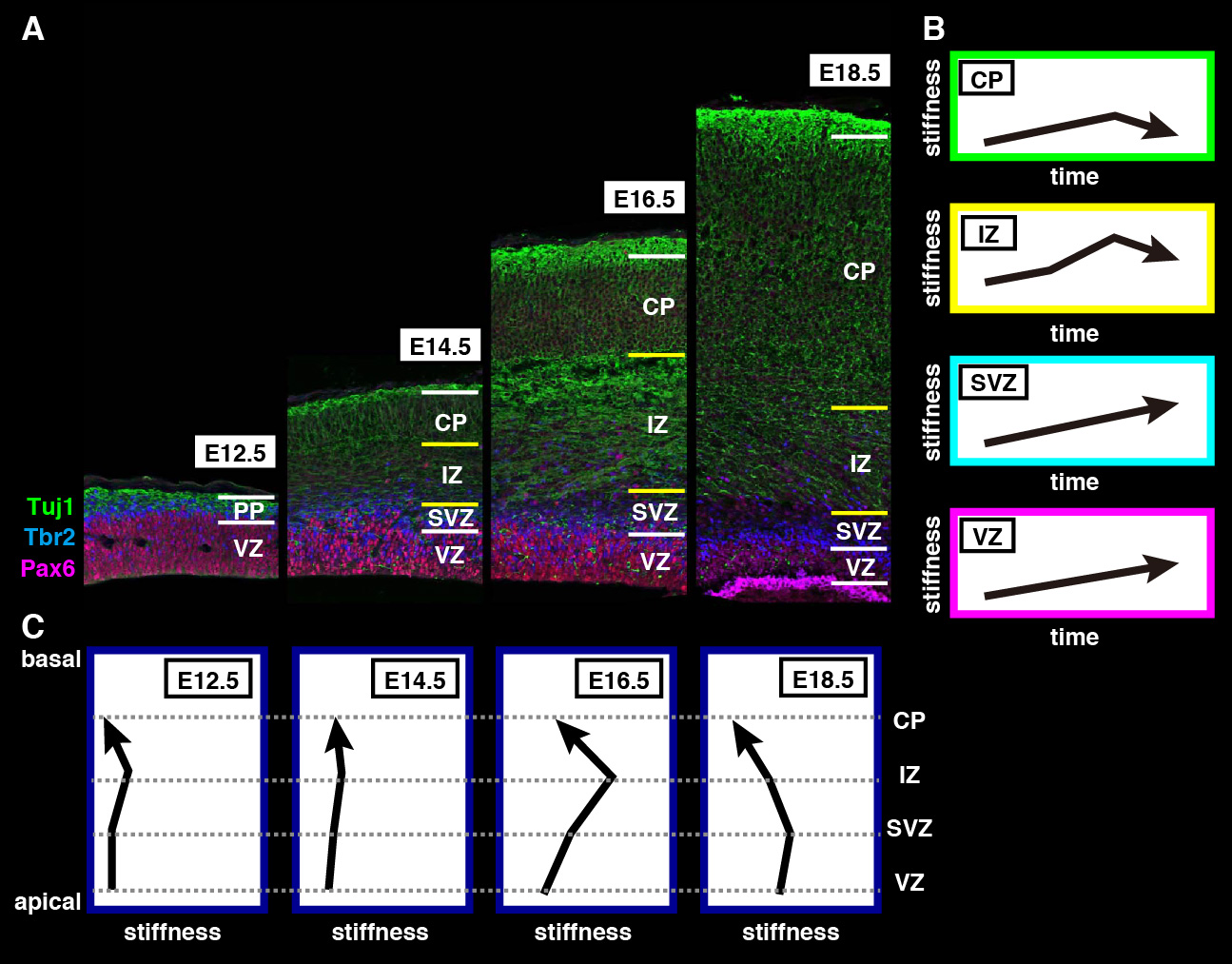
Figure 1. Summary of spatiotemporal measurement using AFM (click image to see a bigger version)
(A) Immunohistological images of developing brains. Cortical layers consist of VZ, SVZ, IZ and CP. Preplate (PP) is a structure transiently appears in early phase of brain development. Red, Pax6; blue, Tbr2; green, Tuj1. This data is modified from Iwashita et al., 2014, Figure 2A.
(B) Schematics of the temporal shifts in stiffness in each layer. The vertical axis shows tissue stiffness. The horizontal axis shows the developmental time course.
(C) Schematics of the spatial shifts in stiffness of cortical layers in each developmental stage. The horizontal axis shows stiffness.
Misato Iwashita and Yoichi Kosodo
Iwashita, M., Kataoka, N., Toida, K., & Kosodo, Y. (2014). Systematic profiling of spatiotemporal tissue and cellular stiffness in the developing brain Development, 141 (19), 3793-3798 DOI: 10.1242/dev.109637
 (4 votes)