9.5 million knockout mouse embryo images now available
Posted by DMDD, on 20 July 2017
A new set of DMDD embryo and placenta data has been released, taking our total dataset to 9.5 million images of around 1300 embryos.
DMDD is a primary screen of embryonic lethal knockout mice, and all data can be freely accessed at dmdd.org.uk. Detailed phenotypes are available for embryos from 73 different knockout lines, and we have phenotyped the placentas from 124 lines. We have also added data on the sex of each embryo.
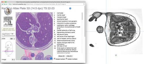
Visitors to our website can now compare HREM embryo images with the closest-matching, annotated histological section from the Kaufman Atlas of Mouse Development. This follows a major project by the eMouseAtlas team at the University of Edinburgh to digitise the Kaufman Atlas at high resolution. The annotated Kaufman sections can be viewed alongside DMDD embryo images to help users who are unfamiliar with the detailed morphological features of a mouse embryo as it develops.
Severe brain phenotypes
Phenotyping of Hmgxb3 knockout embryos revealed severe brain defects, with half of the embryos displaying exencephaly. Embryos from this line also had a range of phenotypes including edema, abnormalities of the optic cup, and defects of the venous system including an abnormal ductus venosus valve and blood in the lymph vessels.
Gene knockout can lead to a huge number of phenotypes
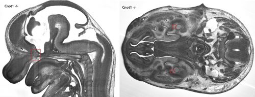
Fifty-five different phenotypes were identified in Cnot1 knockout embryos. These included an absent hypoglossal nerve (which is needed for tongue movement and suckling), abnormalities of the inner ear, testis and thyroid gland, abnormal cell masses that have been classified as embryo tumours, and various heart defects including overriding aortic valve and ventricular septal defects.
The images below show an embryo tumour, and the absence of the mandibular nerve (click the image to view a larger version).

Potential models of human disease
A number of genes studied by DMDD have already been associated with human diseases. For example, Prmt7 mutations have been associated with Short Stature Brachydactyly Obesity Global Developmental Delay Syndrome, an autosomal recessive disease characterised by developmental delay, learning disabilities, mild mental retardation, delayed speech, and skeletal abnormalities. Strikingly, in the Prmt7 knockout embryos studied, the most common phenotypes included neuroma of the motoric part of the trigeminal nerve (a tumour within the skull, affecting the nerve controlling the jaw movements needed for speaking and chewing) and abnormalities of the hypoglossal nerve (which controls movement of the tongue) and the ribs.
Image data has been added for both Cc2d2a and Xpnpep1 knockouts. Mutations of the Cc2d2a gene are known to cause Meckel and Joubert syndromes, while Xpnpep1 has been associated with billiary atresia.
Many of the genes studied by DMDD do not currently appear to be associated with any disease, for example Hmgxb3 or Cbx6. There is potential that careful analysis of the phenotypes from lines such as these could contribute to the identification of new disease models, and our data is freely available in order to encourage this.
A detailed description of normal mouse embryo development
The Atlas of Mouse Development by Professor Matthew Kaufman describes normal mouse embryo anatomy using a series of hundreds of annotated histological sections. Even today, twenty three years after its publication, it is still considered to be the gold standard for describing mouse embryo development. As part of a project to update the book in 2012, the original sections were digitised by the Edinburgh Mouse Atlas Group and made freely available on their eHistology resource.
The images have now been integrated into the DMDD database, and users can directly compare any HREM embryo image with the closest-matching annotated Kaufman section.

This new feature is intended to help users who are not fully confident of the details of mouse developmental anatomy. It means that mutant mouse data can now be explored alongside a fully-annotated wild-type reference point.
A full list of new data
Embryo phenotype data added for: Cnot1, Hmgxb3 and Prmt7
Embryo image data added for: Cbx6, Cc2d2a, Cnot1, Hmgxb3, Prmt7 and Xpnpep1
Placenta images and phenotypes added for: Mir96
To explore the data, visit dmdd.org.uk or for more information please email contact@dmdd.org.uk.


 (1 votes)