An afternoon in the museum with an exploded skull and a 3.5m python skeleton
Posted by Joyce Yu, on 19 March 2024
The people and the research behind the exhibition ‘Growing a backbone’
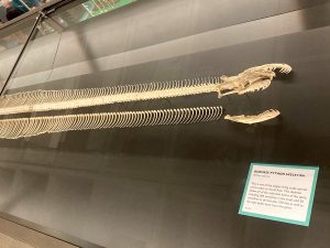
Where can you find a Victorian era ‘exploded skull’ and a 3.5-meter Burmese Python skeleton with every single one of its vertebrae and ribs laid out in a straight line? The answer is the University Museum of Zoology (Cambridge, UK), which recently opened an exhibition ‘Growing a backbone: Rise of the vertebrates’. One Sunday afternoon I decided to pop into the museum to have a look around.
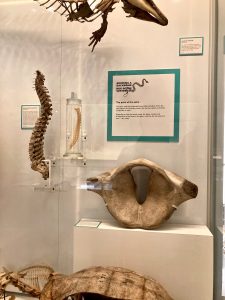
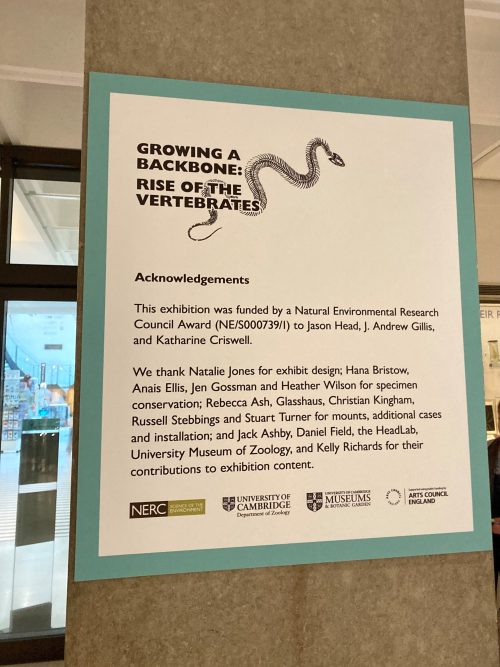
Instead of having a separate space for the exhibition, the exhibits are dotted around the display cases in the museum. Trying to spot the ‘Growing a backbone’ display cards around the museum is like a treasure hunt. From skulls, jaws, teeth, spines, to limbs, visitors are taken along a journey to discover the evolution of the key features in vertebrates that make them unique.
Behind this exhibition is active research being conducted at the University of Cambridge on understanding vertebrate axial skeleton evolution. I reached out to Jason Head, Andrew Gillis and Kate Criswell, researchers working on the project, to find out more about their research on vertebrate evolution and the curation of the exhibition.
Evolution of the vertebrate axial skeleton
The project started with a conversation between Jason, Andrew and Kate. Jason is Professor of Vertebrate Evolution and Ecology and Curator of Vertebrate Palaeontology at the University Museum of Zoology, Cambridge. Before moving to Cambridge, Jason and his colleague Dave Polly had quantitatively analyzed snake skeletal morphology and found that Hox regionalization is maintained in snakes. Then, Jason met Andrew and Kate, who had been working with the little skate (Leucoraja erinacea) as a model system for vertebrate development. “We realize no one had looked in detail at axial skeletal evolution across jawed vertebrates,” said Jason, “and for a long time, people have paid little attention to the diversity of fishes.”
The three of them eventually obtained a grant from the Natural Environment Research Council (NERC), with the aim of integrating anatomical, developmental, and environmental data to reconstruct the evolutionary history of the vertebrate axial skeleton. The experimental aspect of the project was led by Kate, then a postdoc in Andrew’s lab, now Assistant Professor at Saint Francis University.
Kate looked at the different anatomical regions in vertebral columns of cartilaginous fishes (skates, sharks and stingrays) by studying skeletal anatomy and gene expression during development. “Fishes were thought to just have a trunk and a tail. I obtained CT scans of adult skates, created 3D models of each vertebra, and used morphometrics to quantify the shape change along the anterior-posterior axis.” She found that the skate do show some differences in shape in the vertebrae along the axial column, and the expression of certain Hox genes during development matches up with this adult pattern.
Looking beyond the little skate, the team investigated whether fish skeletons more broadly have axial regionalization, by looking at 90(!) fish species. Kate recounted, “a few times I got most of the way through segmenting or landmarking a vertebral column, only to find some defect in a vertebra (this happens a lot in fish!) and have to start from the beginning on another specimen of that same species.”
All the hard work was worth it eventually: the team discovered that the vertebral columns of many cartilaginous fishes have more regions than just a trunk and a tail. Kate said, “I started to get excited after running my analyses a few times on different fish, and consistently getting results suggesting that their vertebral columns were a lot more complicated than the textbooks had indicated. Fish are often treated as primitive or simple, but their anatomy can really surprise us when we look closely!”
Telling the story of vertebrate evolution
Evolutionary developmental biology is fascinating to many people, and creatures like skates and giant snakes often attract public attention. The ‘Growing a backbone’ exhibition in the museum is the perfect opportunity to showcase active research in vertebrate evolution.
Instead of creating the exhibition from scratch, Jason and his team made use of preexisting exhibits in the museum, by adding additional text about the development and evolution of different characteristics in vertebrates. The exhibition also included new specimens from the museum collection. One of Jason’s favorites is the Victorian era exploded human skulls, which were teaching utensils for medical students and zoologists in the mid to late 1800s. “The bones of the displayed skulls are pulled apart,” explained Jason. “The bones are on little armatures so that students could pull them back and forth to see the internal structure and the relationships of the bones.”

Another highlight for Jason is the Burmese Python skeleton. “The Python was completely disarticulated. Our conservator, Natalie Jones, and I re-articulated the skull, and then we laid out the vertebral column and ordinated the ribs in a straight line. We even have one of the pelvic bones still preserved in the specimen.”
But not everything Jason wanted to include in the exhibit ended up being displayed— such as the skull of a sperm whale. “We have a display case about body size, explaining why vertebrates are basically animal giants, in part because we have evolved features allowing us to elevate our metabolism to be very plastic and eat anything that moves. But the sperm whale skull is rather large, so we didn’t really have a place for it.”
Writing museum display texts
Even though Jason has been working in museums for a long time, he acknowledged that it’s sometimes tricky to communicate complex concepts using very simple terminology. “I think it’s very hard to get consensus on how you should be targeting an exhibit for an audience. I tend to increase the expectation of audience comprehension. I think that a good exhibit challenges but doesn’t frustrate — you can introduce new terms and concepts, without blowing people’s minds to the point they can’t understand what you’re talking about. There’s always tension between the people working on the exhibit and the people who did the research. Compromise is how I’d say it works.”

Embedding public outreach into the core of research
On the Sunday afternoon I visited the museum, the gallery was bustling with families and tourists. I was surprised to hear that the museum has not always been accessible by the public. Jason said that historically, the galleries were used for teaching Cambridge students. In recent years, the museum has made an effort to open up the galleries to the public, with temporary exhibitions such as ‘Growing a backbone’ to highlight findings from active research.
Jason thinks researchers should always try to incorporate outreach work as part of their grant proposal, which is what his team did in the NERC grant. “This is a great opportunity to educate the public, tell them the story of vertebrate evolution, and show them the homologies that they might not expect, for example, the fact that our teeth are basically modified scales!” said Jason. “It also gives funding agencies immediate deliverables, to show that our money is spent appropriately, and we can give back to the taxpayers who funded the research. As the exhibition will be here till September, we’ll also be looking at incorporating it into the museum’s public education and outreach programme.”
Filling in the gaps
We are still far from getting a complete picture about the evolution of vertebrate axial skeleton. “As a paleontologist,” said Jason, “I’m dealing with missing data from fossil records. It’s like you’re in a mansion at night: you have to figure out the entire layout of the mansion by looking in the dark with only a small box of matches.” That’s why Jason stressed the importance of expanding taxonomy sampling. “The field has been using the model taxon approach where you take one or two taxa from each major clade. We’ve got a decent sampling across the major divisions of vertebrates, but within the divisions, there’s not much we know. What we need to do is to continue to sample across phyla and look more into the diversity of vertebrates.”
“Most developmental biologists look at developmental processes and then think about what it means in terms of evolution. I’m a paleontologist. I go the other way,” said Jason. “I look at characteristics across phylogeny and then make inferences about the developmental mechanisms that might have led to that.” Kate, also a paleontologist by training studying an extinct group of fossil fishes called placoderms, has since expanded her research focus to include developmental biology: “I think palaeontology and developmental biology are complementary fields that help answer the same evolutionary questions using different approaches.”
Thank you to Jason and Kate for taking the time to explain their research and the process behind curating the exhibition. If you happen to be in Cambridge, UK, do pay a visit to the University Museum of Zoology. The ‘Growing a backbone: Rise of the Vertebrates’ exhibition is running from February to September 2024.


 (2 votes)
I love how the Zoology museum has opened up in recent years – I visit regularly with my toddler, who loves looking at the specimens (though generally keeps me running after him too much for me to have found all the ‘growing a backbone’ exhibits!). It’s a far cry from my earlier experience of the museum, having undergrad supervisions looking at the display cases – which certainly weren’t interrupted by over-excited children or harried mothers…!
Thanks to all the team at the museum for making it such a welcoming place for families, while still keeping the science at the heart of what they do.