A day in the life of a Transgenic Quail lab
Posted by Samara Ranie, on 12 June 2026
Written by Samara Ranie
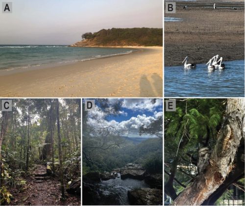
If you drive inland from Meanjin, Australia, also known as Brisbane City, you will find yourself surrounded by subtropical rainforests. Trees and skies are filled with bowerbirds, rainbow lorikeets, cockatoos, my personal favourite, whipbirds, or at night, you may even come across a tawny frogmouth. If you then drive along the coastline, you will meet pelicans, gulls, curlews, spoonbills, oystercatchers, and cormorants sweeping over the ocean surface. If you come to the city, though, and visit The University of Queensland, you may, quite unexpectedly, find the Japanese quail, Coturnix japonica.
At the Institute for Molecular Bioscience (IMB) at The University of Queensland, you will find my research lab, led by Dr Melanie White . In our lab, we use the quail to study spinal cord development and how, when this process goes wrong, neural tube defects (NTDs) develop. As an imaging lab, we work towards beautiful, state-of-the-art imaging of live embryo development, a process that requires us to use fluorescently labelled, transgenic quails (Alvarez et al., 2024).
Quails and human spine development
It may surprise you to learn that human spinal cord development closely resembles that of avian species (Dady et al., 2014). The neural tube begins to develop by week three in humans. A flat sheet of epithelial cells folds and thickens into a tube that will become the brain and spinal cord. The anterior region of this tube, the top, is formed by a process called primary neurulation, where cells fold up into a ‘V’ shape, then bend further to create a closed ‘O ‘- shaped tube. The posterior section, the bottom, of the neural tube is formed by secondary neurulation. In secondary neurulation, the flat sheet thickens before lumens form and fuse to make the hollow neural tube (Copp et al., 2003).
This is a generally agreed-upon set of processes, but it wasn’t until recently that a region bridging these two mechanisms was discovered, junctional neurulation. A region where the primary neurulation process tapers off, and the secondary neurulation process begins to take over (Dady et al., 2014; Wang et al., 2026). This overlapping region, termed junctional neurulation, is unique in that it has not yet been seen in any species outside of humans and avians, making birds arguably the best model for human spine development.
Why use the quail?
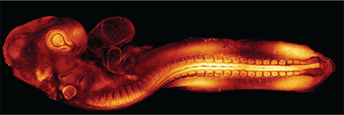
Using an in ovo model, a model that grows in an egg, has the ethical and practical benefits of development outside of a uterus. However, if all avian species develop their spines in this way, then why use the quail? Simply, they grow very fast. A quail will reach sexual maturity at seven weeks old. For creating a transgenic line, this is a huge advantage: you need to breed multiple generations of birds before you have a line suitable for studies, a process which is much faster with a quickly maturing species. It also allows us to work as fast as possible within the lab. When we grow embryos for our studies, we can grow them for as little as 24 hours before they are old enough to begin studying.
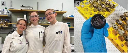
8am: Culturing a quail embryo
A culturing day starts early, and picking up a coffee on the way to the lab is a necessary first step. But then it is time to set up for the day. Agar and albumin (egg white) coated plates are warmed up, and quail eggs are retrieved from the incubator; the eggs having been incubated at 37°C for 24-48 hours beforehand. I often work with embryos after only 24 hours of incubation to catch the earliest stages of neural tube formation.

Very carefully, a window is cut into the delicate shell of each egg to check on the embryos, still smaller than a grain of rice and growing on the surface of each yolk. From here, we can grow them longer, leave them in the egg and provide drug treatment or perform in ovo electroporation, allowing them to grow in their eggs as long as possible; more often, we will culture them out of the egg.
Culturing out a quail embryo means pouring them out of their eggs. The albumin is entirely removed, and the yolk itself is patted dry with tissue. Only now can a culture paper be placed over the embryo; a small square of paper with a rectangle slightly bigger than the embryo itself is placed with the embryo in the space. Scissors are used to cut around the edges of the paper, and the embryo is lifted from the yolk, now suspended within that space. The embryo can then be placed on the warmed agar albumin plates, making sure to place it so that, when the heart begins to develop in another ~6-12 hours, there is room for it to fold and grow before it slowly starts to beat.
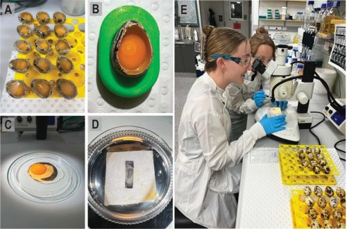
Embryos can then be returned to the warm 37°C incubator to grow for four to five days. But we rarely need an embryo to grow for this length of time. I can also choose to electroporate embryos at this stage or apply a drug treatment to observe if there is any effect on neural tube development.
1pm: Live Imaging
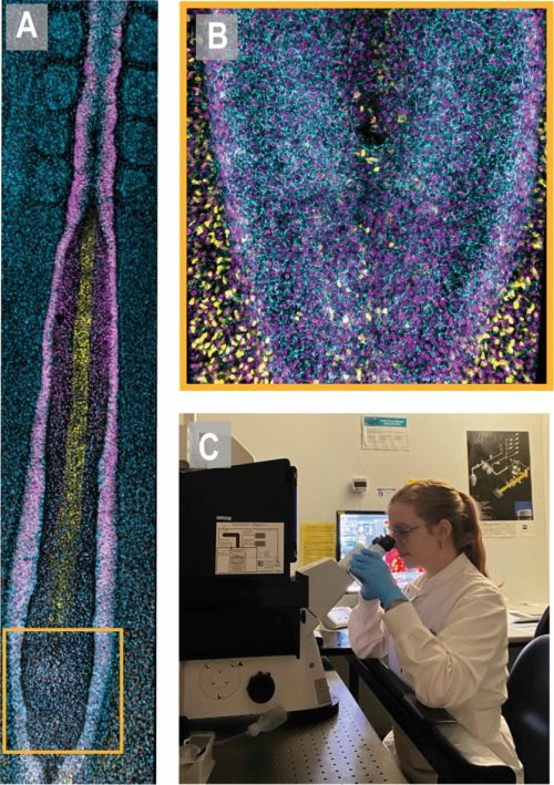
Embryos can be fixed and stained with antibodies for imaging or imaged live. Live imaging of our transgenic quails allows us to study development in real time, recording the symphony of folding and movement that ultimately produces the complete spinal cord and brain. At the IMB , we are lucky to have access to the advanced microscopy facility, with a range of state-of-the-art microscopes and a dedicated microscopy team always willing to troubleshoot any problem.
For live imaging, embryos are mounted on glass-bottom plates with an incredibly thin layer of that Agar-Albumin gel to keep them happy. If, for example, we were using our Lifeact-eGFP embryos in which the actin network is visible through a GFP tag, the embryos would be mounted and then set up on one of our confocal microscopes (Alvarez et al., 2024). After delicately balancing laser power so that it is strong enough to get a signal but not so strong that it bleaches or even damages the embryo itself, you will be able to see the fluorescent, complex, interconnected cabling that actin creates within a single embryo. This beautiful maze of actin will change, move, and grow before your very eyes. Not a bad sight for a Tuesday afternoon.
Live imaging of a single embryo can take anywhere from one hour to 12 hours, depending on which processes you are studying. Development is much slower than it looks when we speed up the final video, but every minute is worth it to unravel these systems. These stunning videos and images don’t just provide us with data; they also allow audiences to connect with our work in unique ways, as visual information drives curiosity. That connection to other scientists and the public drives our research, our passion for the unknown and for this model system.
5pm: Pack-up
By 5 pm, I have worked a long but rewarding day. On some culturing days, you may do everything from egg to image; on others, you might just culture and fix your embryos to work on throughout the week. These big culturing days usually happen once a week, with the rest of the week spent processing your images and data from previous weeks, planning future experiments and prepping in the lab. We have the distinct advantage in the lab of everyone using the same techniques, so our lab “chores” are communal; replace what you use, and everything will be ready for the next person. When everything is restocked and packed away, it’s time to head home, where a cup of tea and a book are waiting for me
Alvarez, Y. D., van der Spuy, M., Wang, J. X., Noordstra, I., Tan, S. Z., Carroll, M., Yap, A. S., Serralbo, O., & White, M. D. (2024). A Lifeact-EGFP quail for studying actin dynamics in vivo. J Cell Biol, 223(9). https://doi.org/10.1083/jcb.202404066
Copp, A. J., Greene, N. D., & Murdoch, J. N. (2003). The genetic basis of mammalian neurulation. Nat Rev Genet, 4(10), 784-793. https://doi.org/10.1038/nrg1181
Dady, A., Havis, E., Escriou, V., Catala, M., & Duband, J. L. (2014). Junctional neurulation: a unique developmental program shaping a discrete region of the spinal cord highly susceptible to neural tube defects. J Neurosci, 34(39), 13208-13221. https://doi.org/10.1523/jneurosci.1850-14.2014
Wang, J. X., Alvarez, Y. D., Tan, S. Z., Ranie, S. N., Stehbens, S. J., & White, M. D. (2026). Quantitative live imaging reveals PRICKLE1 controls junctional neural tube morphogenesis independent of Planar Cell Polarity. Nature Communications, 17(1), 3654. https://doi.org/10.1038/s41467-026-71242-0
 (1 votes)
One thought on “A day in the life of a Transgenic Quail lab”
Leave a Reply
Get involved
Create an account or log in to post your story on the Node.
Sign up for emails
Subscribe to our mailing lists.
Read the latest Development issue
Most-read posts in May
- From comparison to mechanism: decoding heart regeneration
- A day in the life of a Reviews Editor (at Development)
- What does a Reviews Editor do?
- New evo-devo textbook ‘Eco-Evo-Devo: The Environmental Regulation of Development, Evolution, and Health’
- preLighters’ choice – A curated selection of recent preprints
Wonderful! This is the first time I hear of using quail for developmental studies.