

From comparison to mechanism: decoding heart regeneration
Posted by Shih-Lei (Ben) Lai, on 26 May 2026
“Why do some hearts regenerate, while others do not?”
This question has followed me for more than a decade. Not as a single project, but as a thread that kept resurfacing—each time forcing us to rethink what we thought we understood.
The idea of comparing regenerative zebrafish with non-regenerative medaka started as one of several proposals I discussed with Didier Stainier when I joined his lab in 2014. At the time, it felt simple: let biology provide the contrast, instead of trying to extract answers from a single system.
Zebrafish regenerate their hearts. Medaka do not. That difference was too striking to ignore (1).
I was in my second postdoc then, aware that I needed something I could carry forward. Didier encouraged me to pursue it, even though it was clearly high-risk. Looking back, that decision shaped everything that followed.
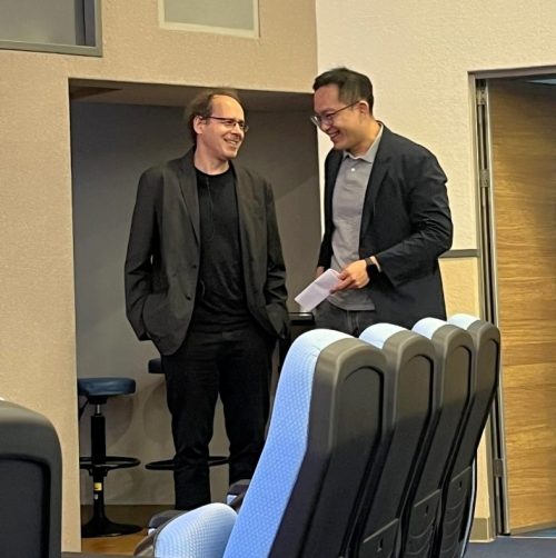
When the data pointed to something unexpected
Together with Rubén Marín-Juez —who later became both a key collaborator and a close friend—we established a cryoinjury model in medaka and generated our first comparative RNA-seq datasets (2).
What we saw was not what we expected.
Instead of cardiomyocyte-centered differences, which at the time were widely viewed as the primary drivers of cardiac regeneration, the strongest signals pointed toward immune responses and angiogenesis (2). I remember hesitating. It felt like we were drifting away from what many would consider the “core” of cardiac regeneration.
But the data were clear, and we decided to follow it.
We used to joke that we were like the Maze Runners—moving forward without knowing what was coming next, or how things would end. Rubén focused on angiogenesis and uncovered how revascularization is an early and essential step in regeneration (3, 4). In parallel, I moved toward immunity—despite being warned, quite accurately, that “immunity is too complicated and messy to work with.”
In hindsight, that hesitation reflects something broader. Work in non-mammalian models is often judged by how directly it translates to human biology, rather than by the clarity of the biological principles it can reveal. Yet it is precisely these systems—and the people willing to pursue them—that allow us to uncover mechanisms that are otherwise difficult to see.
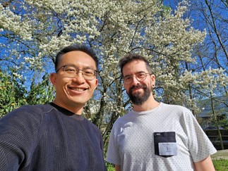
A result we thought we understood—but didn’t
What stood out early was that macrophage infiltration appeared delayed and reduced in medaka (2). So, we asked a simple question: what happens if we delay macrophage recruitment in zebrafish?
Using clodrosome, we transiently depleted macrophages prior to injury and observed impaired regeneration.
At the time, we interpreted this primarily as a timing effect—an early delay that irreversibly disrupts regeneration.
This seemed to fit the data. Macrophages eventually came back, and their numbers recovered within about a week. Yet the heart still failed to regenerate.
We moved forward with that explanation.
But it never fully made sense.
Revisiting the problem with better resolution
It was only years later, after I started my lab at the Institute of Biomedical Sciences at Academia Sinica in Taiwan, that we revisited this question with better tools.
Through temporal single-cell profiling—driven in large part by the careful and persistent work of Ke-Hsuan Wei, one of the first PhD students in my lab—we realized something we had completely missed before: clodrosome was not simply delaying macrophages—it was preferentially depleting the resident macrophage population (5).
These cardiac resident-like macrophage subsets turned out to be essential for heart regeneration—coordinating revascularization, cardiomyocyte survival, debris clearance, and extracellular matrix remodeling.
That realization reframed everything.
Even when we allowed extended recovery, giving circulating, monocyte-derived macrophages ample time to repopulate the heart, regeneration did not recover.
At that moment, the entire story finally made sense.
It was not only about timing. It was also about identity.
What initially appeared to be a delay in macrophage function was, in fact, the loss of a specific and irreplaceable cell population.
Turning a non-regenerative system on
We then asked the opposite question: instead of suppressing the immune response, could activating it change the outcome?
Poly I:C—identified through comparative transcriptomics—enhanced macrophage recruitment and, unexpectedly, enabled de novo regeneration in medaka (2).
That was one of those moments when you don’t immediately trust the data. We repeated the experiments, trying to convince ourselves it wasn’t an artifact.
But it held.
Regenerative capacity began to look less like a fixed property and more like something that could be modulated.
From immune identity to regenerative signal
Our recent paper in PNAS represents the latest step in this progression (6).
Led by Kaushik Chowdhury, this phase of the work brought together comparative analysis, single-cell profiling, and functional experiments to identify a regeneration-associated macrophage population induced by poly I:C.
These macrophages localize to the injury border zone and express Granulin.
What started as a candidate marker became a functional insight. Through a series of carefully executed experiments, the team showed that recombinant Granulin alone is sufficient to promote cardiomyocyte proliferation and reduce scarring—linking immune activation to a concrete regenerative outcome.
Closing the loop, granulin expression is also activated in zebrafish following cardiac injury and is essential for heart regeneration.
Basic science, unexpected translation
I always consider myself a basic scientist and a developmental biologist. None of this work started with a translational goal.
It was driven by curiosity—by a question that seemed fundamental, but not immediately “useful” or “applicable”.
And yet, it led us to a concept that is inherently translational: that regeneration might be induced through immune modulation.
As Didier once put it:
“One never really knows when a basic science finding will transform translational research… CRISPR/Cas9 is just one recent example.”(7)
That perspective has stayed with me throughout this journey. Especially at moments when the work felt uncertain, or when its relevance was not immediately obvious.
Still the same question
If there is one idea that has gradually emerged, it is that regenerative capacity is not fixed.
It is governed, at least in part, by the identity and function of immune cells—and therefore potentially modifiable.
That doesn’t make the problem simple. But it reframes it.
And in many ways, we are still following the same question.
Just with a clearer understanding of what actually matters—and with a team that made it possible to see it.

References
1. Ito K, Morioka M, Kimura S, Tasaki M, Inohaya K, Kudo A. Differential reparative phenotypes between zebrafish and medaka after cardiac injury. Developmental Dynamics. 2014;243(9):1106-15.
2. Lai S-L, Marín-Juez R, Moura PL, Kuenne C, Lai JKH, Tsedeke AT, et al. Reciprocal analyses in zebrafish and medaka reveal that harnessing the immune response promotes cardiac regeneration. eLife. 2017;6:e25605.
3. Marin-Juez R, Marass M, Gauvrit S, Rossi A, Lai S-L, Materna SC, et al. Fast revascularization of the injured area is essential to support zebrafish heart regeneration. Proceedings of the National Academy of Sciences. 2016;113(40):11237-42.
4. Marín-Juez R, El-Sammak H, Helker CSM, Kamezaki A, Mullapuli ST, Bibli S-I, et al. Coronary Revascularization During Heart Regeneration Is Regulated by Epicardial and Endocardial Cues and Forms a Scaffold for Cardiomyocyte Repopulation. Developmental Cell. 2019;51(4):503-15.e4.
5. Wei K-H, Lin IT, Chowdhury K, Lim KL, Liu K-T, Ko T-M, et al. Comparative single-cell profiling reveals distinct cardiac resident macrophages essential for zebrafish heart regeneration. eLife. 2023;12:e84679.
6. Chowdhury K, Huang C-L, Lin IT, Hung Y-J, Lim KL, Liu H-W, et al. Immune modulation promotes heart regeneration through macrophage and Granulin functions in medaka. Proceedings of the National Academy of Sciences. 2026;123(16):e2524705123.
7. Grewal S. An interview with Didier Stainier. Development. 2015;142(17):2861-3.
Get involved
Create an account or log in to post your story on the Node.
Sign up for emails
Subscribe to our mailing lists.