An interview with the 2024 GfE Hilde Mangold Award winner — Daniel Wehner
Posted by the Node, on 2 April 2024

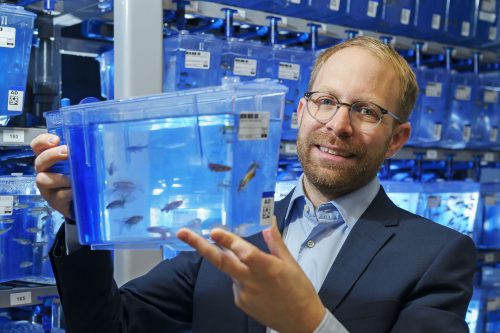
Daniel Wehner is Group Leader at the Max Planck Institute for the Science of Light and the Max-Planck-Zentrum für Physik und Medizin in Erlangen (Germany). At the 2024 joint German and Dutch developmental biology societies meeting in Osnabrück, Daniel was awarded the GfE Hilde Mangold Award for young investigators. After his award lecture at the conference, we chatted about his career background, his transition to becoming a PI, and his research into spinal cord regeneration in zebrafish.
(Image credit: Stephan Spangenberg)
Congratulations on winning the GfE Hilde Mangold Award! What does this award mean to you?
Receiving this award is of tremendous significance to me because it recognizes the efforts and contributions of our research team to the field. The German Society for Developmental Biology is my home turf, so I am deeply honoured to be recognized by this society for our work.
You received the GfE PhD prize back in 2015 as well!
Yes, this makes getting this Hilde Mangold Award even more impactful, as it validates that we are on the right path and encourages me to persist in my research direction. I believe our research has always been somewhat unconventional in that we never opt for the easiest route to tackle scientific challenges. For instance, our lab combines biochemical and physical methodologies to investigate spinal cord regeneration. It’s truly gratifying to see the efforts of our research group acknowledged. I extend my heartfelt thanks to all the members of my group and my past and present mentors who have supported us along the way.
Let’s go back to the beginning, when did you first become interested in science?
I have always had a keen interest in biology and in understanding the fundamental principles of life. However, it was during my final two years of high school in the biology course that my passion truly ignited. That was when I was introduced to the basic principles of cell biology, and I became captivated by the intricacies and sophistication that enable life. I went on to study (medical) biotechnology, yet I found myself unsatisfied with merely learning about established knowledge. The decision to pursue a PhD stemmed from my inherent drive to discover something new.
What did you work on during your PhD?
I completed my PhD under the supervision of Gilbert Weidinger, initially at the Technische Universität Dresden and later at Ulm University. Initially, my research focused on identifying new modulators of the Wnt pathway [1] [2]. Subsequently, I shifted my focus to investigating how Wnt signaling regulates fin regeneration. We adapted the TetON system to examine cell type-specific functions of Wnt signaling in a complex regenerating appendage [3]. This technological advancement led to the concept of Wnt pathway-controlled signaling centers that coordinate regeneration through secondary signals. [4] [5]. In another paper published in Development, we provided evidence that Notch signalling coordinates the proliferation and differentiation of progenitor cells — key events in tissue regeneration [6].
Then you moved to Edinburgh for your postdoc at the Becker lab. What did you work on?
Catherina and Thomas Becker have done pioneering work on spinal cord regeneration in zebrafish. It was an excellent synergy, bringing together the TetON system and their remarkable model system for central nervous system regeneration. It was during this time that we established the principle that after spinal cord injury, fibroblasts modulate the lesion site by depositing a complex extracellular matrix (ECM), but in strong contrast to mammals, in zebrafish this environment promotes regeneration [7].
Is that when you started to find your niche for your own research group?
Yes, I was intrigued by the interspecies difference in fibroblast behaviour because, in mammals, fibroblasts are typically viewed as inhibiting the regeneration of the central nervous system. For instance, research conducted by the Frisén and Göritz labs demonstrated that PDGFRβ-positive fibroblasts of perivascular origin contribute to the formation of inhibitory central nervous system scars [8] [9]. Therefore, the natural question to ask was: do these cells behave differently in response to spinal cord injury in zebrafish? To address this, we developed numerous genetic models to lineage-trace and cell type-specifically interfere with pdgfrb+ cell recruitment, or spatially-confined optogenetic depletion of pdgfrb+ cells. Our findings revealed that in zebrafish, pdgfrb+ fibroblasts are required for regeneration. We discovered that these fibroblasts possess the ability to downregulate specific inhibitory ECM molecules and upregulate regeneration-promoting matrix genes, facilitating axon regeneration. We further demonstrated that the unique biochemical composition of fibroblast-derived injury ECM in central nervous system lesions defines regenerative success, versus inhibitory scar formation in zebrafish and mammals. These findings resulted in the publication of our lab’s first two major papers [10] [11].
How was your experience transitioning to becoming a PI?
I must admit that the transition was not easy. There were moments when I considered leaving academia due to the lack of prospects. This is a challenge many colleagues face, particularly women in the field. As a father of two, I understand the difficulties and challenges of navigating a postdoc while caring for children, and I know firsthand the additional challenges young parents encounter. I consider myself very fortunate that things have ultimately worked out for me.
Following my postdoc in Edinburgh, I joined the Center for Regenerative Therapies at Technische Universität Dresden, working in Michael Brand’s lab with support from a return stipend from the German Research Foundation. Although I applied for an Emmy Noether Fellowship (similar to an ERC starting grant) and didn’t make the final cut, it was during this time that I crossed paths with Jochen Guck, a renowned researcher in tissue mechanics. We discovered excellent synergies by combining physics methodologies with molecular biology tools to investigate spinal cord regeneration in zebrafish. This led to an exciting opportunity to lead my own independent research group at the Max Planck Institute for the Science of Light.
Can you briefly talk about what your lab is working on?
My current research addresses the fundamental questions: i) How can regeneration-permissive lesion environments be established after central nervous system injury? ii) How can central nervous system axon regeneration be promoted in regeneration-limiting environments?
To achieve these goals, we are pursuing several approaches. i) We are investigating the intricate cell-cell interactions that dictate the biochemical and mechanical properties of the regeneration-permissive microenvironment in zebrafish, including the regulation of axon regeneration-permissive injury ECM. ii) Utilizing zebrafish as an in vivo platform, we are dissecting the regeneration-modulating properties of specific components of human scar tissue, along with elucidating their underlying mechanisms. iii) We are also in the process of developing humanized zebrafish models to explore novel strategies for spinal cord repair.
Do you have any advice for people thinking of starting their own lab?
Science thrives on passion and determination, existing within its own realm. Avoid comparing yourself to those outside of this world. Instead, dare to think innovatively and seek out environments that foster support, enabling you to embrace risks and confront challenges.
Speaking of supportive environments, how important do you think mentorship is in navigating an academic career?
A supportive mentor can profoundly influence career development. Navigating the academic path is challenging and fraught with obstacles. Having a great mentor who offers guidance on avoiding these pitfalls can be invaluable, sparing you from unnecessary setbacks.
I count myself fortunate to have benefitted from excellent mentorship. For those seeking PhD or postdoc positions, I recommend considering joining a lab led by a junior PI. Working with a junior PI provides valuable insight into the process of establishing a lab. Junior PIs often prioritize mentorship as it is a mutual responsibility.
My PhD supervisor, Gilbert, was a junior PI at the time. He taught me essential techniques such as microinjections, then entrusted me with the freedom to explore. This supportive environment was echoed during my time at Becker’s lab in Edinburgh, where guidance was readily available, yet I was encouraged to pursue independent research.
What’s your favourite technique? And what are you most excited about in your area?
My preferred technique is in situ hybridization—it’s remarkably reliable! Nevertheless, the method that currently captivates me the most is Brillouin microscopy. This non-invasive, label-free, all-optical approach enables the measurement of viscoelastic properties in vivo using light. Access to Brillouin microscopes is scarce, limited to only a handful of locations worldwide. Having the chance to employ this state-of-the-art technique at the Max Planck Institute in Erlangen to investigate the role of tissue mechanics in spinal cord regeneration is a true privilege. Achieving the initial results that contributed to our latest paper [11] was an overwhelming experience.
Finally, let’s get outside of the lab – what do you like to do in your free time?
Back then, I was deeply immersed in music, playing bass guitar in a band, and engaging in trekking, rock climbing, and mountaineering. However, with the arrival of my two daughters and the commencement of a new lab, I find myself with limited time for hobbies. Nonetheless, I consider science itself a passion—a hobby in its own right!
References
[1] Kagermeier-Schenk B, Wehner D, Ozhan-Kizil G, et al. Waif1/5T4 inhibits Wnt/β-catenin signaling and activates noncanonical Wnt pathways by modifying LRP6 subcellular localization. Dev Cell. 2011;21(6):1129-1143. doi:10.1016/j.devcel.2011.10.015
[2] Özhan G, Sezgin E, Wehner D, et al. Lypd6 enhances Wnt/β-catenin signaling by promoting Lrp6 phosphorylation in raft plasma membrane domains. Dev Cell. 2013;26(4):331-345. doi:10.1016/j.devcel.2013.07.020
[3] Wehner, D., Jahn, C., Weidinger, G. Use of the TetON System to Study Molecular Mechanisms of Zebrafish Regeneration. J. Vis. Exp.(100), e52756, doi:10.3791/52756 (2015)
[4] Wehner D, Cizelsky W, Vasudevaro MD, et al. Wnt/β-catenin signaling defines organizing centers that orchestrate growth and differentiation of the regenerating zebrafish caudal fin [published correction appears in Cell Rep. 2014 Feb 27;6(4):777-8]. Cell Rep. 2014;6(3):467-481. doi:10.1016/j.celrep.2013.12.036
[5] Wehner D, Weidinger G. Signaling networks organizing regenerative growth of the zebrafish fin. Trends Genet. 2015;31(6):336-343. doi:10.1016/j.tig.2015.03.012
[6] Bartholomäus Grotek, Daniel Wehner, Gilbert Weidinger; Notch signaling coordinates cellular proliferation with differentiation during zebrafish fin regeneration. Development 1 April 2013; 140 (7): 1412–1423. doi: 10.1242/dev.087452
[7] Wehner D, Tsarouchas TM, Michael A, et al. Wnt signaling controls pro-regenerative Collagen XII in functional spinal cord regeneration in zebrafish. Nat Commun. 2017;8(1):126. Published 2017 Jul 25. doi:10.1038/s41467-017-00143-0
[8] Dias DO, Kim H, Holl D, et al. Reducing Pericyte-Derived Scarring Promotes Recovery after Spinal Cord Injury. Cell. 2018;173(1):153-165.e22. doi:10.1016/j.cell.2018.02.004
[9] Göritz C, Dias DO, Tomilin N, Barbacid M, Shupliakov O, Frisén J. A pericyte origin of spinal cord scar tissue. Science. 2011;333(6039):238-242. doi:10.1126/science.1203165
[10] Tsata V, Möllmert S, Schweitzer C, et al. A switch in pdgfrb+ cell-derived ECM composition prevents inhibitory scarring and promotes axon regeneration in the zebrafish spinal cord. Dev Cell. 2021;56(4):509-524.e9. doi:10.1016/j.devcel.2020.12.009
[11] Kolb, J., Tsata, V., John, N. et al. Small leucine-rich proteoglycans inhibit CNS regeneration by modifying the structural and mechanical properties of the lesion environment. Nat Commun 14, 6814 (2023). https://doi.org/10.1038/s41467-023-42339-7
 (1 votes)
Get involved
Create an account or log in to post your story on the Node.
Sign up for emails
Subscribe to our mailing lists.