Echinoderm development on film
Posted by Bruno Vellutini, on 6 December 2010
“I also here salute the echinoderms as a noble group especially designed to puzzle the zoologist.”
Libbie Hyman, 1955
Echinoderms are fascinating creatures. They have extensive regenerative capabilities, a mutable connective tissue that dynamically (and deliberately) changes its stiffness, and a complex system of hydraulic canals involved in the circulation of internal fluids and locomotion.
However, the most notable feature of echinoderms is the pentamerous symmetry of their bodies, derived from a bilateral ancestor. These exclusively marine deuterostomes are mostly bottom dwellers with a biphasic life cycle, where the adult tissues develop inside a bilateral planktonic larva (swimming in the water column) and metamorphose into a benthic juvenile.
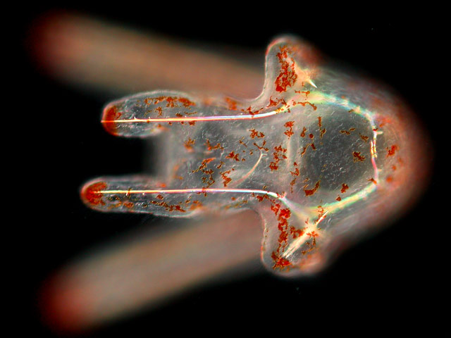
A planktonic pluteus larva of a sea biscuit.
During my master’s project at University of São Paulo, Brazil, I studied the development of a different kind of sea urchin, a sea biscuit. Sand dollars and sea biscuits belong to a lineage of urchins that developed a secondary bilateral symmetry. Also, during their evolution, around 55 million years ago, the adult morphology changed in association with the occupation of sand beds; more specifically, the body flattened, the spines got shorter, the number of tube feet increased, and their feeding apparatus (lantern of Aristotle), which was absent in other adult irregular urchins, was retained into adulthood.
Since I was interested in the developmental origins of such changes in morphology I documented the embryonic, larval, and juvenile development of a sea biscuit species, Clypeaster subdepressus. After gathering all data, I compiled it into a science outreach video showing a resumé of the life cycle of this species, from fertilization to the first steps of the juveniles. Hope you enjoy it:
We collected adults from sand beds of São Sebastião Channel (São Sebastião, SP, Brazil) and induced gamete release (eggs and sperm). We did the fertilization in vitro and followed the embryonic development in the laboratory, under light microscopy. Embryos become swimming larvae, approximately 0.2 mm wide, which we fed with microalgae until metamorphosis. A diminute sea biscuit grows inside the larva. When the minuscule podia and spines are formed the larva sinks and undergoes metamorphosis. The juvenile sea biscuit reabsorbs the larval tissue and begins to explore its new habitat, between sand grains. [Numbers on the upper right corner show how much the scene was accelerated.]
The video is available to download here, feel free to reuse it and share it around! If you want further details on sea biscuit development (including images and video footage) the official description was published earlier this year.
 (9 votes)
5 thoughts on “Echinoderm development on film”
Leave a Reply
Get involved
Create an account or log in to post your story on the Node.
Sign up for emails
Subscribe to our mailing lists.
Nice!
Good to see you on The Node, Bruno!
Beautiful video Bruno, thanks for posting it.
Developmental Biology is definitively a Science, but with movies like this one, it also has to be considered as pure Art (and still very informative…). As a mouse embryologist, I am sometimes (often?) jealous of these developmental biology models!
Thanks for posting this movie!
Stephane
Thanks! Forgot to say that we are almost finishing a longer and more complete version of this video. Should be available at the beginning of 2011!