From our sister journals- June 2015
Posted by the Node, on 22 June 2015
Here is some developmental biology related content from other journals published by The Company of Biologists.

Elucidating pulmonary hypoplasia in ciliopathies
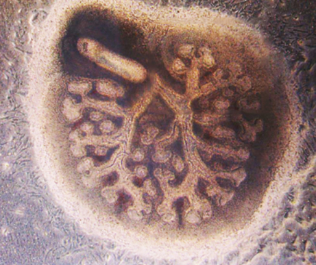 Ciliopathies are developmental disorders caused by mutations in components of the primary cilium (a microtubule-based mechanosensor organelle present in many mammalian cells), and are usually characterised by multi-organ abnormalities. Congenital lung malformation (pulmonary hypoplasia) often occurs, and is considered the leading cause of death in Meckel-Gruber syndrome (MKS), a lethal ciliopathy associated with mutations in the transmembrane protein 67 gene, Tmem67. To investigate mechanisms of pulmonary hypoplasia in MKS, Colin A. Johnson’s group characterised Tmem67–/– knockout mutant mice and used biochemical methods to further elucidate TMEM67 function. The group found that TMEM67 interacts with Wnt5a and receptor tyrosine kinase-like orphan receptor 2 (ROR2), two components of non-canonical Wnt signalling. Tmem67–/– embryos and pups manifest pulmonary hypoplasia phenotypes and, consistent with other available data, these are mediated by mutations of any component in the Wnt5a-TMEM67-ROR2 axis. Interestingly, pharmacological targeting of downstream effectors of this axis is able to rescue pulmonary abnormalities in cultured lungs from Tmem67–/– mice. These results implicate the dysregulation of the Wnt5a-TMEM67-ROR2 axis in ciliopathies and suggest that its downstream modulation can prevent pulmonary hypoplasia in these diseases. Read the paper here (Open Access).
No sperm without miRNAs
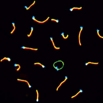 The microRNA (miRNA) pathway is known to be required for completion of murine spermatogenesis. However, the exact functions of miRNAs and the identity of their targets that are crucial for spermatogenesis are unclear. In their study, Paula Cohen, Andrew Grimson and colleagues unravel an essential role for miRNAs in the regulation of DNA damage repair to prevent sex chromosome defects during meiosis in males. The authors analysed mice with conditional knock-outs (cKO) of DGCR8 and DICER, which are essential for pri-miRNA and pre-miRNA processing, respectively, and found abnormal pairings of sex chromosomes or their fusion to autosomes. They also showed that levels of ataxia telengiectasia mutated (ATM) kinase were elevated in both cKO lines and that the phosphorylated ATM substrate mediator of DNA damage checkpoint-1 (MDC1) was mislocalised in DICER KO spermatozoa. As shown here, the Atm 3ʹUTR functionally interacted with miRNA (miR)-18, miR-16 and miR-183, suggesting a role of these miRNAs in the regulation of spermatogenesis. Importantly, the authors demonstrated that the DNA damage repair protein RNF8, whose localisation is partly controlled by ATM-MDC1 signalling, was redistributed from sex chromosomes to autosomes. Taken together, these results indicate that ATM is regulated by miRNAs to ensure the proper localisation of its substrates that are involved in maintaining chromosomal stability during spermatogenesis. Read the paper here (Open Access).
Dendrite arborisation – it has to be NudE
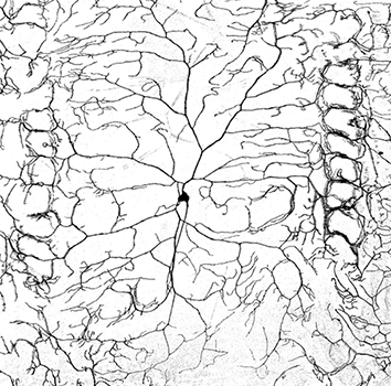 Dynein and kinesin molecular motors play important roles in neuronal morphogenesis, but the precise mechanism of their action is still unclear. Nuclear distribution E (NudE) proteins are involved in neuronal proliferation and migration, but they are also known to function as dynein cofactors and, as such, could be involved in neurite outgrowth. Taking advantage of the fact that only one NudE protein is present in Drosophila melanogaster, Jill Wildonger and colleagues investigate the role of NudE in dendrite arborisation of class IV neurons in 3rd instar larvae. They found that NudE colocalised with vesicular cargo and Golgi outposts in wild-type larvae, whereas its depletion resulted in shorter dendrites with fewer branches, increased microtubule dynamics and altered microtubule polarisation in the axon. Confirming a role for NudE in dendritic arborisation as a dynein cofactor, NudE ablation also enhanced the severity of a mild dynein-mutant phenotype. The authors found that the C-terminus of NudE was inhibitory to its activity in promoting dendrite arborisation, as a C-terminus-deletion mutant rescued the dendrite arborisation defect in NudE-depleted neurons to a greater extent than the full-length protein. Importantly, overexpression of another dynein cofactor, Lis1, rescued NudE neurons, but no rescue was observed when NudE was prevented from interacting with Lis1. Based on their results, the authors conclude that the function of NudE in promoting dendrite growth is to stabilise the dynein–Lis1 interaction. Read the paper here (Open Access).
Role of traction force in definitive endoderm specification
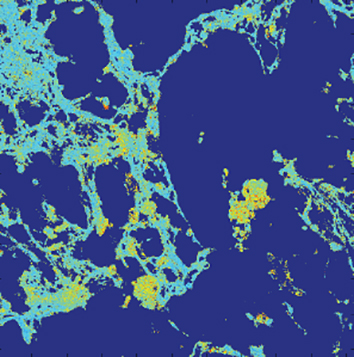 Differentiation of embryonic stem cells (ESCs) into definitive endoderm requires signalling by both growth factors – such as activin A and Wnt3a – and extracellular matrix (ECM) components – such as fibronectin (FN) and laminin. Although ESCs do both, exerting traction forces and responding to the mechanical properties of the ECM, little is currently known about how soluble factors induce biochemical and physical changes in the ESCs and their associated matrix, when added to differentiate ESCs. Here, Adam Engler and colleagues used a force-sensitive FN matrix assay in mouse ESCs to understand the crosstalk between mechanical factors and chemical signals in ESCs and the ECM in directing developmental cues. Addition of the myosin inhibitor blebbistatin inhibited ESC differentiation, indicating that traction forces are necessary for the formation of definitive endoderm. This effect is possibly exerted through TGF-β signalling because this treatment transiently prevented the nuclear translocation of the TGF-β effector phosphorylated SMAD2. The authors then showed that extracellular laminin-111 regulated differentiation; its binding to α3β1-integrin inhibited the SMAD2 inhibitor SMAD7 and also decreased FN-induced matrix strain that acts through α5β1-integrin. Taken together, this study shows that soluble factors that induce ESC differentiation activate traction forces, which – in turn – utilise integrins and ECM remodelling to feedback and support growth-factor-activated signalling cascades. Read the paper here.
Pipefish fathers do not boost embryos’ oxygen
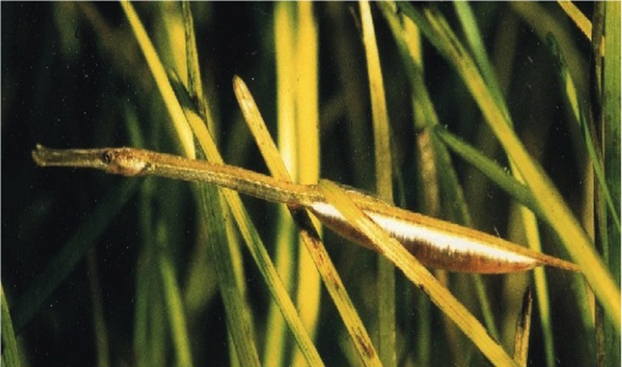 The pipefish brood pouch presents a unique mode of parental care that enables males to protect, osmoregulate and nourish the developing young. Lower availability of oxygen in water was believed to naturally limit the size of fish eggs and, as pipefish eggs are relatively large for the fish’s size, it was assumed that the males somehow provided an abundant supply of oxygen. Using a very fine O2 probe, Braga Goncalves and colleagues assessed the extent to which males of the broad-nosed pipefish oxygenate the developing embryos and are able to maintain pouch fluid O2 levels when brooding in high and low oxygen concentration. Their results show that male pipefish are unable to boost the oxygen supply that they provide to their precious cargo and the mystery of why pipefish eggs are so large remains. Read the paper here.
Bubbles create cosy environment for developing embryos
 Some amphibians breed in terrestrial environments and make bubble nests to lay their eggs in. It was long assumed that the bubbles helped protect developing eggs from predators, reduced egg dehydration and improved the oxygen supply to the developing embryos. Steve Portugal highlights a paper by Méndez-Narváez and colleagues recently published in Physiological and Biochemical Zoology, identifying an additional role for terrestial bubble nests- insulating the developing embryos from extreme fluctuations in ambient temperatures. Read this Outside JEB feature here.


 (No Ratings Yet)