“Here’s looking at -oids, kid”: meeting report – Vienna ISSCR symposium, December 2023
Posted by Alexandra Bisia, on 19 December 2023
Picture credits: https://wordhistories.net/2018/03/21/heres-looking-at-you/;
Vincent van Batenburg, Hubrecht Institute
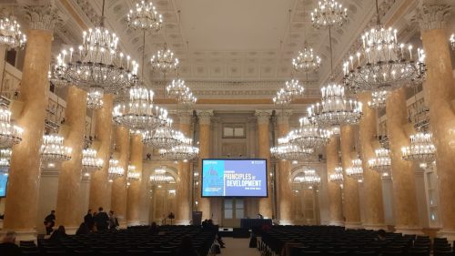
One does not easily pass up the opportunity to attend an ISSCR conference, especially when said conference is in Vienna, in the leadup to Christmas, and the venue is the Hofburg palace. I was really lucky, therefore, to have the opportunity to attend the meeting, held in Vienna this month, from 4-6 December. The theme of the meeting was “Elucidating Principles of Development with Stem Cells,” and boy, were those principles elucidated. With a suite of stem cells from various organisms available to developmental and stem cell biologists, research groups are now developing and refining ever more numerous and sophisticated ways to represent aspects of embryonic development with them. Accordingly, across the three days of the meeting, we were showered with a variety of talks covering every modality of -oid imaginable.
As suggested by the title of the meeting, a big focus was the use of stem cell-based or -derived models (with various -oid names, such as gastruloid, chimeroid, organoid, blastoid, cardioid, axioloid…). These are increasingly sophisticated and are being manipulated and studied in a variety of ways, and it is possible they may be able to supplement to a great extend the requirement for real embryos in research. I find it fascinating that we are now so familiar with the various signalling regimes that are required during different stages and in different tissues in development that it’s possible to replicate synthetically specific stages or tissues of development, starting from pluripotent stem cells (and sometimes extraembryonic stem cells too) – for example only preimplantation or post-implantation development, or axial elongation, or heart development, or neural tissues, or the gut. One of the many benefits of these systems is, of course, that these -oids can be reproducibly generated in great numbers, giving researchers an abundance of material on which to carry out experiments.
The inaugural talk of the meeting was delivered by Denis Duboule, an excellent speaker, whose lab’s extensive work on Hox genes is revealing increasing detail on the workings of the Hox cluster. Specifically, his lab has been using “stembryos” that recapitulate Hox gene expression along the anterior-posterior (or rostro-caudal) axis of the embryo. His use of a rope with interspaced knots and clothespins to represent Hox genes and the CTCF sites that ensure the sequential and appropriately-timed expression of genes along the cluster was a personal highlight. And it was still only 9:30 on the Monday!
The meeting had a strong showing of groups that work on ectodermal/neural tissue models. As someone who is not very familiar with these cell types from my own research, I was nonetheless impressed by how interesting I found them. Among others, there were talks by Paola Arlotta, Barbara Treutlein, Madeleine Lancaster, Thomas Vierbuchen, Anna Kicheva, Sharad Ramanathan, Elly Tanaka, Akanksha Jain, and Jürgen Knoblich. A topic which stood out for me was Joanna Wysocka’s talk on DNA-guided transcription factor cooperativity and its functions in cranial neural crest cells (CNCCs). Briefly, her lab identified a novel long consensus DNA motif, dubbed the “Coordinator,” containing a homeobox and an E-box binding motif. The Coordinator is specifically bound by the bHLH transcription factor Twist1 and homeobox factor Alx4. These interact only when bound to the Coordinator via a tiny segment of a loop only found on Twist1 and no other bHLH factors, thus promoting expression of a host of genes associated with CNCC behaviour.
Another talk on a much less common model organism was by Ali Elagoz, a doctoral student who works on the embryonic development of octopus nervous systems, in a broader effort to identify the evolutionary mechanisms by which cephalopods (octopuses, nautiluses, cuttlefish, and squids) have succeeded in evolving the largest nervous system of all invertebrates. For those not familiar with octopus nervous systems, you might be delighted to find out that only about 1/3 of octopus neurons are actually found in their brains, which, by the way, are wrapped around their oesophagus. Rapid reogranisation of the cephalopod genome and expansion of their suite of protocadherin genes may have contributed to the innovations that permitted the cephalopod brain to dramatically expand in size.
That said, fans of other germ layers were not disappointed, with talks on mesodermal and endodermal specification and derivatives by André Diaz from Universitat Pompeu Fabra (gastruloids), Sasha Mendjan (cardioids), Aryeh Warmflash (2D ESC patterning to study Wnt and Nodal/Activin signalling gradients), Cantas Alev (axial development), Katharina Sonnen (timing of somitogenesis), and Sarah Bowling (hematopoietic stem cells).
Another thematic arc highlighted during the meeting was the novel quantitative aspects that developmental and stem cell biology are being explored from, and which, until very recently, were almost exclusively assessed from a qualitative point of view. Ewa Paluch’s lab, for instance, is studying how epithelial-to-mesenchymal transition, a process essential in multicellular development, can be approached from the perspective of cell shape. Her lab has developed a “morphospace analysis” pipeline whereby cell shape is segmented and many variables quantified, finally undergoing dimensionality reduction to create a 2D “grid” of possible cell shape phenotypes. Cell shapes were then quantified before, during, and at the end of EMT, with cells undergoing EMT having a much “noisier” cell shape than epithelial cells, in other words exploring more of the morphospace. This suggests an instance of noise-driven transitions, where noise may actually help to overcome a barrier, in this case from an epithelial to a mesenchymal state.

Finally, I would be remiss if I didn’t highlight some methods development talks that really captivated me. Alexdander van Oudenaarden detailed a new method, developed by his postdoc Michael VanInsberghe, for profiling both single cells’ full complement of RNA (as single-cell RNA-seq already does), and their ribosome-bound RNA (like single-cell Ribo-seq) in a single experiment, in order to elucidate gene regulation at the transcriptional vs translational level. This is achievable by titrating MNase (micrococcal nuclease), an enzyme that can digest single-stranded nucleic acids, and that can function both as an endonuclease and exonuclease in a concentration-dependent manner. It’s amazing how such a simple concept can yield such rich data at the single-cell level!
I was also blown away by Kate McDole’s talk on imaging in developmental biology. Her unparalleled skills in custom-building microscopes are a boon to developmental and stem cell biologists who want to image samples that are rapidly growing, undergoing morphogenesis and constantly changing optical properties. Her “event-driven microscopy” platform is designed to assist users in imaging their organisms or organoids in a way that accounts for changes in the sample that are going to be happening in the future, without requiring the constant direct supervision of the user.
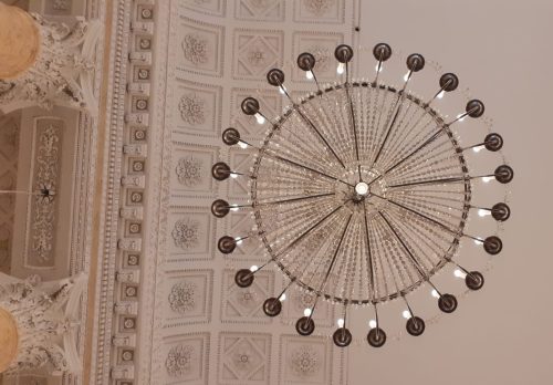
There was a broad and interesting array of posters at the meeting, and as always at a conference it was great to have the opportunity to meet researchers at all stages of their career, from many institutes and countries. As I mentioned, the venue was spectacular, and since it was literally a room in the former imperial palace, it was quite easy to become distracted by the numerous chandeliers, marble columns, and the stuccoed ceiling. As a bonus, every morning what I assume was a military band would parade past one of the windows, inadvertently serenading the first speaker of the day with some drumming.
Overall, the conference was a great experience. Nothing quite beats the excitement of hearing scientists talk about their work in person, especially about results that are often fresh out of the oven. And it’s always inspiring to meet new scientists with different interests and approaches to conducting research – you never know where a novel idea for your own work may come from, and which technique or new finding will form the basis for a new avenue of scientific exploration!
 (3 votes)
Get involved
Create an account or log in to post your story on the Node.
Sign up for emails
Subscribe to our mailing lists.