

How do sea anemones make babies?
Posted by Stefanie Williams, on 24 October 2026
Inspiration for scientific research projects can come from many unexpected places. With the publication of my second PhD research paper [1], I am looking back at my sources of inspiration as well as some hurdles I had to overcome to see this work come to fruition.
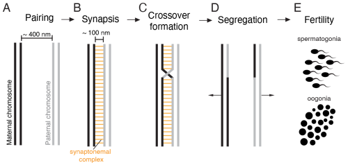
As a PhD student at the Stowers Institute for Medical Research in Kansas City, I was going through the first semester trying to absorb as much information as possible while deciding on my rotation labs. I had decided on my first rotation in the lab of Dr. Scott Hawley very quickly after joining the program. The esteemed Drosophila geneticist was particularly enthralled by the synaptonemal complex, a multi-protein structure that assembles between the maternal and paternal copy of each chromosome in early meiosis (Fig. 1). This structure is crucial for the formation of crossovers between these homologous chromosomes and thereby ensures proper chromosome segregation and fertiltiy (Fig. 1). Apart from this fundamental function, I was fascinated by the fact that this massive proteinaceous structure is able to assemble across the entire length of chromosomes.
As is common at many research institutes, we regularly heard seminar talks from researchers coming to us from all over the world. One of these presentations was given by Dr. Catriona Munro, a scientist who studies meiosis in the jellyfish Clytia hemisphaerica [2]. Previously, I hadn’t really considered the possibility of doing research with unusual organisms like jellyfish. For my bachelor’s thesis in Germany, I did strictly in vitro work. For my year-long research stint at Purdue University in the US, I worked with Drosophila melanogaster. All fairly standard research systems. Seeing that someone performed systematic studies using a jellyifsh was truly fascinating and unexpectdely paved the way for my own future research.
Shortly after this talk, I heard from Dr. Matt Gibson at the Stowers Institute about his lab’s research using the sea anemone Nematostella vectensis. His research team investigates various kinds of fundamental biological questions ranging from how stinging cells in cnidarian species function [3] to neuroscience [4] and Hox-mediated segmentation during embryogenesis [5]. What particularly caught my attention was the fact the Nematostella is relatively large and that each adult has eight gonads that took up the majority of the animal’s body mass. Immediately, I thought that this would make a pretty good meiosis model organism. Scott had previously told me that most meiosis models come with the difficulty that meiotic material is fairly limited. This certainly wouldn’t be a problem with Nematostella.
After I went home for the day, I googled whether anyone was doing research on Nematostella meiosis and its synaptonemal complex. I was surprised to find that research on cnidarian meiosis in general was quite sparse. When it came to the synaptonemal complex, almost 10 years ago a research team in Germany had identified specific protein homologs in the related cnidarian species Hydra vulgaris [6]. In addition, Catriona had looked at the synaptonemal complex in Clytia briefly as part of her research [2]. Other than that, there was no systematic research being done in this area. Considering the importance of the synaptonemal complex for the maintenance of an organisms’ fertility, I was convinced that we needed to understand how it functions in cnidarians. Particularly, as cnidarians like corals are facing tremendous challenges to their fertility. I gathered my scientific curiosity and courage to email Scott and Matt, proposing a research project on studying the conservation of synaptonemal complex proteins in Nematostella. The answers came quickly: Scott absolutely loved the idea. Considering his lab’s newest research with the silk moth Bombyx mori [7] which had quite an unusual synaptonemal complex, he practically couldn’t wait to learn about what novelties the Nematostella equivalent could reveal. Matt was similarly intrigued by this new research direction and my enthusiasm for cnidarian research.
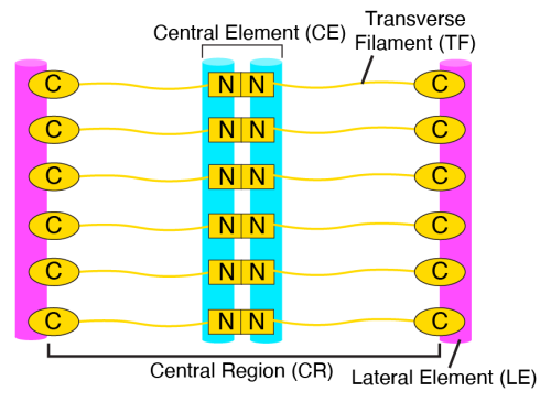
Once I completed my three lab rotations, I decided to pursue my PhD thesis research co-mentored by Scott and Matt. My particular focus was to study whether the architectural functions of Nematostella synaptonemal complex proteins were conserved. Curiously, synaptonemal complex proteins exhibit rapid sequence divergence, despite their crucial function in ensuring meiotic success throughout eukaryotes [8]. As a result of this divergence, it was actually surprising how easy the identification of protein homologs of vertebrate synaptonemal complex proteins in Nematostella was. Most protein homologs were already identified by other researchers but I was able to expand these findings [1]. Now we needed to try and figure out whether these proteins actually function in the same way as they did in other organisms. Considering that the synaptonemal complex consists of three distinct structural elements (Fig. 2), I wanted to generate three individual mutants each disrupting one of these elements. This would allow us to understand the functional importance of each element as well.
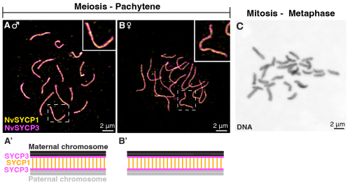
While Nematostella has quickly become my favorite laboratory model, one of the major downsides of working with it is the relatively long generation time of roughly 3 months. This meant that I was raising animals and making crosses for over a year with mutants I had no idea would show any interesting phenotypes. In the meantime, I established necessary tools to study meiosis and the synaptonemal complex in this model, allowing me to investigate the wild-type structure. Some techniques that I had to adapt was preparing chromosome spreads of meiotic and mitotic stages (Fig. 3). During this time, I also worked on a second project investigating the synaptonemal complex in Drosophila species, which you may learn more about here.
Unfortunately, in my case, this long waiting period also meant that my co-supervisor Scott passed away only one month before I was able to start working with the homozygous mutant adults I had established and raised. His death was not only felt deeply because of the sudden loss of scientific support but also because he was so much more than just a scientific supervisor to so many people – including me.
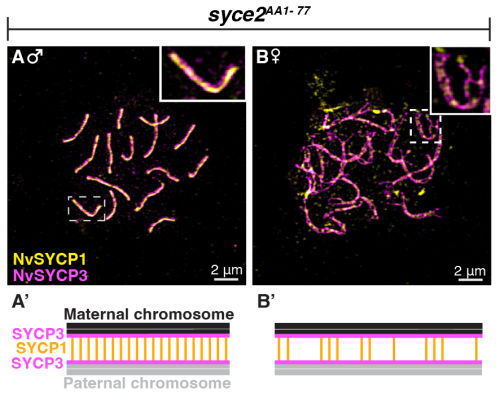
Once I was able to exit the sudden stupor that Scott’s death led me into, I began investigating my homozygous mutants. The three Nematostella proteins I mutated shared approximately 30% of sequence identity with their vertebrate orthologs. While this may seem very low, it is worth emphasizing that orthologous proteins between vertebrates and Drosophila, for example, share no identifiable sequence homology. Thus, I hypothesized that the Nematostella synaptonemal complex proteins and their architectural functions are fully conserved in comparison to vertebrates. First and foremost, this meant that each individual protein component is essential for the successfull assembly of the synaptonemal complex. Thus, I definitely thought I must have made a huge mistake when my first microscopy session on one of my mutants’ chromosome spreads showed no significant assembly defects (Fig. 4). As it turns out, it was not a mistake but the fact that this particular protein is not essential for synaptonemal complex assembly in Nematostella males. Looking at all three of my synaptonemal complex mutants, I was able to reveal that some architectural functions of synaptonemal complex proteins in Nematostella are distinct from those found in vertebrates and, additionally, exhibit dramatic sexual dimorphisms.
While these findings by themselves were deeply fascinating, I also wanted to understand the functional relevance of the synaptonemal complex and its protein components on overall fertility and chromosome segregation. While a TUNEL assay was supposed to provide me with a quick answer to the question whether spermatocytes were undergoing increased rates of cell death (hinting at meiotic checkpoint activation), this experiment turned into a 6-months long ordeal of troubleshooting and protocol optimizations. Nonetheless, I persevered and was able to confirm that even those proteins that appeared to be non-essential for synaptonemal complex assembly were necessary to ensure proper meiotic progression. Taken together, it became clear that the synaptonemal complex in Nematostella was quite distinct in its architectural composition when compared to vertebrates. The sexual dimorphisms I observed further complicated this picture but made it all the more intriguing.
When I envisioned this research project, I could have never foreseen where this work would go. Practically all my hypotheses were proven wrong. In addition, I discovered a research interest that follows me into my current postdoc and likely beyond. By establishing Nematostella vectensis as a model for investigating meiosis, this work now opens the door to exploring how the mechanisms that ensure reproductive success have evolved across animals. As this work is the first systematic investigation into how cnidarian synaptonemal complexes funciton mechanistically, this also paves the road to understand how fertiltiy is ensured in these basal metazoans. Considering that Nematostella is closely related to corals, this work further has important implications for coral sexual reproduction. Altogether, this research paper is the product of a lot of sweat and tears but I could not be prouder and happier to see its publication and I cannot wait to see what else I will learn about cnidarian reproduction.
References
[1] Stefanie Williams, Jennifer Gardner, Stephanie H. Nowotarski, Matthew C. Gibson. The Nematostella synaptonemal complex mediates divergent and sexually dimorphic meiotic programs. Science Advances Volume 12, Isse 43, eaei0758 (2026). https://doi.org/10.1126/sciadv.aei0758
[2] Catriona Munro, Hugo Cadis, Sophie Pagnotta, Evelyn Houliston, Jean-René Huynh. Conserved meiotic mechanisms in the cnidarian Clytia hemisphaerica revealed by Spo11 knockout. Science Advances Volume 9, Issue 4, eadd2873(2023). https://doi.org/10.1126/sciadv.add2873
[3] Ahmet Karabulut, Melainia McClain, Boris Rubenstein, Keith Z. Sabin, Sean A. McKinney, Matthew C. Gibson. The architecture and operating mechanism of a cnidarian stinging organelle. Nature Communications Volume 13, 3494 (2022). https://doi.org/10.1038/s41467-022-31090-0
[4] Ruohan Zhong, Chris W. Seidel, Anna M. L. Klompen, Matthew C. Gibson. Morphological, molecular, and functional evidence for a CNS-like oral nerve ring in the sea anemone Nematostella vectensis. bioRxiv 2026.01.28.701697; doi: https://doi.org/10.64898/2026.01.28.701697
[5] Shuonan He, Florencia Del Viso, Cheng-Yi Chen, Aissam Ikmi, Amanda E. Kroesen, Matt Gibson. An axial Hox code controls tissue segmentation and body patterning in Nematostella vectensis. Science 361,1377-1380 (2018). https://doi.org/10.1126/science.aar8384
[6] Johanna Fraune, Miriam Wiesner, Ricardo Benavente. **The Synaptonemal Complex of Basal Metazoan Hydra: More Similarities to Vertebrate than Invertebrate Meiosis Model Organisms. Journal of Genetics and Genomics, Volume 41, Issue 3, 107-115 (2014) https://doi.org/**[10.1016/j.jgg.2014.01.009](https://doi.org/10.1016/j.jgg.2014.01.009)
[7] Youbin Xiang, Dai Tsuchiya, Zulin Yu, Xia Zhao, Sean McKinney, Jay Unruh, Brian Slaughter, Cathleen M. Lake, R. Scott Hawley. Multiple reorganizations of the lateral elements of the synaptonemal complex facilitate homolog segregation in Bombyx mori oocytes. Current Biology, Volume 34, Issue 2, p352-360. (2024). https://doi.org/[10.1016/j.cub.2023.12.018](https://doi.org/10.1016/j.cub.2023.12.018)
[8] Stefanie Williams, R. Scott Hawley. From conservation to adaptation: understanding the synaptonemal complex’s evolutionary dynamics. Current Opinion in Genetics & Development. Volume 93. (2025) https://doi.org/[10.1016/j.gde.2025.102349](https://doi.org/10.1016/j.gde.2025.102349)
Get involved
Create an account or log in to post your story on the Node.
Sign up for emails
Subscribe to our mailing lists.