

The little beach hopper that could
Posted by Billy Hinchen, on 6 August 2026
How Parhyale hawaiensis went from mundane beach critter to lab celebrity across biology, chemistry and even physics.
Pop down to the beach, pick up a handful of seaweed and you’re likely to be greeted by dozens of little crustaceans exploding out from underneath, frantically trying to hop away for the nearest bit of cover. You probably never gave them much attention because you were busy hunting for crabs or just hoping to fling some seaweed at a sibling. And that’s completely fair as they’re far from the most attractive critters on the shore.
But if you’d looked a little closer, you’d find that those little crustaceans were beach hoppers, also known as sand fleas, which, let’s be honest, doesn’t help win them any additional popularity. These beach hoppers are a group of crustaceans we call amphipods. And one in particular, Parhyale hawaiensis, inadvertently became a bit of a biology celeb.
Coming in at under 2 cm, Parhyale lives in intertidal rubble and rotting leaf litter around warm coastlines. For those who decided to keep these little critters, it became apparent they breed every two or three weeks, all year-round, from the comfort of a standard plastic tray filled with artificial seawater. Oh and they’ll happily eat a slice of carrot.
Rather mundane to warrant the title ‘celeb’, you might say, but its academic CV is nonetheless impressive. Over twenty-five years this animal has turned up in developmental biology, evolutionary theory, regeneration research, soft-matter physics, neuroanatomy, the study of biological clocks, immunology, pollution monitoring, biofuel enzymology and aquaculture. So, not too shabby for something you’d shake out of your beach towel without a second thought. Without any more rambling from me, here’s how that all happened.
It began with an egg
Not a transparent one, as you might expect of an animal we chose specifically for watching. The Parhyale egg is about half a millimetre wide and looks like a dense ball of white(ish) yolk, which you can’t see into at all. What you can see is the surface, where a bunch of large cells sit, each identifiable by position, and all visible under an ordinary microscope. So far so good.
In 2002 researchers dyed the cells of the egg, which we’ll correctly call an embryo from here on out, at a point when the whole thing was made of just eight cells. Four smaller ones on top, and four larger ones beneath. These eight cells of the very early embryo were followed, with studious researchers carefully mapping out where each one ended up, and found the whole future animal was already neatly parcelled out between them (1): three would build skin and nervous system, three the muscle and other internal tissues, one the gut lining, and one the germ line (the cells that make things like gametes for reproduction). These identities, or fates, were settled before there’s anything you’d call an animal. Locking in fates itself was interesting because this tends to happen much later in most other animals. Importantly, it opened the door for lots of intriguing questions around how these predestined cells related to each other.
The obvious step was to see what happens if we removed one of those early cells. Do neighbours pick up the slack and chip in to build our little beach hopper, or is it destined to be without a particular type of tissues, such as muscle, and end up distinctly more beach ‘crawler’ than hopper? Well, it turns out if you remove one of the three internal-tissue founders early enough, the neighbours do indeed cover for it, but nothing crosses between lineages (2), i.e. a muscle precursor removed meant only muscle neighbours helped compensate. Lose the germ-line founder and the embryo carries on building itself, but reaches the end of embryogenesis with no germ cells to be found, and nothing else steps in to supply them (3), and they still hatch! (4)
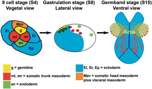
Surprisingly, if you strip out the little patch of cell goop (officially called the cytoplasm) that carries the germ-line instructions in the one-cell embryo, you get no germ cells as expected, but the embryo also never makes it as far as an early-but-important stage of development called gastrulation (the point at which the ball of cells starts folding itself into something with an inside and an outside). Those two points turn out to be unrelated, because an embryo that loses its germ-line cell three divisions later, at the eight-cell stage, gastrulates perfectly happily (5). This suggests the germ plasm is doing something quite separate from just specifying germ cells, which is cool and intriguing. What’s super weird is that the Patel lab reports, though not yet in print, that some animals go on to rebuild a germ line from non-germ line tissue and reach maturity fully fertile. “Nature, erm, finds a way.”
Sorting all this out required tools, and a shared staging system was the logical starting point (1). Transgenics and then CRISPR leapt in to answer more detailed questions (6), and then a genome of roughly 3.6 billion letters, which is a little bigger than ours, came along to help link things together (7). Each new step was developed to answer another question about development and each new step handed the animal and knowledge to someone else.
Where insect wings came from
As a crustacean, Parhyale isn’t an insect (obviously), but it is an arthropod and you’d be forgiven for noting the similarities. Like insects, crustaceans have an impressive array of appendages; they usually come equipped with claws, gills, paddles, walking legs, jumping legs and mouthparts, all of which form from the same repeating segments. And once the genes that assign those jobs (the Hox genes) had been mapped (8) they could be switched off one at a time. With some clever molecular work, you can show all this with experiments where you get walking legs where an abdomen should be, or missing gills or an animal that gives up on specialisation altogether and builds the same generic limb over and over.
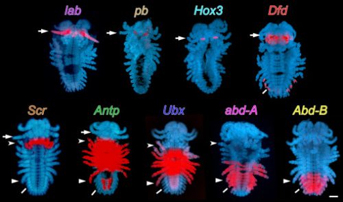
But back to insects and an argument running since the nineteenth century about where insect wings came from: a fresh outgrowth on the back, or a bit of ancestral leg carried upward as the body wall swallowed the leg’s base? Unanswerable in insects, who absorbed the relevant leg segments long ago and conveniently destroyed the evidence.
But Parhyale never absorbed them. In 2020 two groups attacked this from opposite ends and published back to back (9, 10). One knocked out leg-patterning genes and compared the wreckage to insects, while the other went looking for the genetic signature of a wing in crustacean tissue. They came to different conclusions about the route (one that the wing descends from an outgrowth on that ancestral leg segment, the other that it is a merger of two tissues, back and leg) but they agree an insect’s flank corresponds to a crustacean’s topmost leg segment, and that the wing is built partly from what used to be leg. Later work chased the same ancient tissue across to a very distant crustacean cousin, where it turns up as the shell of the water flea (11).
Neither settled it and the argument grinds on, with Paleozoic fossils now thrown in. But a question fought over for a century mostly by staring at insects, who had destroyed the evidence, finally had an animal you could do genetics in.
Legs that come back
We all like legs. They’re great for getting to and from places, I always find. Losing one is therefore somewhat of a bother. Unless you happen to be our favourite beach hopper of course, in which case if you lose a leg, you can regrow it in about a week (12). We know this because the limb’s shell is stiff and clear, so you can glue it to a microscope coverslip and film the whole thing in an awake animal, using the exoskeleton as splint and window at once (13). Turns out the new tissue comes from cells already committed to a fate rather than some reserve of blank ones, so, muscle from muscle lineages, skin from skin. And the muscle is rebuilt by satellite-like cells, which are a back-up group of cells lying dormant, wedged between the muscle fibre and its surrounding membrane, until there’s some damage. Up until this work, satellite cells had only ever been found in chordates, and if they really are the same cell type, the machinery was already there in the common ancestor of pretty much every animal with a front and a back! The skin, however, isn’t lucky enough to have these standby cells, and instead ordinary cells near the cut just divide, work out where they now are, and get on with it.
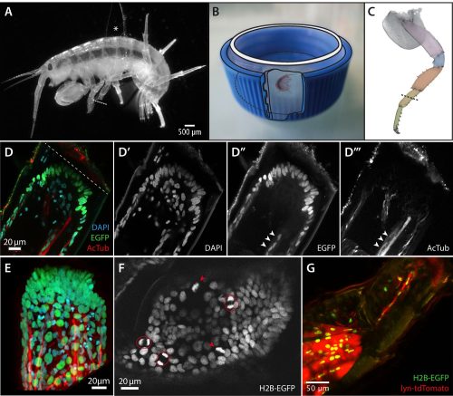
Compare what an embryo does when building a leg with what an adult does regrowing one and the two schedules simply refuse to line up (14). Yet when you take a look at the regrown leg properly, its sensory hairs, whether they work, what cell types it contains, and you’d never be able to tell it apart from one that was never cut off, other than being a little on the small size (15).
Squares
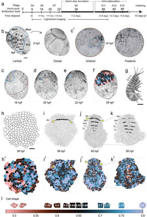
Cells in a sheet normally pack into hexagons, or close enough, like bubbles or honeycomb. Parhyale embryos use tidy rows of squares (16), and since rows of those squares become the segments of the adult, this is likely more than just for show and could be tied to cell identities and the movements needed for development. But right now it’s a bit of an open question. It does however form a nice bridge between a biology problem and a physics one, because dividing cells should really mess up any tidy arrangement. Filming whole embryos, however, showed it’s the divisions themselves keeping things tidy: waves of oriented division sweep through, and faults get swept out to the edge (17, 18), maintaining a tidy, ‘global’ pattern across the embryo. And so now, Parhyale has a whole new audience in physics journals, which nobody planned.
A brain and some rubbish eyes
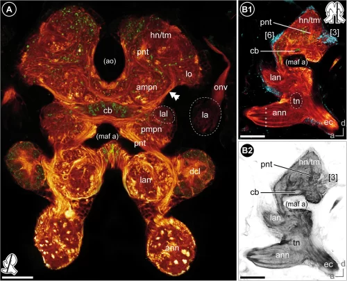
You probably don’t immediately think of a brain when you think of tiny crustaceans, hopping around the beach. But it’s there, in a way, mostly in the form of a concentrated nervous system and that nervous system went undescribed for an awkwardly long time (especially in considering how much attention invertebrate neurobiology has had over the years!). Thankfully, it eventually got the recognition it deserved, and it now has a proper brain atlas (19) and even some rather splendid maps of its signalling molecules (20). The eyes are an especially cool part, each with about fifty facets, five light-sensitive cells apiece rather than the usual eight, wired into the brain differently from related animals (21). Okay, that’s the cool part, well, to me at least, but the reality is they’re a little bit rubbish: fine for working out roughly where you are, but not for seeing anything. But hey, it’s good to know and have genetic access to understand them, especially since nearly everything we know about arthropod eyes comes from one fly.
Two clocks
I keep an eye on the tides because they’re going to influence whether I can escape my office and get out for a surf. I imagine if I was tiny beach hopper, living between the tidemarks, I’d probably keep a closer eye on them since whether I ate or starved, lived or died, may depend on it. But tide times move, so you need to track the tides as well as the day, and tides run on a 12.4-hour cycle that drifts against the 24-hour one. Everyone knew intertidal animals anticipate tides, but nobody could find the machinery, because there simply weren’t tidal animals that could be genetically manipulated to poke around for answers.
Parhyale to the rescue. It’s a tenacious little swimmer and can swim in impressive 12.4-hour bursts that persist without tides; you can train them to swim to ‘fake’ tides in the lab and they keep to their hardcore timed swimming sessions even when temperatures shift. With some genetic jiggery pokery, you find that if you knock out a gene called Bmal1, you knock out the ordinary daily clock (the circadian one) and their sense of tidal rhythm (the circatidal one), too. This is first hard evidence the two clocks share parts (22). Later work also found separate clusters of clock cells in the brain, some following light, others sticking to the tides regardless (23), and very recent research suggests the remaining clock genes matter for both rhythms (24). If you give the animals irregular tides, some abandon tidal time for daylight altogether, which may be how one species colonised coastlines with wildly different tides.
Everything else, quickly
It doesn’t even stop there! But since I’m sure you have more to do with your day than read a mini-thesis about beach fleas, I’m going to whizz through some of the additional, but not less important, topics since they cover an impressive range of subjects!
Pollution
Tropical marine ecotoxicology has always badly needed a standard test model organism since most protocols were built for temperate species. A Brazilian group supplied one in 2017 in the form of, you guessed it, Parhyale hawaiensis, complete with a ‘how to look after your very own beach hopper ‘n the lab’ guide and a short toxicity test in 96-well plates (25). Since then, the ecotox world has cottoned on to how useful Parhyale might be and now we have DNA damage assays, reproduction and growth protocols to look at chronic toxicity (26), the finding that males and females respond to the same contaminants differently and evidence that hydrocarbons and microplastics wreck feeding, mating and moulting well below lethal doses (27). Some studies now use limb regeneration itself as the readout.
Immunity
Parhyale’s blood cells were finally described in 2023 and they come in three types, all of which will happily eat E. coli in a dish (28). Since farmed prawns and shrimp are close relatives with worsening disease problems, an immune system we can actually study in detail is worth having. Proof that no research ever evades immunology forever!
Wood
Bet you didn’t expect to see anything about wood in a marine crustacean article? Well, weirdly, the Parhyale genome carries a full set of enzymes for breaking down plant fibre, as do those of related crustaceans (7). I don’t think they’ll be any competition for beavers – no one’s watched one digest a log… yet – but what the data turned up was the genetic capacity in a group where wood-eating was thought to be a ‘niche speciality’, shall we say.
Heat
In case you hadn’t noticed, it’s been rather hot of late, so having more ways to understand how heat affects marine life is always going to be important. Folk have now grown Parhyale at 20, 23, 26 and 29oC to work out what suits them, albeit mostly with an eye on aquaculture (29). Growth peaks when warm, survival peaks when slightly cooler and once you dip below about 26oC they take so long to breed that a generation didn’t finish inside the study. It’s worth pointing out that this is aquaculture data, not climate data, but a hardy tropical intertidal animal with a genome, a brain atlas, a whole molecular toolkit and a known thermal preference is an obvious candidate for asking what warming seas do to the things living in them. That work is mostly still to come.
What actually did it for Parhyale?
It’s tempting to say the animal is special but, in the grand scheme of things, it’s really not. Amphipods are among the most species-rich groups in Crustacea and most are all much of a muchness. As for suitability in the lab, that happened by accident, truth be told; just researchers happening to stumble across them doing very well in an aquarium. So rather than uniqueness, the thing that did it for Parhyale really comes down to legibility: you have a few dozen big cells on the outside of a yolk ball, each one easy to spot, each one nameable and each one watchable for days, and that was a very useful position to start from.
And from there, every piece of the toolkit turned out to be good for something other than its original purpose. Reporter lines built to trace lineages were what was needed to light up photoreceptors, long-term filming built for embryology was what a physicist needed, gene editing built to test how segments get their identity was what a chronobiologist needed to break a clock. Each new tool, each new piece of understanding, passed to the next scientist asking a new question. And none of the answers were visible from the beach, where there’s just a small, rather boring-looking crustacean under a pile of old seaweed, keeping time with the tides.
References
1. M. Gerberding, W. E. Browne, N. H. Patel, Cell lineage analysis of the amphipod crustacean Parhyale hawaiensis reveals an early restriction of cell fates. Development 129, 5789–5801 (2002).
2. A. L. Price, M. S. Modrell, R. L. Hannibal, N. H. Patel, Mesoderm and ectoderm lineages in the crustacean Parhyale hawaiensis display intra-germ layer compensation. Dev. Biol. 341, 256–266 (2010).
3. C. G. Extavour, The fate of isolated blastomeres with respect to germ cell formation in the amphipod crustacean Parhyale hawaiensis. Dev. Biol. 277, 387–402 (2005).
4. A. R. Nast, C. G. Extavour, Ablation of a Single Cell From Eight-cell Embryos of the Amphipod Crustacean Parhyale hawaiensis. J. Vis. Exp., 51073 (2014).
5. T. Gupta, C. G. Extavour, Identification of a putative germ plasm in the amphipod Parhyale hawaiensis. EvoDevo 4, 34 (2013).
6. A. Martin, J. M. Serano, E. Jarvis, H. S. Bruce, J. Wang, S. Ray, C. A. Barker, L. C. O’Connell, N. H. Patel, CRISPR/Cas9 Mutagenesis Reveals Versatile Roles of Hox Genes in Crustacean Limb Specification and Evolution. Curr. Biol. 26, 14–26 (2016).
7. D. Kao, A. G. Lai, E. Stamataki, S. Rosic, N. Konstantinides, E. Jarvis, A. D. Donfrancesco, N. Pouchkina-Stancheva, M. Sémon, M. Grillo, H. Bruce, S. Kumar, I. Siwanowicz, A. Le, A. Lemire, M. B. Eisen, C. Extavour, W. E. Browne, C. Wolff, M. Averof, N. H. Patel, P. Sarkies, A. Pavlopoulos, A. Aboobaker, The genome of the crustacean Parhyale hawaiensis, a model for animal development, regeneration, immunity and lignocellulose digestion. eLife 5, e20062 (2016).
8. J. M. Serano, A. Martin, D. M. Liubicich, E. Jarvis, H. S. Bruce, K. La, W. E. Browne, J. Grimwood, N. H. Patel, Comprehensive analysis of Hox gene expression in the amphipod crustacean Parhyale hawaiensis. Dev. Biol. 409, 297–309 (2016).
9. H. S. Bruce, N. H. Patel, Knockout of crustacean leg patterning genes suggests that insect wings and body walls evolved from ancient leg segments. Nat. Ecol. Evol. 4, 1703–1712 (2020).
10. C. M. Clark-Hachtel, Y. Tomoyasu, Two sets of candidate crustacean wing homologues and their implication for the origin of insect wings. Nat. Ecol. Evol. 4, 1694–1702 (2020).
11. H. S. Bruce, N. H. Patel, The Daphnia carapace and other novel structures evolved via the cryptic persistence of serial homologs. Curr. Biol. 32, 3792-3799.e3 (2022).
12. N. Konstantinides, M. Averof, A Common Cellular Basis for Muscle Regeneration in Arthropods and Vertebrates. Science 343, 788–791 (2014).
13. F. Alwes, C. Enjolras, M. Averof, Live imaging reveals the progenitors and cell dynamics of limb regeneration. eLife 5, e19766 (2016).
14. C. Sinigaglia, A. Almazán, M. Lebel, M. Sémon, B. Gillet, S. Hughes, E. Edsinger, M. Averof, M. Paris, Distinct gene expression dynamics in developing and regenerating crustacean limbs. Proc. Natl. Acad. Sci. 119, e2119297119 (2022).
15. A. Almazán, Ç. Çevrim, J. M. Musser, M. Averof, M. Paris, Crustacean leg regeneration restores complex microanatomy and cell diversity. Sci. Adv. 8, eabn9823 (2022).
16. B. L. Steinert, L. Blondel, C. Kuyyamudi, E. Stamataki, A. Pavlopoulos, C. G. Extavour, Lineage domains and cytoskeletal cables organize a cellular square grid in a crustacean. bioRxiv, 2025.08.31.673345 (2026).
17. C. Wolff, J.-Y. Tinevez, T. Pietzsch, E. Stamataki, B. Harich, L. Guignard, S. Preibisch, S. Shorte, P. J. Keller, P. Tomancak, A. Pavlopoulos, Multi-view light-sheet imaging and tracking with the MaMuT software reveals the cell lineage of a direct developing arthropod limb. eLife 7, e34410 (2018).
18. D. J. Cislo, F. Yang, H. Qin, A. Pavlopoulos, M. J. Bowick, S. J. Streichan, Active cell divisions generate fourfold orientationally ordered phase in living tissue. Nat. Phys. 19, 1201–1210 (2023).
19. C. Wittfoth, S. Harzsch, C. Wolff, A. Sombke, The “amphi”-brains of amphipods: new insights from the neuroanatomy of Parhyale hawaiensis (Dana, 1853). Front. Zool. 16, 30 (2019).
20. S. Raspe, K. Kümmerlen, S. Harzsch, Immunolocalization of SIFamide-like neuropeptides in the adult and developing central nervous system of the amphipod Parhyale hawaiensis (Malacostraca, Peracarida, Amphipoda). Arthropod Struct. Dev. 77, 101309 (2023).
21. A. P. Ramos, O. Gustafsson, N. Labert, I. Salecker, D.-E. Nilsson, M. Averof, Analysis of the genetically tractable crustacean Parhyale hawaiensis reveals the organisation of a sensory system for low-resolution vision. BMC Biol. 17, 67 (2019).
22. E. R. Kwiatkowski, Y. Schnytzer, J. J. C. Rosenthal, P. Emery, Behavioral circatidal rhythms require Bmal1 in Parhyale hawaiensis. Curr. Biol. 33, 1867-1882.e5 (2023).
23. A. Oliphant, C. Y. Sia, C. P. Kyriacou, D. C. Wilcockson, M. H. Hastings, Expression of clock genes tracks daily and tidal time in brains of intertidal crustaceans Eurydice pulchra and Parhyale hawaiensis. Curr. Biol. 35, 2802-2815.e5 (2025).
24. V. Louis, Z. Bellido, A. Helfenbein, J. J. C. Rosenthal, P. Emery, Core circadian clock genes control molecular and behavioral circatidal rhythms in Parhyale hawaiensis. bioRxiv, 2026.02.27.708297 (2026).
25. M. C. Artal, A. dos Santos, T. B. Henry, G. de A. Umbuzeiro, Development of an acute toxicity test with the tropical marine amphipod Parhyale hawaiensis. Ecotoxicology 27, 103–108 (2018).
26. A. dos Santos, G. de A. Umbuzeiro, Proposal of a chronic toxicity test using the tropical epibenthic amphipod Parhyale hawaiensis. Mar. Pollut. Bull. 194, 115375 (2023).
27. A. dos Santos, M. T. Botelho, M. Vannuci-Silva, M. C. Artal, F. I. Vacchi, G. R. Magalhães, V. Gomes, T. B. Henry, G. de A. Umbuzeiro, The amphipod Parhyale hawaiensis as a promising model in ecotoxicology. Chemosphere 307, 135959 (2022).
28. A. dos Santos, M. T. Botelho, W. R. Joviano, V. Gomes, J. R. M. C. da Silva, G. de A. Umbuzeiro, Characterization of hemocytes from the marine amphipod Parhyale hawaiensis (Dana 1853): Setting the basis for immunotoxicological studies. Invertebr. Biol. 142 (2023).
29. S. Laramore, E. Albright, Influence of temperature on production of the amphipod Parhyale hawaiensis. BMC Zool.10, 6 (2025).
Featured image by Longhua Guo from the Embryology Course at the Marine Biological Laboratory. Imaged on a Zeiss LSM 780.
Get involved
Create an account or log in to post your story on the Node.
Sign up for emails
Subscribe to our mailing lists.