How to eradicate an organ
Posted by Manuel Stemmer, on 11 February 2015
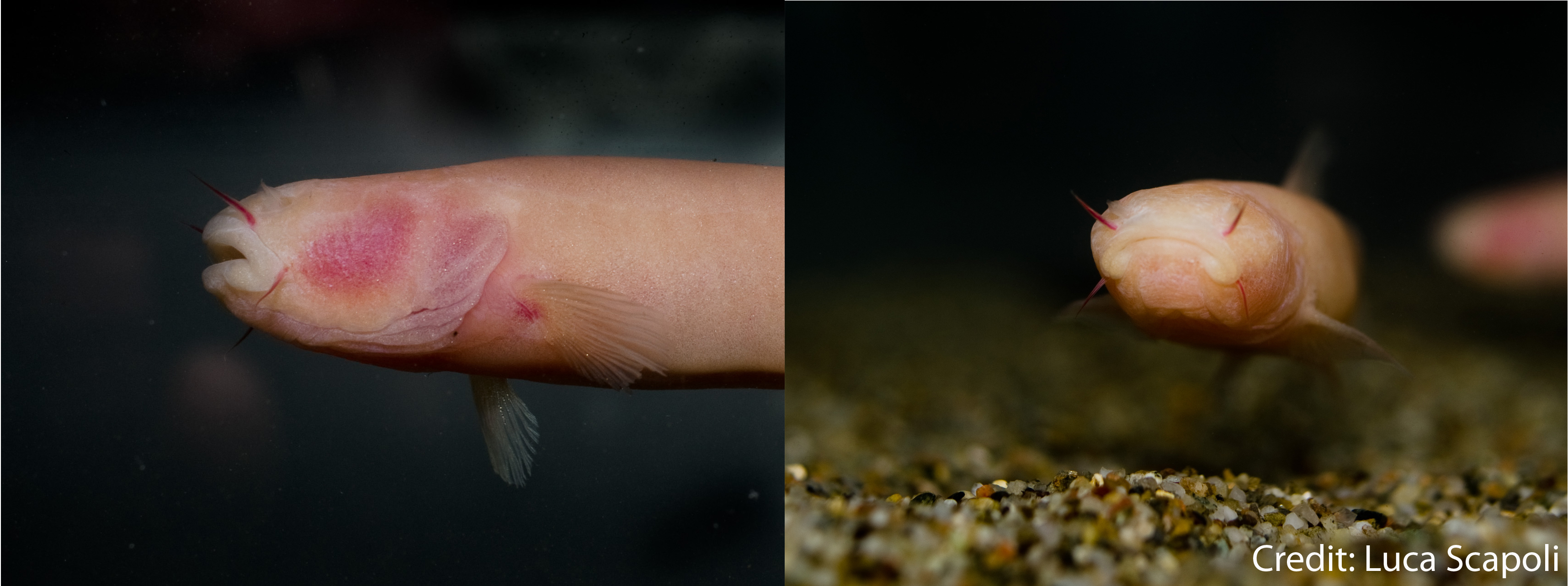
Phreatichthys andruzzii, lateral view (left), frontal view (right)
Adaptations of some fish species to their environment can be most peculiar, especially within cave dwelling kinds. The so called troglomorphisms slowly turn these fish into almost grotesque looking creatures with no eyes, lost pigments and no scales on the one hand, but with enhanced alternative sensory systems on the other hand.
Cavefish of different orders have been found in cave systems from Mexico to China, all revealing different degrees of adaptations to their life underground. One astonishing example for extreme troglomorphism is represented by the species Phreatichthys andruzii. These fish have been living for about 2 million years in complete darkness in a place, where one would not expect swimming fish at all: under the Somalian desert. Due to this total isolation from surface streams, adult P. andruzzii exhibit full regression of eyes, scales and pigments (see pictures). Remarkably, and similar to the well studied Mexican cavefish Astyanax mexicanus, eyes start to develop during embryogenesis following the stereotypic patterning of eye field determination and subsequent evagination, forming the optic cups. Despite the effort, apoptosis is initiated in later stages and ultimately leads to the loss of the organ. Both species have clearly been evolving separately from each other in two different continents, but concerning the eye loss, the outcome is very similar.
Thus in order to investigate, whether the development is similar on the molecular level as well, or if there maybe are differences, we started an intensive in situ hybridization (ISH) study of key transcription factors and components of regular vertebrate eye development (based on zebrafish and medaka). In the cavefish Astyanax it has been shown that midline signalling of shh (sonic hedgehog) is expanded in early developmental stages, when the eye field is forming, which leads to dysregulation of subsequent transcription factor expression and guidance. When we analysed the early developmental stages of P. andruzzii, we could not detect any obvious deviation of transcription factor patterning and corresponding eye formation. Moreover, when we looked at later stages, we even detected an establishing ciliary marginal zone (CMZ), the teleost stem cell niche of the eye. This observation is further supported by anti-PCNA immunohistochemistry, revealing active proliferation in the CMZ. Taken together, until the onset of further differentiation, the early patterning and formation of the Phreatichthys eyes proceed as in surface species like D. rerio.
From this stage onwards, normal morphogenesis of the vertebrate eye is accompanied by highly stereotypic differentiation of retinal progenitor cells (RPCs), which results in the characteristic layering of the eye. Retinal ganglion cells (RGCs) are born first, followed by horizontal, amacrine and bipolar cells and the late-born rod and cone photoreceptors, as well as non-neuronal Müller glia cells. Since no discrete layering of the cavefish retina has been observed at any time point, we studied this developmental phase by addressing cell type specific transcription factor genes with ISH. Our data revealed that only the first born RGCs are established, but not maintained. Detailed analysis of processes of these neurons also revealed no connection to the optic tectum, rendering them functionless. This disruption of the typical differentiation cascade consequently inhibits generation of further cell types and no layering occurs.
Furthermore, we detected massive apoptotic events spreading over the entire neuroretina from this time point onwards, which might very well be triggered by the dysregulated differentiation, in order to protect the eye against aberrant proliferation. Thus, we speculate that a simple differentiation block building on intrinsic control mechanisms elegantly eliminates the Phreatichthys retina.
In comparison to Astyanax, a different strategy is being followed to eradicate the eye during embryogenesis, which specifically blocks differentiation and layering of the neuroretina. In several Astyanax populations this layering proceeds, but apoptosis eventually intervenes, too. By studying these diverse modes of eye degeneration within cavefish species, light can be shed on the different developmental checkpoints and how they are controlled in normally developing vertebrate eyes.
Photographs courtesy of Luca Scapoli.
Stemmer, M., Schuhmacher, L., Foulkes, N., Bertolucci, C., & Wittbrodt, J. (2015). Cavefish eye loss in response to an early block in retinal differentiation progression Development, 142 (4), 743-752 DOI: 10.1242/dev.114629
 (7 votes)
One thought on “How to eradicate an organ”
Leave a Reply
Get involved
Create an account or log in to post your story on the Node.
Sign up for emails
Subscribe to our mailing lists.
Hallo Manuel!
I’m Leo, and we met in Allende’s lab during 2010. I came across your paper as I’m also working on eye development (but not from an evolutionary point of view).
Nice story!
It would be nice to catch up and discuss at some point!
Best Fishes!
Leo