PhD Studentship in Stem Cell Biology, Galway, Ireland
Posted by uri.frank@universityofgalway.ie, on 22 April 2013
Posted by uri.frank@universityofgalway.ie, on 22 April 2013
Posted by Rada-Iglesias, on 22 April 2013
Closing Date: 15 March 2021
The Center for Molecular Medicine Cologne (CMMC) is a multidisciplinary center at the University of Cologne providing a forum that brings together physician scientists with basic researchers from the Faculty of Medicine and the Faculty of Mathematics and Natural Sciences to perform competitive basic, disease-oriented research. The mission of the CMMC is to advance the understanding of the underlying molecular and cellular mechanisms as a prelude to improving prevention, diagnosis and treatment of many common health problems. For further information, please visit http://www.zmmk.uni-koeln.de.
The Center for Molecular Medicine Cologne invites applications of bright and motivated individuals for the Junior Research Group “Developmental Genomics” of Dr. Alvaro Rada-Iglesias.
One full time Post-doctoral fellow
The position is immediately available for 24 months with the possibility of extension. The position is based on the German TV-L salary conditions. The CMMC places strong emphasis on gender equality and seeks to increase the proportional representation of women in this field. Applications from female scientists are especially welcomed; suitably qualified women will be given preferential consideration unless other candidates clearly demonstrate superior qualifications. We also welcome applications from disabled candidates, who will also be given preferential consideration over applicants with comparable qualifications.
What we look for:
The ideal candidate should hold a PhD and demonstrated research experience in developmental biology, preferentially in one or more of the common vertebrate model organisms (mouse, chicken, frog or zebrafish). Experience with routine molecular biology techniques is also required. In addition, candidates with previous experience in stem cell or chromatin biology are also encouraged to apply. We look for highly motivated candidates willing to expand their previous expertise and interests as part of a multidisciplinary and collaborative scientific environment. The successful candidate will receive mentoring and will be given the opportunity to develop his/her own projects in preparation for a future independent career.
Research overview:
During mammalian embryogenesis, cell fates are acquired according to highly defined spatiotemporal patterns, which involves the deployment of cell type specific gene expression patterns as developmental programs unroll. Focusing on the crosstalk between transcription factors and epigenetics, our group aims to better understand the transcriptional regulatory principles that orchestrate the earliest steps of mammalian development. In doing so, our ultimate goal is to characterize the vast non-coding genomic space of the human genome and to investigate how genetic variation within non-coding regulatory elements can contribute to common and congenital human diseases.
In order to gain a global as well as mechanistic understanding of these biological processes, our lab uses a multidisciplinary approach where in vitro (i.e. human and mouse embryonic stem cells) and in vivo developmental models are combined with biochemical and genomic approaches. Please visit http://zmmk-ari.uni-koeln.de/Home.html to learn more about our previous work and interests.
Research environment:
CMMC along with its partner institutes such as the Cologne Cluster of Excellence in Cellular Stress Responses in Aging-associated Diseases, Cologne Center for Genomics and Max Planck Institute for Biology of Ageing research all located in a same campus provides a vibrant scientific community. Therefore, the CMMC`s JRG in “Developmental Genomics” is placed perfectly at an environment where the basic research meets its cutting edge of translational science.
If you have further questions please do not hesitate to contact Dr. Alvaro Rada-Iglesias: aradaigl@uni-koeln.de
Please forward your complete application including CV, a brief statement of scientific interests and three reference letters (in a single pdf document) until 31.05.2013 by e-mail, quoting the reference number e129 to Dr. Alvaro Rada-Iglesias: aradaigl@uni-koeln.de and in CC. to Dr. Debora Grosskopf-Kroiher (Managing Director, Center for Molecular Medicine Cologne) (zmmk-office@uni-koeln.de).
 (No Ratings Yet)
Posted by Rachael Inglis, on 19 April 2013
Last week, I was distracted somewhat by a palaeontology article in Nature: Reisz and colleagues reported their discovery of some fossilised dinosaur embryos. Not exactly relevant to my research, but very cool nonetheless…
The remains that they unearthed in southern China are from the early Jurassic period, almost 200 million years old, and are thought to belong to a Lufengosaurus species. This was a sauropodomorph dinosaur: a group distinguished by their large size, with a very long neck and tail and a small head. The most famous of the sauropodomorphs were probably the Diplodocus species.
These fossils are so unusual, and so informative, because they include embryos at a range of developmental stages. The majority of fossilised dinosaur embryos discovered to date have been single clutches of eggs, all synchronised in their development, which provides only a snapshot of development in that particular species. Finding a whole collection of samples from the same species, but at different stages, gives a rare insight into the dynamics of development in an extinct animal.
The authors focused on the growth of the thigh bone, analysing 24 femurs that ranged in length from 12 to 22 mm. Using sectioning and histological techniques, they showed that these bones were highly vascularised at all stages, so they think that these giant dinosaurs began life with rapid embryonic growth.
They also observed that the dinosaur femurs became thicker on one side as they grew larger, and developed a prominent fourth trochanter (an outgrowth to which the main thigh muscle attaches). In living tetrapods, asymmetrical bone thickening and the growth of skeletal features at muscle attachment sites depends on the muscles being active during embryonic development. This suggests that these ancient embryos also used their muscles to move around inside their eggs, and that these movements were an important part of their development too.
I was really amazed by how much information could be gleaned from these tiny fossilised remains. Geology rocks! In evo-devo, we use observations from extant species to make inferences about their common ancestors, but if palaeontology can provide insights into the embryonic development of extinct animals, it might help us to think about the evolution of some developmental processes from a different, and very interesting perspective.
Reisz, R.R. et al (2013) Embryology of Early Jurassic dinosaur from China with evidence of preserved organic remains, Nature 496: 210-214.
 (4 votes)
Posted by Katherine Brown, on 18 April 2013
A couple of days ago, the University of Chicago Development, regeneration and stem cell journal club posted their first piece on the Node – a write-up of the discussion they’d had in their recent journal club meeting. It’s a great summary and analysis of a recent Development paper and its context within the field, and I’d encourage you to read it!
 This piece marks the first in what we hope will be an irregular series of journal club reports from the Chicago group, and we’d like to find other developmental biology or stem cell journal clubs out there interested in writing for the Node. After all the effort of reading, analysing and discussing a paper for a journal club, we’d like to give you the opportunity of sharing that discussion with a wider community, and not just with your immediate colleagues.
If you’re interested in contributing to this, please get in touch at thenode[at]biologists.com. We’d love to hear from you!
(10 votes)Posted by UChicagoDRSB_JC, on 16 April 2013
![Figure 1 Schematic of the clock model as proposed by Thorogood (1991). (A) The bold arrow represents the timing of the AER-to-AF transition in the developmental process. (B-D) Hypothesized representations of fin/limb development in the clock model (above) with endochondral skeletal patterns of the fin/limb (below,). (B) Fin development in a teleost, demonstrating a short period of time with AER signaling prior to the AER-to-AF transition. (C) Fin development in lobe-finned fishes, showing a longer relative time with AER signaling prior to AF transformation. (D) Limb development in a tetrapod, in which AER signaling persists throughout limb development. Figure modified from Yano et al. [3]; based on Thorogood [2]; with fossil form representations in C-D from Long et al. [4].](https://thenode.biologists.com/wp-content/uploads/2013/04/node1.png)
The initial steps of limb development are basically identical in fish and tetrapods: a combination of signals in the lateral plate mesoderm creates a limb-forming region where a bulge of mesodermal cells form the first visible sign of a limb, the limb bud. In both fish and tetrapods, a ridge of ectodermal tissue, the apical ectodermal ridge (AER), initially forms at the distal apex of the bud. The AER persists throughout limb outgrowth in tetrapods, acting both to maintain a zone of proliferating mesodermal cells at the distal end of the limb and to provide important patterning signals. In fish, however, the AER is later transformed into a different structure, the apical fold (AF), which is morphologically distinct from the AER.
One model for the evolution of limbs, the clock model (fig. 1), suggests that a heterochronic shift in timing of the AER-to-AF transition may have been the main developmental process driving the fin-to-limb transition [2]. The AF is thought to pattern fin ray outgrowth whereas the AER is thought to regulate endoskeletal patterning and promote endochondral outgrowth.
To make a limb from a fin, one needs to do two important things (among others, of course): lose fin rays and gain well-patterned endochondral elements. A trend of less time with an AF structure, while maintaining the AER for a longer amount of time would produce the limb-type morphology. And in fact, the AER-to-AF transformation occurs relatively early in the ray-finned fishes, at a later time point in lobe-finned fishes, and not at all in the tetrapods.
The basic idea is that ray-finned fish have a limited amount of developmental time spent with an AER, thus the endoskeletal region is relatively short. Then the AF drives the majority of limb outgrowth, resulting in elaborated fin rays. In the lobe-finned fishes, the signals from the AER are maintained for a longer amount of time, resulting in elaboration of the endoskeletal pattern. Subsequent AF formation results in some fin rays. In tetrapods, the AER is maintained through the entirety of limb outgrowth and an AF never forms, resulting in an elongated endoskeleton and a limb with no fin rays (see fig. 1). While this model fits the fossil evidence for the fin-to-limb transition well, little evidence from embryological and developmental studies support this hypothesis. A recent paper in Development sought to change this.
Yano et al., [3] described the structure of the AF in detail and investigated the timing of the AER-to-AF transformation in the zebrafish. They found that after AF formation, the majority of pectoral fin outgrowth resulted from growth of the AF region; the endoskeletal region only modestly increases in length. These data support the idea that endoskeletal outgrowth is mainly moderated by the AER; when the AER is no longer around to signal, the endoskeleton grows little. To investigate this further, Yano et al. took advantage of microsurgical techniques in the zebrafish to remove the AF from developing limb buds. Zebrafish have amazing regenerative capabilities and upon removal of the AF the endoskeletal region slightly increased in length and an AER was regenerated within six hours. The surgically manipulated fin then underwent the normal AER-to-AF transformation and outgrowth proceeded normally.
![Figure 2 - Repeated apical fold removal caused excessive elongation of the endoskeletal region compared to control (non-removal) fin. Zebrafish larva (7 days post-fertilization) after AF removal was performed three times on the left side pectoral fin bud; right side is control fin. Black brackets indicate the endoskeletal region. Scale bar: 200µm. From Yano et al., [3].](https://thenode.biologists.com/wp-content/uploads/2013/04/node2.png)
When these fish completed limb development, the endoskeletal region of the removal fin was altered, losing some normal fin bones and in some cases gaining structures distally. These alterations can be interpreted to be more limb-like, but much more work should be done characterizing these phenotypes to make such a claim. Even so, these data do suggest that exposure to AER signaling controls the outgrowth and morphology of the endoskeleton. The AER-to-AF transition might indeed restrict the growth and shape of the endoskeletal region.
This study is a great example of evolutionary developmental biology designed to experimentally test a proposed model. The clock model rests on the assumption that the AER and AF have different functions patterning distinct morphological structures. This paper demonstrated different skeletal morphologies for fins exposed only to endogenous AER signals versus those exposed to the AER for a longer amount of time, lending embryological support to the clock model. We may not be able to literally turn back time to examine the common ancestor, but by experimentally “manipulating the clock” we may be able to get a pretty good idea as to how extant representatives gained their characteristic limb features. Read the paper here.
WORKS CITED
1. Coates, M.I. (1994). The origin of vertebrate limbs. Dev Suppl, 169-180.
2. Thorogood, P. (1991). The development of the teleost fin and implications for our understanding of tetrapod limb evolution. In Developmental patterning of the vertebrate limb. Springer, pp. 347-354.
3. Yano, T., Abe, G., Yokoyama, H., Kawakami, K., and Tamura, K. (2012). Mechanism of pectoral fin outgrowth in zebrafish development. Development 139, 2916-2925.
4. Long, J.A., Young, G.C., Holland, T., Senden, T.J., and Fitzgerald, E.M. (2006). An exceptional Devonian fish from Australia sheds light on tetrapod origins. Nature 444, 199-202.
This post results from the discussion of Yano et al., 2012 by the Development, Regeneration, and Stem Cell Biology Journal Club at the University of Chicago. It was authored by Haley K. Stinnett, Department of Organismal Biology and Anatomy, University of Chicago, Chicago, IL 60637.
(10 votes)Posted by Katherine Brown, on 13 April 2013
The results of the latest image competition, this time featuring five beautiful stem cell images, are in! In what rapidly turned into a two horse race between the corn snake dental organ, and the mouse hippocampus, it was the confocal image of the adult mouse hippocampus that eventually came out on top.
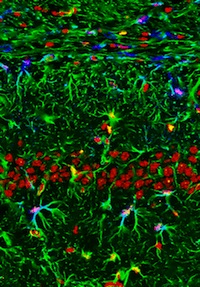
Taken by Lulu Xing of the University of Melbourne and titled “The Garden of Memory”, this striking image will be appearing on a cover of Development in the coming weeks.
Many thanks to all who submitted an image for this competition – especially those who made the Final Five – and to everyone who voted. You’ve definitely proved that stem cells can be just as visually stunning as the tissues, organs and organisms you’re more used to seeing on the cover of the journal!
(2 votes)Posted by Erin M Campbell, on 11 April 2013
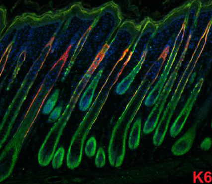 Monday is tax day for most of us on the American side of the pond. That ought to cause massive hair loss for many folks, but we have amazing hair follicles that constantly regenerate hair throughout our adult lives (well, at least for those of us without male pattern baldness). A recent paper in Development helps us understand the hair follicle stem cells involved.
Our hair follicles maintain hair growth throughout our adult lives, and go through different predictable phases of growth (anagen), regression (catagen), and rest (telogen). Each follicle is a little homeostatic system with adult stem cells that have the ability to self-renew and generate all of the cells required in the hair follicle. It is not completely understood how all of these mechanisms function together to achieve long-term homeostasis, but a recent paper helps identify the lineage progression of the different cell types in the follicle, and the relationships between them. Takeda and colleagues identified the marker Hopx in adult hair follicle stem cells. Lineage-tracing experiments showed that Hopx+ cells give rise to all cell types in the fair follicle. Takeda and colleagues also identified a novel population of Hopx+ cells in the lower hair bulb of follicles in anagen. Later in telogen, these newly-identified cells differentiate into the K6+ inner bulge layer in the stem cell niche, where they regulate the quiescence of nearby hair follicle stem cells. In the image above, Hopx (green) is found in several regions of a hair follicle in anagen. K6+cells (red) are the innermost cells surrounding the hair follicle shaft.
For a more general description of this image, see my imaging blog within EuroStemCell, the European stem cell portal.
![]() Takeda, N., Jain, R., LeBoeuf, M., Padmanabhan, A., Wang, Q., Li, L., Lu, M., Millar, S., & Epstein, J. (2013). Hopx expression defines a subset of multipotent hair follicle stem cells and a progenitor population primed to give rise to K6+ niche cells Development, 140 (8), 1655-1664 DOI: 10.1242/dev.093005
Takeda, N., Jain, R., LeBoeuf, M., Padmanabhan, A., Wang, Q., Li, L., Lu, M., Millar, S., & Epstein, J. (2013). Hopx expression defines a subset of multipotent hair follicle stem cells and a progenitor population primed to give rise to K6+ niche cells Development, 140 (8), 1655-1664 DOI: 10.1242/dev.093005
 (4 votes)
Posted by Steff Knappe, on 11 April 2013
In this second part of my BSDB/BSCB spring meeting report, I will attempt to paint a picture of the scientific content of the conference in broad brush strokes. I have grouped talks into overarching topics and will highlight some of my favourites in each category.
It is only on rare occasions that a presentation makes you go “wow” and forces you to reconsider your thinking about the most fundamental element of our bodies, the cell, and how it works. I experienced one of these moments as I was listening to Mike White (University of Manchester, UK) talk about his work on cell to cell heterogeneity. He presented fascinating live imaging data demonstrating that several molecules of the NFkB pathway periodically change location within the cell at distinct rates. Based on this, he suggested that the position of proteins could be just as important in determining the cellular response to a stimulus as the signal itself. This would add a whole new dimension to consider in the study of cell biology, which seems both exciting and daunting at the same time.
I was also particularly impressed by Martin Howard’s (John Innes Centre, UK) research, which combines experimentation and mathematical modelling to investigate epigenetics in plants. By equating histone modifications with a binary system, his group found that genes can only exist in a state of silenced (“0”) or active (“1”) state. What seems like a smooth transition from one state to the other at an organismal level is in reality caused by a change in the fraction of cells that are “0” or “1”. He also showed preliminary experimental data to support this model. Granted, the main reason I was so awed by this talk was most likely my limited understanding of mathematics, much less mathematical modelling. Regardless, I believe it illustrated the value of using mathematics to formulate elaborate hypotheses which can then be tested and validated through laboratory science.
Other recurring topics throughout the meeting included RNAi and epigenetic reprogramming in early development. Barabara Pernaute (National Institute of Medical Research, UK) presented her work on the role of miRNAs in naive embryonic stem cells compared to epiblast cells in post-implantation embryos. Similarly, Juriaan Holzenspies’ (University of Copenhagen, Denmark) presentation focused on histone marks and RNA polymerase phosphorylation states in blastocyst and epiblast. As part of the post-graduate symposium, Siyao Wang (University of Manchester, UK) presented her PhD research on the role of MLL in germ line cells of the C. elegans embryos and Richard Kaschula (University of Sussex, UK) talked about ubx-targeting miRNAs in Drosophila. Finally, Danesh Moazed (Harvard Medical School, USA) focused on his work into the mechanisms of RNAi-mediated silencing, specifically the signals initiating this process and the machinery which retains siRNA at its target site.
Upon browsing the preliminary programme of speakers about two weeks before the start of the meeting, I noticed with great delight that a number of lectures would be focused on regeneration. I especially looked forward to Elly Tanaka’s (Technische Universitaet / Center for Regenerative Therapies Dresden, Germany) talk on limb regeneration in two species of salamander, axolotl and newt. She highlighted some fundamental differences in the mechanisms of limb replacement in these two organisms after amputation. I was especially struck, and perhaps a little confused, by the fact that axolotl and newt both belong to the order of salamanders, yet seem to use different mechanisms to regenerate lost appendages.
Focusing in particular on skin, Ben Simons (University of Cambridge, UK) talked about his inquiry into the mechanisms by which skin stem cells maintain their tissue. His lab’s previous work on this topic challenged the accepted theory of skin maintenance at the time. As he explained the validation and alternate experiments they carried out to confirm that their hypothesis was substantiated by the evidence, I became more and more impressed with his work. I felt that it needed a lot of confidence and courage to challenge long-standing theories and a great amount of perseverance to convince other people of the validity of your research. He also talked about the role these skin stem cells in injury repair and in the progression from papilloma to cancer.
With great relevance to my own work, I was also excited to hear about Jyotsna Dhawan’s (National Centre for Biological Sciences, India) research into the factors which mediate reversible quiescence in muscle progenitor cells. She proposed a completely new way of thinking about the G0 stage in cell cycle, suggesting that quiescent stem cells are poised for proliferation or differentiation rather than simply lying dormant. I find particularly intriguing that this may not only be the case in muscle progenitors, but also in other tissue stem cells which remain quiescent in the absence of activation stimuli. She also implicated chromatin-modulating proteins, which may act as a molecular switch.
Further talks included Cathrin Brisken (Ecole polytechnique federale de Lausanne, Switzerland), who presented hear team’s research on the role of progesterone in adult mammary gland maturation and also addressed the significance of hormone mimetics in breast cancer. Moreover, both Anna Bigas (Institut Hospital del Mar d’Investigacions Mediques, Spain) and Sarah Bray (University of Cambridge, UK) talked about the role of Notch signalling in blood cell precursor specification in mouse and Drosophila, respectively.
Another highlight for me was Olivier Pourquie’s plenary lecture about the generation and differentiation of the paraxial mesoderm. I had already been familiar with several aspects of his work, but I was surprised to hear a wealth of new findings which looked at the paraxial mesoderm from as many angles as possible. He addressed the oscillatory expression of several genes as somitogenesis takes place as well as remarkable metabolic signatures which differ between anterior and posterior paraxial mesoderm. Further, he spoke about his quest to differentiate induced pluripotent stem cells into paraxial mesoderm in vitro.
The use of unconventional organisms to get to the bottom of evolutionary questions has always fascinated and inspired me. I was thus very interested when listening to Patrick Lemaire (Centre de Recherche de Biochimie MacromoléculaireMontpellier, France) and Robb Krumlauf (Stowers Institute for Medical Research, USA). Patrick presented his work using different genera and species of ascidians, with which he investigates how morphological characteristics can stay stable across time despite major genomic changes. Robb talked about his lab’s research into the evolutionary origins of the Hox clusters using the lamprey. In both their presentations, the speakers highlighted the problems of working with such unconventional organisms and I was particularly intrigued by the fact that many of the conventional techniques in molecular biology can be adapted for these exotic organisms.
Additional talks looked at the migration of precursor cells and their differentiation. Stephen Fleenor (University of Oxford, UK) presented his PhD project on the role of G-coupled proteins in craniofacial ganglion precursor proliferation, migration and differentiation. Moreover, James McColl talked about the role of several signalling molecules in the migration of heart progenitor cells. Lastly, Ferenc Mueller (University of Birmingham, UK) addressed the complexity of promoters and enhancers, which his team investigates through the maternal-zygotic transition of transcription in the early embryo.
In this last section, I will focus on several talks which I have not managed to assign any particular topic to. One of the last talks of the meeting was also one I considered among the most compelling. Gero Miesenbock (University of Oxford, UK) presented his work on the role of circadian and homeostatic components in sleep control in a most captivating manner. Using a Drosophila model with sleep deficits, he described his team’s experiments step by step to expose the cause of insomnia. I particularly appreciated the use of a wide range of techniques, including molecular biology, optogenetics and electrophysiology.
As part of the post-graduate symposium, Daniel Hayward (University of Exeter, UK) focused on his research on centrosome-mediated and chromatin-mediated microtubule nucleation. With a focus on moesin and actin, Nelio Rodriguez (University College London UK) presented his work on cell shape changes throughout the cell cycle. Finally, Andrei Luchici (University College London, UK) explored the role of forces in contact inhibition.
In my opinion, the BSDB/BSCB spring meeting 2013 was a great success, with a wealth of fascinating talks and posters along with great social events which stimulated conversation and networking. I feel like I have seen and heard a lot of interesting, novel and exciting science and have had the privilege of listening to many inspiring people. This being my first “big” conference experience, I couldn’t have asked for a better meeting to go to and I am already looking forward to next year’s BSDB/BSCD spring meeting.
(1 votes)Posted by Patricia Gongal, on 11 April 2013
Retinoic acid is one of the most important signaling molecules during development, and that the embryo gets the right levels of this small molecule is critical. Too much or too little, and the basic patterning of the nervous system and many other organs goes terribly wrong. Indeed, you have to think for a bit to find an organ whose development isn’t affected by retinoic acid levels.
It’s been thought for a long time that retinoic acid acts through a morphogen gradient. However, because the molecule is small, extremely labile, and not protein-based, it’s been difficult to actually measure its levels in the early embryo. The best visualization of a gradient has come from transgenic zebrafish lines. Perz-Edwards et al (2001) established a line in which the expression of YFP is controlled by a trio of retinoic acid response elements (RARE; a regulatory motif that is activated by a complex of retinoic acid and its receptors). This line was nicely quantified by White et al (2007), and in the hindbrain, where retinoic acid is important for anterior-posterior patterning, you can see a gradient of fluorescent signal. However, the signal is only visible from about 22-24 hours of development, well after basic patterning is already established. Even stringing together 12 RAREs to boost the level of fluorescence isn’t sufficient to make the signal visible much earlier (Waxman and Yelon, 2011).
A new paper in Nature by Shimozono and colleagues uses a clever approach to more directly measure retinoic acid levels during zebrafish gastrulation, when the basic patterning of the nervous system and somites is set up. They took the ligand-binding domain of a retinoic acid receptor and fused it with both CFP and YFP. When the ligand-binding domain binds retinoic acid, its conformation changes and there is a FRET event. Measuring FRET therefore gives you a read-out of retinoic acid levels. They show clearly that a two-tailed gradient is established during gastrulation, but the anterior and posterior sides of the gradient (ie. in the hindbrain versus the somites) differ in their dynamics, shape, and regulation.
A useful aspect of their system is that while previous transgenic lines measure the signaling capacity of retinoic acid, as they depend on transcriptional activity, this method measures the molecule’s absolute levels. Comparing quantities and activity could be useful for the study of how retinoic acid is processed, sequestered, and regulated. Another advantage of their system is that they created versions of the sensor protein that have different affinities for retinoic acid- allowing the measurement of both higher and lower levels of the molecule, depending on what part of the embryo you’re interested in.
Overall, this new tool will be extremely valuable to the research community, and will allow labs to study this key signaling molecule more precisely and directly.
——–
References:
Perz-Edwards, A., Hardison, N., Linney, E. (2001) Retinoic acid-mediated gene expression in transgenic reporter zebrafish. Developmental Biology, 229(1):89–101.
Shimozono, S, Iimura, T., Kitaguchi, T, Higashijima, S., Miyawaki, A. Visualization of an endogenous retinoic acid gradient across embryonic development. Nature (2013), published online April 7, 2013.
Waxman, J. and Yelon, D. (2011) Zebrafish retinoic acid receptors function as context-dependent transcriptional activators Developmental Biology, 352(1):128–140.
White, R., Nie, Q., Lander, A., Schilling, T. (2007) Complex regulation of cyp26a1 creates a robust retinoic acid gradient in the zebrafish embryo. PLoS Biology, 5(11): e304.
(1 votes)Posted by Prof. Dr. Brand Michael, on 10 April 2013
Closing Date: 15 March 2021
Technische Universität Dresden (TUD) is among the top universities in Germany and Europe: strong in research, offering first-rate programs with an overwhelming diversity, with close ties to culture, industry and society. The TUD is one of the eleven German universities that were identified as an “elite university” in June 2012. As a modern full-status university with 14 faculties it offers a wide academic range making it one of a very few in Germany. TUD is the largest technical university in Germany.
The DFG Research Center for Regenerative Therapies Dresden, CRTD (www.crt-dresden.de) and Cluster of Excellence forms a network of more than 90 research groups working in the areas of Haematology, Diabetes, Neurodegenerative diseases as well as bone regeneration. The CRTD offers a position for an outstanding applicant with international scientific qualification as a
Junior Research Group Leader for Retina Research (up to E 15 TV-L)
This position is available for up to 5 years. The period of employment is governed by the Fixed Term Research Contracts Act (Wissenschaftszeitvertragsgesetz – WissZeitVG).
The aim of the junior research group leader is to pursue basic research into the cellular and molecular mechanisms of retinal degeneration and regeneration using vertebrate animal models like zebrafish or mouse and/or tissue culture. You will join a community of researchers working towards a better understanding of cellular and molecular mechanisms of retinal disease, and to develop cell based methods for restoring retinal function. The successful applicant will be invited to apply, together with the CRTD, for junior group leader funding, e.g. from the Deutsche Forschungsgemeinschaft (Emmy Noether program) or the European Union (ERC starting investigator program). If successful, funding for the Junior Research Group would typically run for 5 years. The period of employment is governed by the Fixed Term Research Contracts Act (Wissenschaftszeitvertragsgesetz – WissZeitVG). The new research group will be housed in a new state-of-the-art building equipped with excellent core facilities located on the Life Science Campus, next to the Biotechnology Centre (www.biotec.tu-dresden.de), the Max-Planck-Institute for Molecular Cell Biology and Genetics (www.mpi-cbg.de), as well as the “Dresden International Graduate School of Biomedicine and Bioengineering” (www.digs-bb.de) and the Dresden University Hospital Carl Gustav Carus (www.uniklinikum-dresden.de).
TU Dresden seeks to employ more women in leadership positions. Hence we should particularly like to encourage qualified women to apply. Applications from disabled candidates or those with additional support needs are welcome. TU Dresden is a family-friendly university and offers a dual career service.
Applicants must have a university degree in natural or medical sciences, a doctoral degree and an outstanding international scientific track record.
Please send your application forms, including a CV, a publication list, description of past and future research activities, acquired third-party funding, and two letters of recommendation by 16.05.2013 (stamped arrival date applies) preferred via e-mail (PDF) to sabine.matthiae(at)tu-dresden.de (Please note: We are currently not able to receive electronically signed and encrypted data) or to TU Dresden, DFG-Center für Regenerative Therapien Dresden, Cluster of Excellence, Director CRTD, Prof. Dr. Michael Brand, Fetscherstrasse 105, 01307 Dresden, Germany.
(No Ratings Yet)