Present and future of quantitative developmental biology in Les Treilles
Posted by Jean-Léon Maître, on 1 September 2022
Update 10/1/23: more on this meeting in The Lonely Pipette “Why do we go to conferences?“
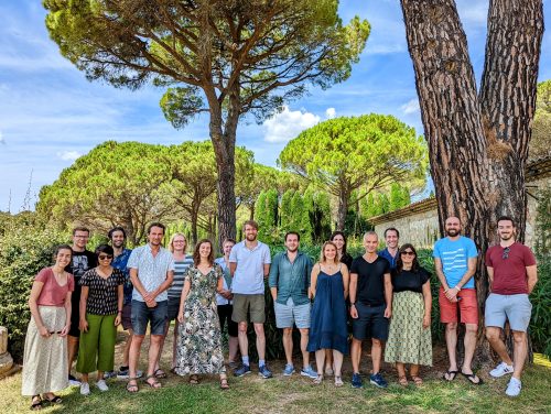
What an exciting time to be studying embryonic development! Emerging experimental systems, methods and analyses allow addressing a whole new set of fascinating questions, as well as revisit older ones. In particular, the field of patterning and morphogenesis, which investigates how embryos establish their body plan with the correct cell types and shape, currently experiences rapid technological and conceptual transformations. For example, microfluidics and microfabrication now permit manipulating the culture conditions of developing embryos with extended flexibility; organoid systems allow the reconstitution of tissues and structures entirely from stem cells; microscopy methods such as light sheet microscopy, live sensor of signalling pathways, optogenetic tools allow dissecting developmental mechanisms over unprecedented temporal and spatial scales. Together, these new possibilities will be key to structure the field of patterning and morphogenesis over the next decades. This research will be carried out, in part, by a new generation of emerging leaders. From July 25th to 29th of 2022, scientists who recently established their labs in Europe gathered in Les Treilles to discuss the emerging quantitative approaches to study embryonic development. From nematodes, flies, sea urchin, frogs, zebrafish, cow and mouse to human, a broad variety of developmental processes were covered from fertilization to organogenesis.
Observing embryonic development is key to understand it.
Technological developments in light sheet microscopy have allowed imaging of large-scale morphogenetic events while maintaining cellular resolution. Kate McDole, who locked herself in a room during the pandemic to build a new light sheet microscope, presented never-seen-before cell movements during mouse gastrulation, shedding light on the origin of cells constituting the different germ layers. Rita Mateus also used light sheet microscopy to image the patterns involved in the formation of the zebrafish pectoral fins and better characterise how those patterns scale during growth and regeneration. As the fins emerge, other organs, like the heart, also form complex structures such as the muscular trabecular ridges that enable proper heart contraction. Using quantitative live imaging and clonal analysis, Rashmi Priya explained how complex tissue architecture is built during trabecular morphogenesis. Zebrafish was also at the heart of Diana Pinheiro’s work, as she reported on a mechanism explaining the coordination of mesoderm progenitors migration and specification by Nodal signalling during gastrulation. When missing, Nodal signaling causes characteristic phenotypes that expert zebrafish embryologists can easily recognize. Patrick Müller leveraged deep learning analyses to develop algorithms that outperform embryologists at identifying the phenotypes of zebrafish gastrulation mutants.
Together, these studies show how imaging large-scale shape changes and quantitatively tracking them can help understand the global patterning and morphogenesis of embryos.
Down to the sub-cellular scale, high resolution microscopy reveals the mechanisms employed by cells to shape embryos. In particular, Anne-Cécile Reymann looked into the influence and distribution of maternally deposited regulators of the actin cytoskeleton during the early cleavages of c elegans embryos. In contrast, Tommaso Cavazza reported on how microtubules help coordinate chromosome segregation in cow zygotes before the nuclear envelope of the parental pronuclei breaks down. Later during mammalian development, Jean-Léon Maître described how protrusions help cells control the formation and positioning of a fluid-filled lumen that helps setting the first axis of symmetry of the embryo. A different set of protrusions was introduced by Jakub Sedzinski to explain how cells can insert themselves within epithelia and increase the complexity of tissues. Finally, peering into fly embryos, Timothy Saunders quantified the scaling of Bcd gradients by combining fluorescence correlation spectroscopy and mutants affecting egg size.
Altogether, bridging the sub-cellular scale to embryonic changes is key to understand the molecular and physical regulation of embryonic development.
Probing and challenging embryos in novel ways
Exploring how cells change their chemical composition and physical properties during development will help understand their behaviour. Mariaceleste Aragona quantitatively analysed the effect of tissue stretching in vivo using clonal analysis and single cell sequencing to understand how cells change composition and fate after a long-term physical perturbation. To understand how cells sense mechanical perturbations, Verena Ruprecht investigated the short-term response of cells to compression by combining high resolution microscopy, microfabrication and optical tweezers. Tweezers were also on the menu for Nicolas Minc who developed magnetic tweezers strong and precise enough to move the entire mitotic spindle within cells to induce division asymmetries and explore the mechanics of the cytoplasm.
Understanding how embryos develop requires the need to finely perturb this process in controlled ways. Romain Levayer leveraged the powerful genetics of Drosophila and optogenetics to induce the death of a precise number of cells and determine the range of robustness of epithelia to cell delamination. Another way to control in space and time the molecular regulation of embryonic development is microfluidics: using precise oscillations of chemical compound, Ina Sonnen could explore the coupling of biological clocks during somitogenesis. Finally, Elias Barriga measured endogenous electric fields and applied electric fields of similar magnitude to steer the migration of neural crest cells in Xenopus embryos.
Together, discussing the current research and future projects of young European scientists revealed the promises and challenges of applying quantitative methods to developmental biology. On the one hand, quantitative methods allow dissecting developmental mechanisms with unprecedented precision. On the other hand, the integration of multiple spatial and temporal scales, and different source of biological information (such as chemical, mechanical and electrical signals) remain extremely challenging. Moreover, tackling these problems when most biologists lack the appropriate training in mathematics and coding can be challenging. A consensus appeared regarding the need to include more biology-oriented maths in university programmes, as this will be key to anchor biology to the realm of modern science. Importantly, Renaud Pourpre, who carried out several initiatives to bring microscopy data to the public, reminded us how microscopy images constitute a straightforward avenue to communicate around science to a lay audience (see CellWorlds documentary). By the same token, there is no doubt that the beauty of developing embryos will continue motivating biologists and other scientists to expand their toolbox to decipher the mechanisms of development.
 (6 votes)
Get involved
Create an account or log in to post your story on the Node.
Sign up for emails
Subscribe to our mailing lists.
Read the latest Development issue
Most-read posts in May
- From comparison to mechanism: decoding heart regeneration
- A day in the life of a Reviews Editor (at Development)
- What does a Reviews Editor do?
- New evo-devo textbook ‘Eco-Evo-Devo: The Environmental Regulation of Development, Evolution, and Health’
- preLighters’ choice – A curated selection of recent preprints