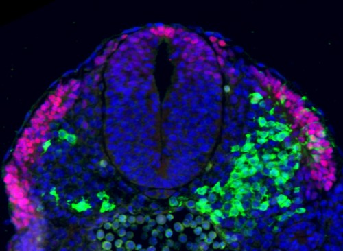
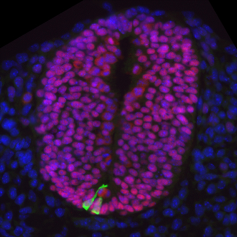
Researchers grow ‘seed’ of spinal cord tissue in a dish
Posted by MRC Press Office, on 3 September 2014

Medical Research Council (MRC) scientists have for the first time managed to turn stem cells into the specialised cells that go on to form spinal cord, muscle and bone tissue in the growing embryo. Their discovery could lead to a new way of studying degenerative conditions such as spinal muscular atrophy, which affects the nerve cells in the spinal column, and may pave the way for future treatments for this and other neuromuscular conditions.
During normal embryo development the spinal cord, muscle and skeleton all form from a group of cells called NMPs (neuro-mesodermal progenitors). This process is driven by a series of carefully timed chemical signals, which instruct NMPs to turn into the different cell types in the growing embryo.
By carefully studying and then mimicking this process in a petri dish, researchers at the MRC National Institute for Medical Research and the MRC Centre for Regenerative Medicine, at the University of Edinburgh, were able to coax mouse and human embryonic stem cells into becoming NMPs and then spinal cord cells.
Dr James Briscoe, who co-led the research from the MRC National Institute for Medical Research, said: “There have been some great advances in the field of stem cell research in recent years, with scientists being able to grow liver, heart and even some brain tissue in the lab. The spinal cord, however, has remained elusive because the NMP cells have largely been overlooked – even though they were first discovered more than 100 years ago.
“The real breakthrough for us was realising that we had to coax the stem cells into this intermediate ‘stepping stone’ cell type before turning them into spinal cord and muscle cells. We can’t yet produce the tissues themselves, but this a really big step. It’s like being able to make the bricks and raw materials but not yet build the house.”
Researchers have previously been able to grow some types of nerve, muscle and bone cells in the lab by converting them directly from stem cells. But this is the first time the intermediate NMP cell type, which acts like a ‘stepping stone’, has been created from stem cells. The advantage provided by guiding cells through the routes used in normal development is that the resulting cells may bear closer resemblance to those that occur naturally in the body. This may help any future therapy utilising these cells by providing positional cues to allow them to better integrate with the surrounding tissue.

In the near-term being able to grow NMP cells in the lab will allow researchers to learn more about normal human development in a part of the embryo that is otherwise difficult to study. In future the method could also be refined to allow scientists to grow tissue from patients with diseases that affect the spinal cord, muscles, or the motor neurones that connect muscles to the brain and spinal cord. This would provide a powerful new tool to study in a dish how these diseases progress and take hold in the body.
Prof Val Wilson, the co-leader of the research from the MRC Centre for Regenerative Medicine, at the University of Edinburgh, added: “NMPs are important because they’re the source of the spinal cord and most of the bones and muscles in our body. But they have been like Cinderella cells. Although recognised for more than a century in the embryo, they’ve tended to be ignored by scientists trying to make these cell types in a dish. We hope this work will bring them out of obscurity and highlight their importance.”
Dr Rob Buckle, Head of Regenerative Medicine at the MRC, said: “This study is a fantastic example of how combining different branches of science can lead to new discoveries. While there have been many important advances in reprogramming stem cells, it’s important that we explore all the possible routes to generating the specific cell types best suited to clinical development. Incorporating detailed knowledge of early developmental processes is likely to play an important role in providing the fine tuning required to achieve this.”
This article was first published on the 26th of August 2014 in the news section of the MRC website.
The paper, entitled ‘In vitro generation of neuromesodermal progenitors reveals distinct roles for Wnt signalling in the specification of spinal cord and paraxial mesoderm identity’, by Gouti et al, is published in the journal PLOS Biology. Further information available from the MRC press office: press.office@headoffice.mrc.ac.uk


 (1 votes)
Nice findings.
A general comments about this way of reporting:
I am not a fan of such ‘promoting’ articles on the Node, writen by press offices. If the Node wants to be a community, articles about new findings or about new publications should be written by the researchers themselves and not by more distant press officers. I mean, the ‘researchers’ in the titles (Wilson, Briscoe) we all know, right?
We are the incrowd people; we are not the general society that wants to see the possible applications for fundamental science. No press stories anymore, please
Thank you for your comment Charles. I understand your concerns regarding the presence of press releases on the Node. As you say, the Node is a community blog, so ideally we would want most of the posts about research to be written by the authors of the work. However, spontaneous posts about research are not frequent. We often contact researchers whose papers we think may be of interest to the community, but often the authors don’t have the time to post on the Node. You are right that press releases are written by non-scientists, but they are prepared in close collaboration with the researchers behind the work. As such, we think that the occasional press release is a good way to highlight recent research that would otherwise not feature on the Node.