

Results of the YEN 2026 Image Competition
Posted by Ceren Canse, on 6 July 2026
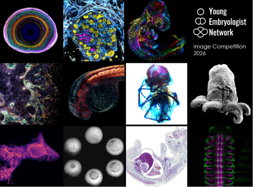
We are delighted to announce the winners of the Young Embryologist Network Image Competition held as part of the YEN 2026 Conference, which took place on the 15th June at the Francis Crick Institute. A huge thank you to all 30 participants who submitted their beautiful images.
The winning image, Lamination in the Zebrafish Retina by Jack Nicholls, will be featured in the YEN 2027 promotional materials. Congratulations to all of our shortlisted entrants!
Winning image: Jack Nicholls – Lamination in the Zebrafish Retina
Runner-up: Ipek Gassaloglu Guler – The First Contact: Interaction Between an Embryo Model and Maternal Tissue
Runner-up: David Arancibia-Altamirano – Aristotle’s Egg Through a New Lens
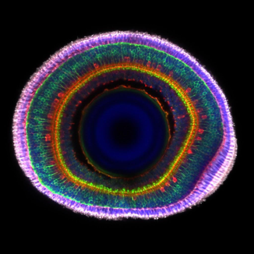
1. Lamination In The Zebrafish Retina
Jack Nicholls, City St George’s, University of London
Immunohistochemistry of an 8-day post-fertilisation larval zebrafish retina stained with anti-ChAT in red (star burst amacrine cells), anti-PKCα in green (ON bipolar cells) and grey (photoreceptors), with a DAPI counterstain. The anti-PKCα signal was manually separated into the bipolar and photoreceptor cell regions and coloured in green and grey to highlight the boundary between the two layers. Imaged on a LSM800 confocal microscope.
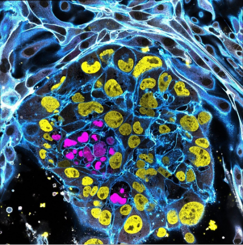
2. The First Contact: Interaction Between an Embryo Model and Maternal Tissue
Ipek Gassaloglu Guler, Yale University
This image portrays a stem cell-derived human embryo model (blastoid) cultured on endometrial epithelial cells. This experiment aimed to visualize the initial contact between the blastoid and endometrial epithelium, to capture one of the earliest stages of embryo-maternal interaction.
Within the blastoid, the epiblast is shown in magenta (SOX2) and the trophectoderm in yellow (GATA3). To highlight the interaction between the blastoid and maternal cells, integrin B1 (ITGB1) is shown in white. F-ACTIN, which outlines cellular architecture is shown in blue. The image was acquired using a Leica STELLARIS 5 confocal microscope in tile-scan mode with a 63x objective.
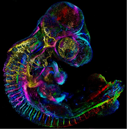
3. Aristotle’s Egg Through a New Lens
David Arancibia-Altamirano, University College London, Universidad Mayor
Since Aristotle’s studies in embryology, the chicken embryo has captivated the imagination of scientists and spurred exploration of epigenesis and preformation. Echoing classic experiments with India ink, fluorescent ink injection combined with tissue clearing and light-sheet microscopy now allows us to study the beauty and complexity of vascular networks in 3D and with high resolution. Sample displayed by depth colour-coding across 3.1mm; prepared by me and my friend, Jesus Juarez.
4. Dancing Ghosts
Ornella Clara, Aix-Marseille University
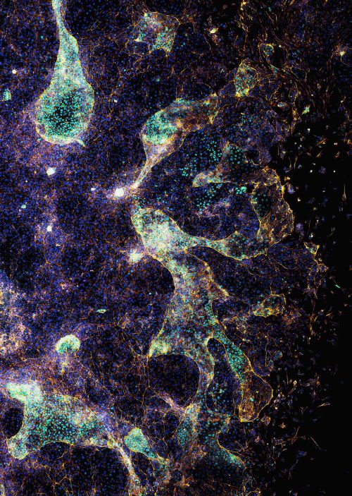
At first glance, the image resembles a ghostly choreography suspended in a cosmic landscape. In reality, it captures the collective cell migration during the early formation of a gut-like tissue structure. These gastruloids spread on a laminin-coated substrate, self-organizing into patterns reminiscent of embryonic development. The transcription factor CDX2 (light blue) marks intestinal identity, while phalloidin staining (yellow-orange) highlights the actin cytoskeleton that drives cell movement, and nuclei are shown in blue-violet.
This image was acquired using confocal microscopy. It illustrates how coordinated cellular behaviour gives rise to complex tissue architecture.
Experimental work by Dalia El Arawi; staining and confocal imaging by Ornella Clara.
5. Growing Into Form
Matyas Bubna-Litic, University College London
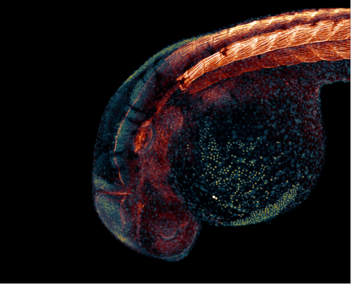
A one-day old zebrafish embryo already has recognisable features such as the early forms of the eye and ear as well as the segmented backbone, which will go on to form vertebrae and muscle. Cell nuclei and filamentous actin are visualised in this stained fixed sample. Imaged using a Zeiss LSM980 with an Airyscan2 detector in multiplex mode.
6. Down to the Bone
Alexandra Lion, Brigham and Women’s Hospital and Harvard Medical School
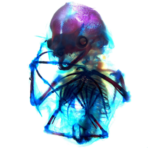
A beautiful network of bones and cartilage, which in life would provide support, protection and facilitate movement of the body. The image shows a short-tailed fruit bat (Carollia perspicillata) embryo at embryonic stage 22 which has been cleared and stained with alcian blue for cartilage and alizarin red for bone. This staining allows for visualization of the still-ossifying bones of the bat autopod, and most strikingly of the skull, which appears to smile with clearly visible canine teeth. Imaged during the 2024 Embryology Course at the Marine Biological Laboratory using transmitted light on a Zeiss Axio Zoom.V16 microscope, then further processed using FIJI.
7. Octopus Embryo
Ailen Cervino Len, Baylor College of Medicine
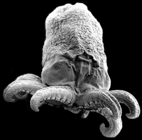
Octopus embryo taken with Scanning Electron Microscopy (SEM).
8. Wiring Diagram
Ryan Cheng, Centre for Developmental Neurobiology, King’s College London
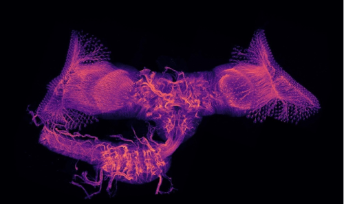
Maximum intensity projection image of the central nervous system of Drosophila melanogaster during metamorphosis. This sample was collected at around 24 hours after puparium formation and stained with an anti-Neuroglian antibody to visualize the neurite tracks. Multiple z-stacks were imaged on a Zeiss LSM800 and reconstructed in FIJI.
9. The Phases of Gastrulation
Hoang Anh Le, University College London
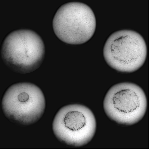
A Xenopus laevis embryo was imaged from the ventral side showing the different phases of gastrulation, from the formation of the blastopore lip to its closure and the beginning of neurulation. The embryo was imaged with an upright brightfield microscope.
10. beCOWming
Noemi Monferini, Developmental Biology Institute of Marseille
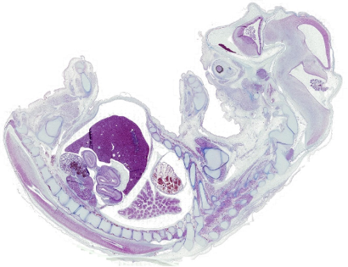
A bovine foetus in histological section stained with Azan trichrome, revealing the delicate architecture of foetal tissues.
We would also like to highlight the winner of the public vote, which has received 128 out of the 773 votes cast:
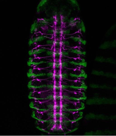
Weaving a Nervous System
Lamiya Dohadwala, Tata Institute of Fundamental Research
A confocal view of the developing central nervous system in a Drosophila embryo. Green marks engrailed-expressing segmental compartments, while magenta highlights Fasciclin II-positive nerve fibres, tracing the intricate network of axon pathways that form the embryonic nerve cord.
Get involved
Create an account or log in to post your story on the Node.
Sign up for emails
Subscribe to our mailing lists.