Vote for your favourite image for the Node postcards
Posted by the Node, on 11 May 2023
One of the crowd-favourite giveaways here at the Node is our collection of postcards. With our supplies dwindling, we are planning to reprint some of the postcards, and take this opportunity to add some more #devbio favourites to our collection.
Thank you to all the researchers who have submitted their images to the Node postcard competition back in April. We have now narrowed down to the final 8, and would like to see which ones the community like to feature on our postcards. The top 4 will be printed on our postcards, and the winner will be also be featured on the cover of a ‘Development’ issue in 2023.
Thank you and good luck to the following researchers for their contributions:
Daniel Rios, Elio Escamilla, Elisabeth Kugler, Jessica Marin, Margot Smit, Nick Gatford, Özge Özgüç, Valerie Tornini
And a big thank you to everyone who submitted their images to the competition. There were many good quality submissions that it was very difficult to narrow down the selection!
Please vote for your favourite image at the bottom of the page. The voting will close on Sunday 4 June 11:59pm.
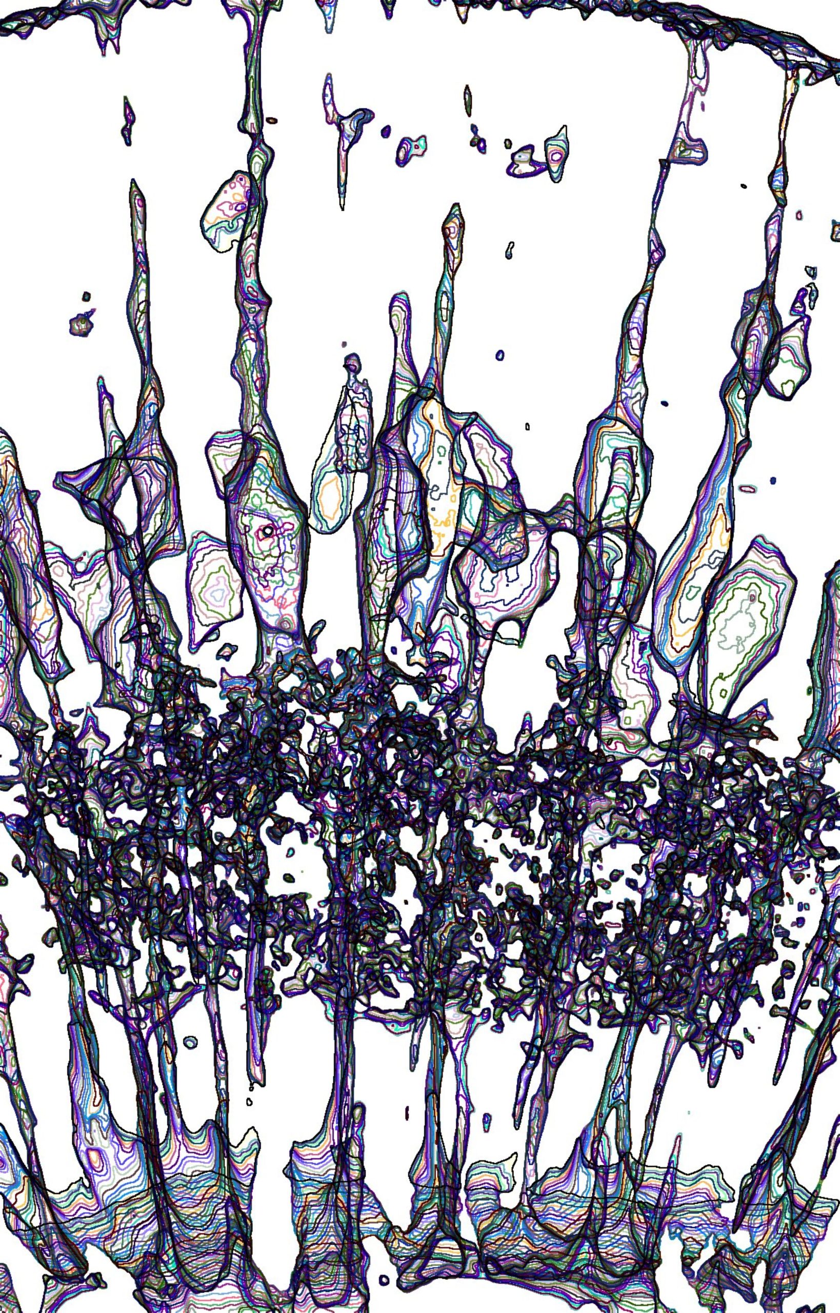
“Geographical Maps“
Depicted is a digital section of the developing eye in a zebrafish. Specifically, these are support cells of the eye called glia cells, which have undergone 3D data processing to extract the surface of these cells. Different colours represent different depths in the studied eye. Technique: The image was acquired with the Zeiss LSM 900 AiryScan microscope, using a 40x water-immersion LD C-Apochromat (NA 1.1) objective. Processing was conducted using Fiji. Following image rotation in 3D, data were segmented and the surface extracted. Using depth-coding and application of different colour-palettes achieved the different colorations seen.
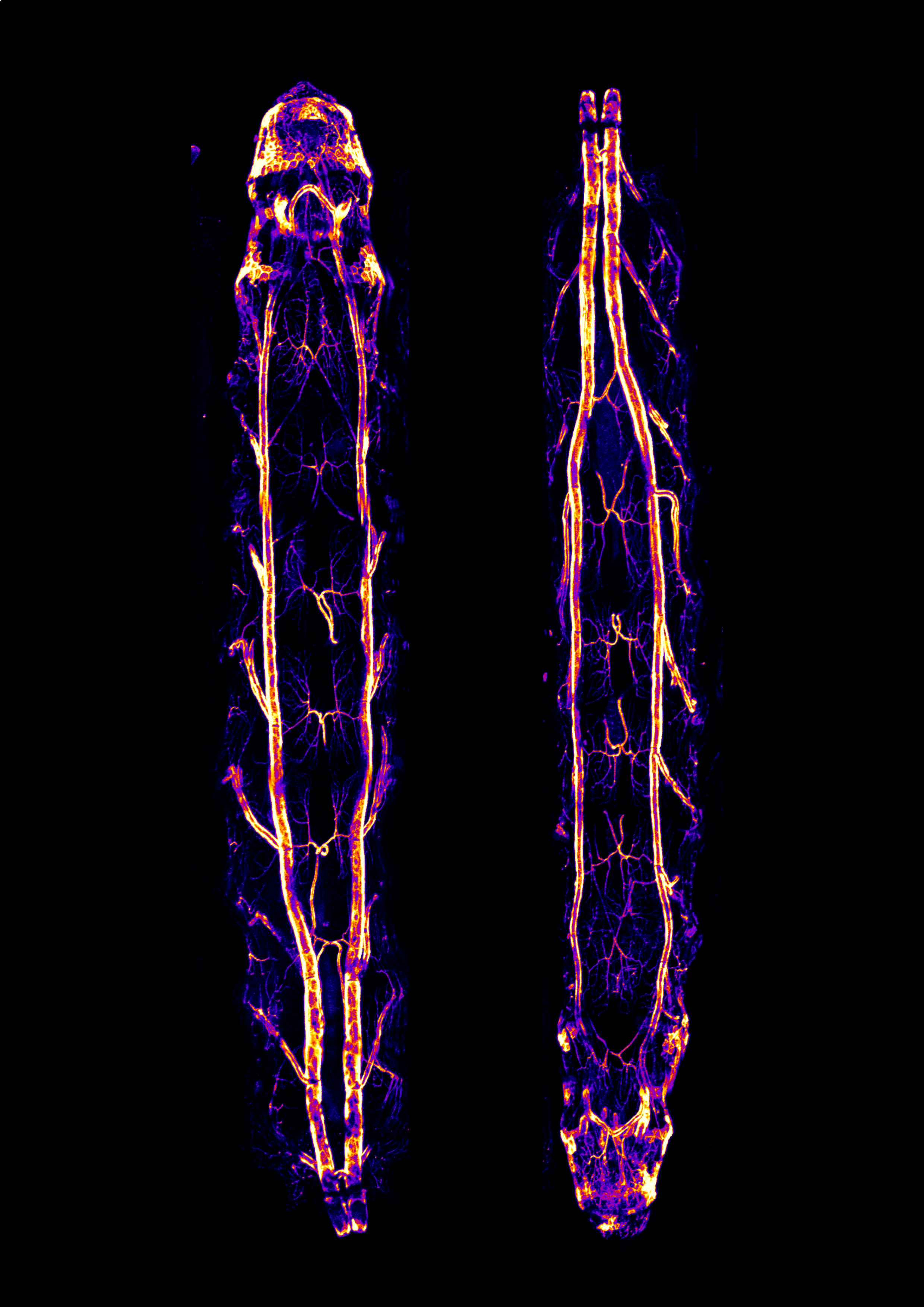
Drosophila larvae
This shows heat-fixed Drosophila larvae expressing an infrared fluorescent protein (IFP) in the tracheal system using the Gal4/UAS system. Images were acquired in a confocal microscope (Nikon A1R+) with a 10x objective and using the mosaic modality. Stitching was done using the microscope’s software (Nikon NIS-Elements). The images were Z-projected and pseudocolored in Fiji and further processed using Inkscape.
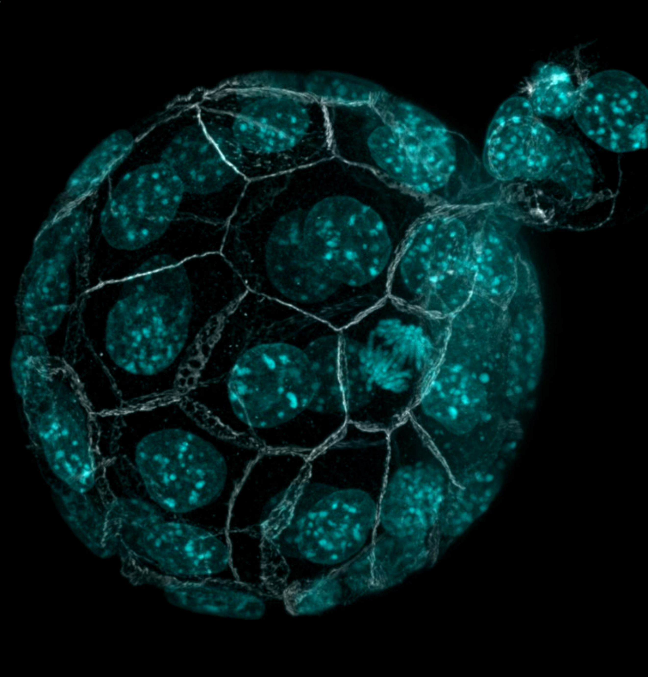
Hatching embryo
Immunostaining of a hatching mouse blastocyst imaged with confocal microscopy
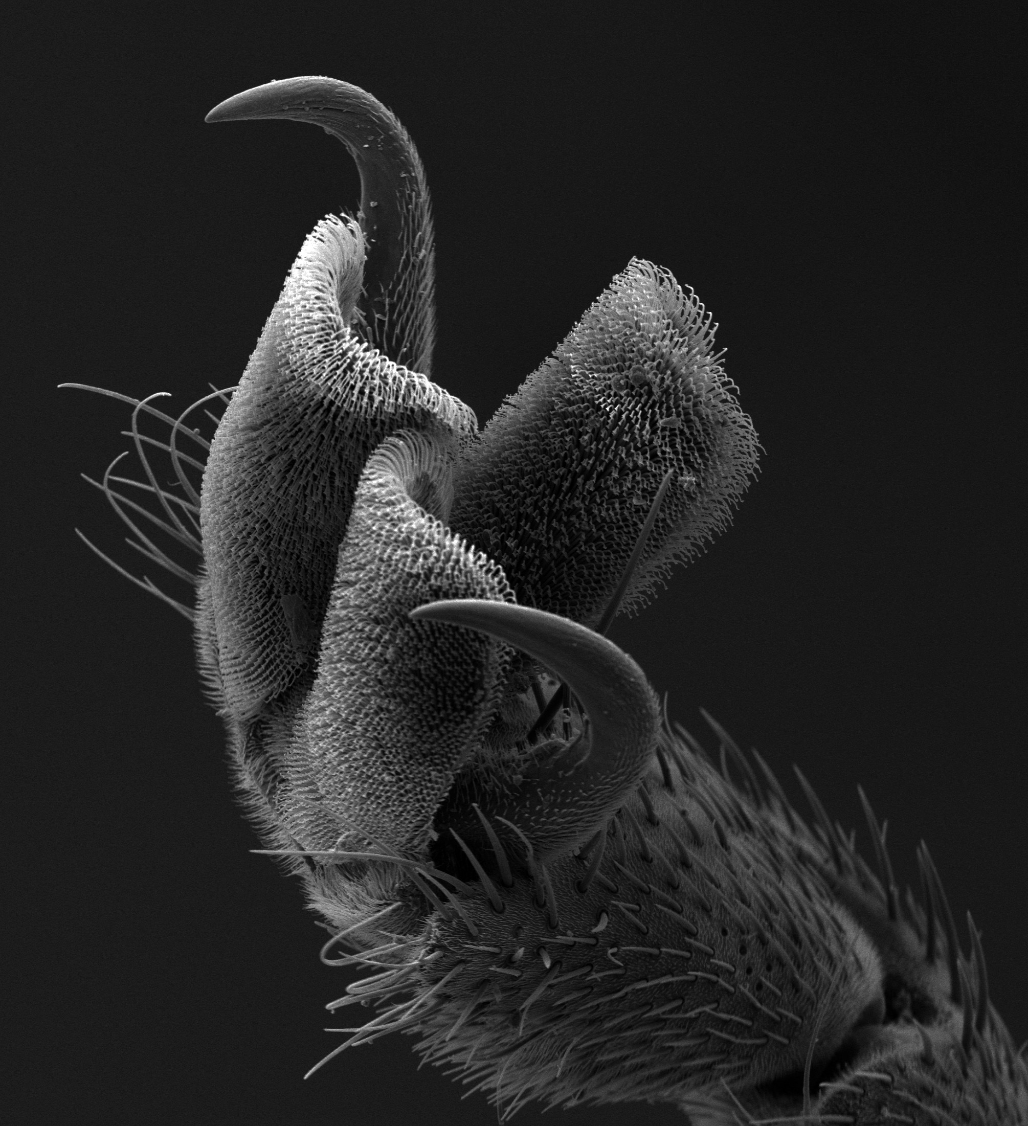
Tarsal Claw
This scanning electron microscope (SEM) image of the tarsal claw of the horsefly (Tabanus sulcifrons) juxtaposes the complexity and simplicity of “nature’s Velcro.” The menacing sturdiness of the tarsal claws contrasts with the delicate nature of the tarsal pad, with fine, hooked hairs that allow the fly to hold on to animal fur.
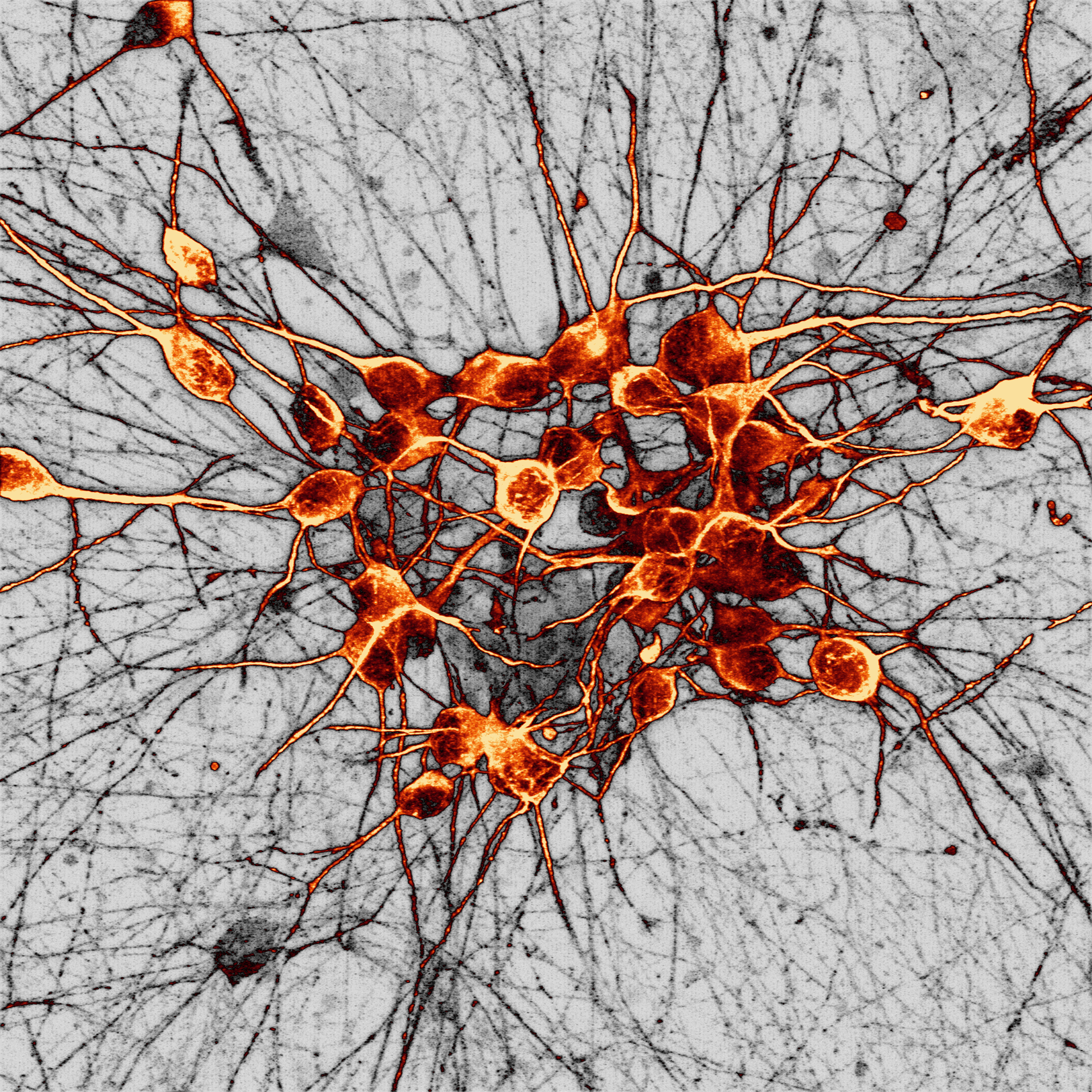
Human dopaminergic neurons
This image shows a culture of human dopaminergic neurons generated from human stem cells acquired via super-resolution Airyscan confocal microscopy at the University of Oxford Micron facility. Dopaminergic neurons are the main cell type that deteriorate in Parkinson’s disease, partly due to toxic build-up of a protein called alpha-synuclein.

Arabidopsis leaf
Cells on the epidermis of a 3 day old Arabidopsis leaf. This is an adaptation of a linocut print created based on a microscopy image.
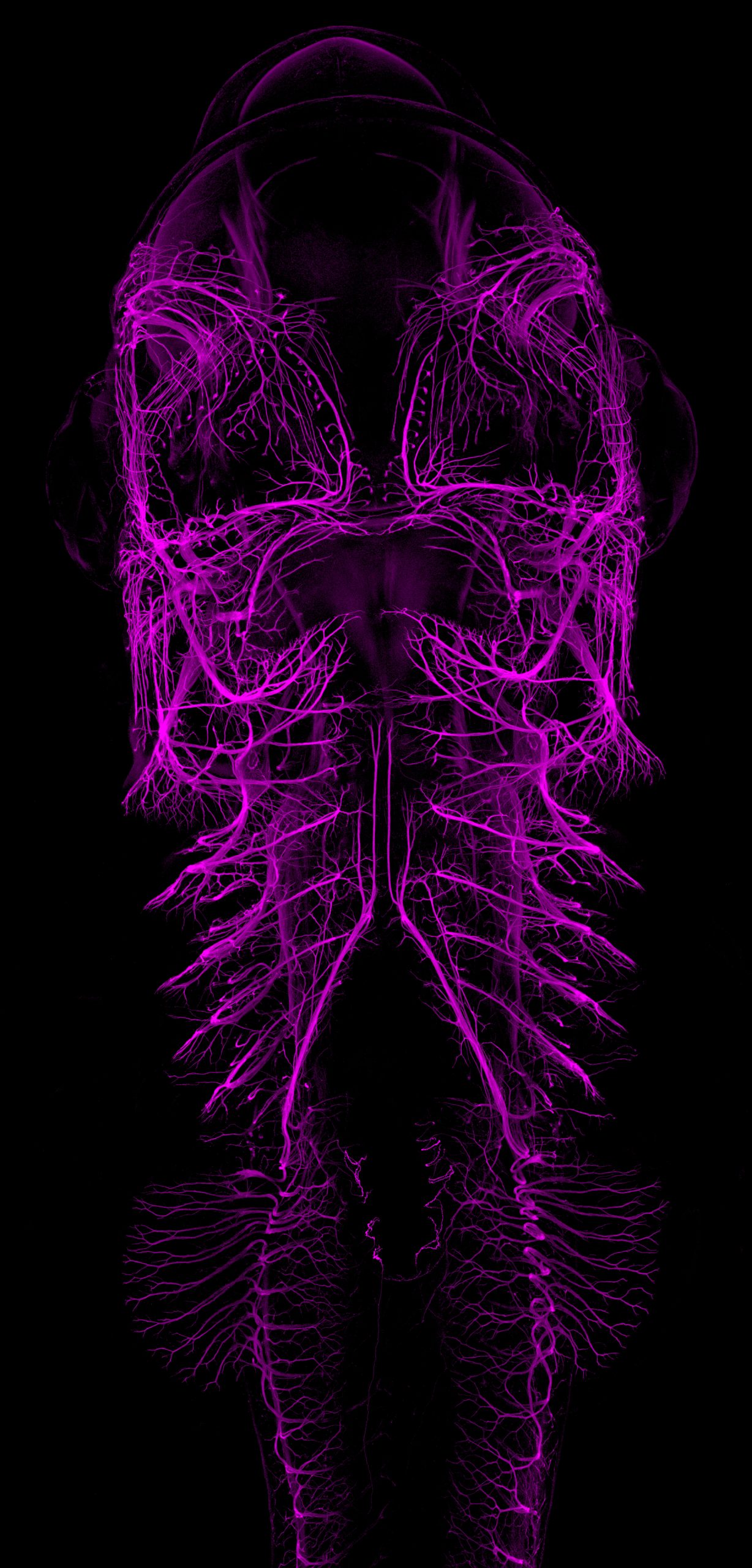
Catshark embryo
Ventral view maximum intensity projection from an immunofluorescence staining labeling the developing nervous system (primarily nerves and ganglia) of a stage 30 small-spotted catshark embryo (Scyliorhinus canicula). The image was acquired using a ZEISS LSM980 with Airyscan2 confocal microscope, stitched and processed using ZEN software from the same microscope.
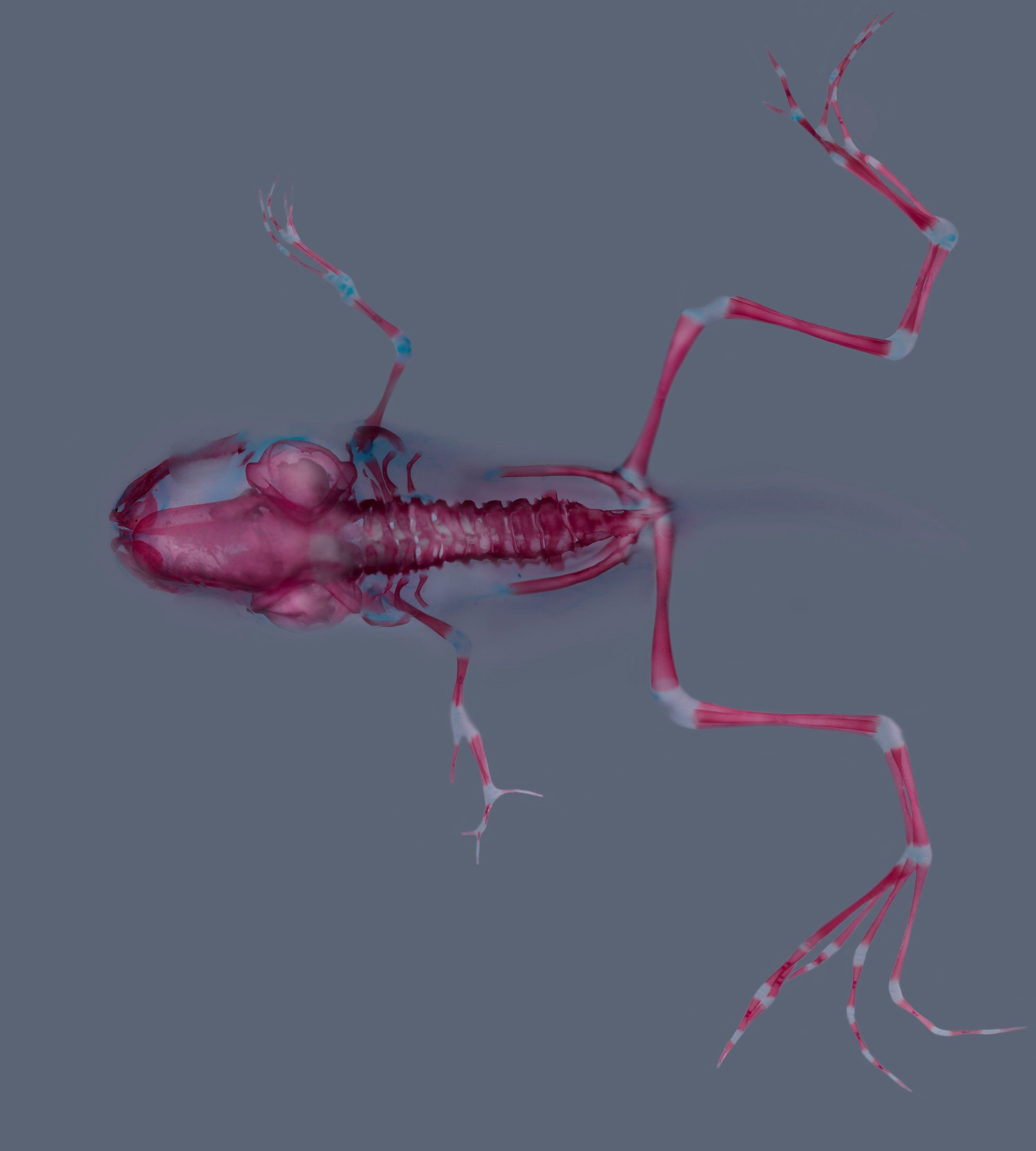
Xenopus laevis skeleton
Skeletal staining (alizarin red and alcian blue) of a Xenopus laevis at stage 62. Stage 51 larva was treated with a Cyp26a inhibitor during forelimb regeneration. Notice proximo-distal duplication in the left forelimb.
[poll id=”6″]
 (35 votes)
12 thoughts on “Vote for your favourite image for the Node postcards”
Leave a Reply
Get involved
Create an account or log in to post your story on the Node.
Sign up for emails
Subscribe to our mailing lists.
Very good
Amazing!
Original
Magnifica idea, muy original.
Es una maravilla. Enhorabuena al autor.
Geographical Map!
The phenotype is very interesting
Que hermosa fotografía, felicidades!
Imágenes sorprendentes
Amazing work Elisa…it’s a master piece
Excelente work Dan Rios!
Muy bien, me parece muy interesante, gracias por fomentar la investigación!!!👏