Woods Hole Images round 3 – vote for a Development cover
Posted by the Node, on 7 June 2013
It is time for round 3 of last years’ Woods Hole embryology course images! The regenerated planarian won the last round , but who will you vote for this time? Below are 4 great images, and you can decide which one will feature in the cover of Development. To see bigger versions, just click on the image.
Voting will close noon GMT on June 27th.
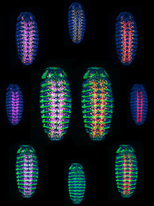
1. Ventral view of a Drosophila melanogaster embryo (stage 12/13) fluorescently stained for Repo (yellow; nuclei of glial cells), axons (red, anti-HRP), Hedgehog (green; Hh-GFP), Elav (pink, nuclei of neurons), and nuclei (blue, DAPI). The different maximum projection images show different combinations of the color channels. This image was taken by Davon Callander (Oregon State University).
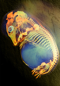
2. Color inverted image of a skeleton preparation of a pig (Sus scrofa domesticus) embryo. This image was taken by Marina Venero Galanternik (University of Utah), Rodrigo G. Arzate-Mejía (Universidad Nacional Autonoma de Mexico), Jennifer McKey (Universite Montpellier) and William Munoz (The University of Texas MD Anderson Cancer Center).
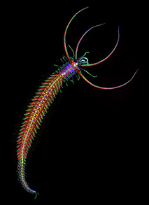
3. Male stolon (ventral view, anterior up) of the annelid, Proceraea sp., fluorescently stained for acetylated tubulin (green), serotonin (yellow), F-actin (red; phalloidin), and nuclei (blue; DAPI). Confocal z-stacks were viewed as maximum projections and tiled together to cover the entirety of the animal (body length approximately 7.5 mm).This image was taken by Eduardo Zattara (University of Maryland, College Park).
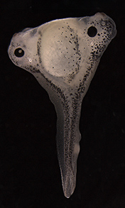
4. Generation of a secondary body axis resulting from an organizer graft in Xenopus laevis. The graft was performed at stage 10 and the tadpole was photographed at stage 41. This image was taken by Elsie Place (MRC National Institute of Medical Research).


 (2 votes)