Zürich to Dresden and back: of small fish and big data
Posted by Karin Prummel, on 9 February 2016
In Spring 2015, just a couple of months into my PhD, I started to settle with my new surroundings in Zürich, making friends in my PhD lab of Dr. Christian Mosimann, and learning the fine details of early zebrafish development. That is when suddenly one morning Christian casually asked me how I’d feel about moving to Dresden in Germany for a while…
My key interest is the molecular control of cell migration and rearrangements. In our lab, we use the zebrafish (Danio rerio) to investigate the cell fate determination of the lateral plate mesoderm (LPM). The LPM is a fascinating mesoderm lineage that gives rise to the heart, blood, vessels, kidneys, and limbs. After gastrulation, the LPM in zebrafish arranges in tiers of cells at the lateral edge of the developing embryo. How the LPM originally arises from the remaining mesoderm, what common molecular program links its vast array of cell fates, and how early LPM cells arrange at their lateral positions remain unclear. I am particularly interested in the cell migration dynamics and migration control of the early LPM. To visualize the emerging LPM, the Mosimann lab has generated several novel transgenic zebrafish strains that mark the LPM cells at all stages of development. Nonetheless, traditional confocal imaging only enables imaging of small parts of the LPM and not of the whole embryo in its entirety.
A great solution to image the entire zebrafish embryo is selective plane illumination microscopy (SPIM), also called lightsheet microscopy. While we do have a local SPIM setup, processing the huge amounts of data accumulated is time-consuming. The lab of Jan Huisken, at the Max Planck Institute (MPI-CGB) in Dresden (Germany) is pioneering the development of high-speed fluorescence microscopy to enable systematic studying of developmental processes. Their selective plane illumination microscopy (SPIM) allows imaging several zebrafish embryos at the same time, overnight, and enables visualizing the whole zebrafish early development.
Gopi Shah, a senior grad student in the Huisken lab, had given a great presentation on panoramic SPIM at the European Zebrafish meeting in 2013, and Christian was immediately hooked. After several discussions between Jan and Christian and trading postdoc stories from their times at opposite coasts in the USA, Jan generously offered to have me come to his lab in Dresden and use their unique lightsheet setup to image our unique transgenic zebrafish lines. I was immediately enthusiastic about this great opportunity as I could learn a new technique, apply it to fascinating biology, and learn more about cell migration dynamics in vivo.
As the whole panoramic SPIM setup is too bulky to quickly ship to Switzerland, I moved for a month to Dresden. My arrival in the lab was warm and welcoming. Gopi introduced me into their lightsheet setup and supported me greatly with the imaging. Also the rest of the lab unconditionally helped me out during my stay. Although research with zebrafish is always an adventure, my zebrafish cooperated really well and I could gather more data than I had hoped for: I successfully managed to image over 60 zebrafish embryos.
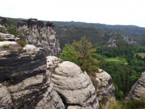
Despite the many hours spend “behind” (or rather next to and around) the SPIM setup and on data processing, I also had the opportunity to fully explore beautiful Dresden, visit the Elbe Valley with its castles, and hike in the surrounding nature, especially in the national park referred to as Sächische Schweiz (Saxonian Switzerland).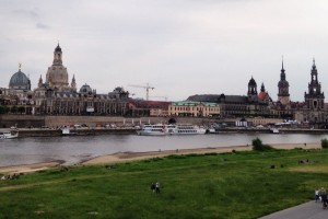
Already just looking at the imaging provides us with new insights about the emerging LPM and early zebrafish development. For the first time, can we see the migration and organization of the whole LPM from its onset during gastrulation. This unique data set is a key fundament for my future studies of LPM patterning and for deciphering the cell migration dynamics in the emerging LPM. We have already lined up follow-up experiments coordinated between our both labs, and I continue to receive great help from the Huisken lab with analyzing all the data.
My collaborative visit was a definite success based on the amount of data I collected, the new ideas and immediate follow-up experiments, and the new contacts and friends that I made. It was a great experience to be in a lab focused on a different discipline, but with the same greater goal in mind: studying the molecular and cellular processes of early development. I am very grateful to Dr. Jan Huisken for giving me the possibility to use his lab’s infrastructure and hospitality, and to The Company of Biologists for the support in form of a Development Travelling Fellowship.
 (10 votes)
Get involved
Create an account or log in to post your story on the Node.
Sign up for emails
Subscribe to our mailing lists.