Embryosafari: an attempt at illustrating development
Posted by KalinDN, on 12 September 2017
Hello, Community! In July, I revealed to the world my attempts at scientific illustration. Aidan (The Node’s Community Manager) took a keen interest in my work and was very kind to share it on social media. Here is my long-promised post about my biology art (which can be viewed at www.embryosafari.com).
Before I delve into the illustrations, let me introduce some background information about myself:
1st Instar (Science). Like many of us, I have been enthralled by biology since an early age. The specific focus of this fascination fluctuated through the years (from paleontology, through molecular virology to immunology), but I finally settled on developmental biology (in October 2006, to be precise), as I saw it was the science which tackled the most wonderful of all subjects: how bodies form (and evolve). Eventually, this lead to my PhD in developmental genetics in the labs of Anne-Gaelle Borycki and Phil Ingham, where I studied the expression of an ECM (extracellular matrix) gene using mouse, chicken and zebrafish embryos.
2nd Instar (Art). Similar to my fascination with science, I have enjoyed art (particularly clay modelling and pencil sketching) for as long as I can remember. As a child, I used to grab a block of clay and start sculpting (with arguable success) sci-fi creatures and characters that had grabbed my imagination on screen. Unlike science, I have never had any formal training in the arts (I don’t count art classes in primary school as such). In my later years, when I was already studying embryos, I frequently resorted to clay modelling when I needed to understand the three-dimensional anatomical organisation of a body region or organ. Subsequently, this “hobby” evolved into a digital form, the results of which I present below.
“Building The Brain”
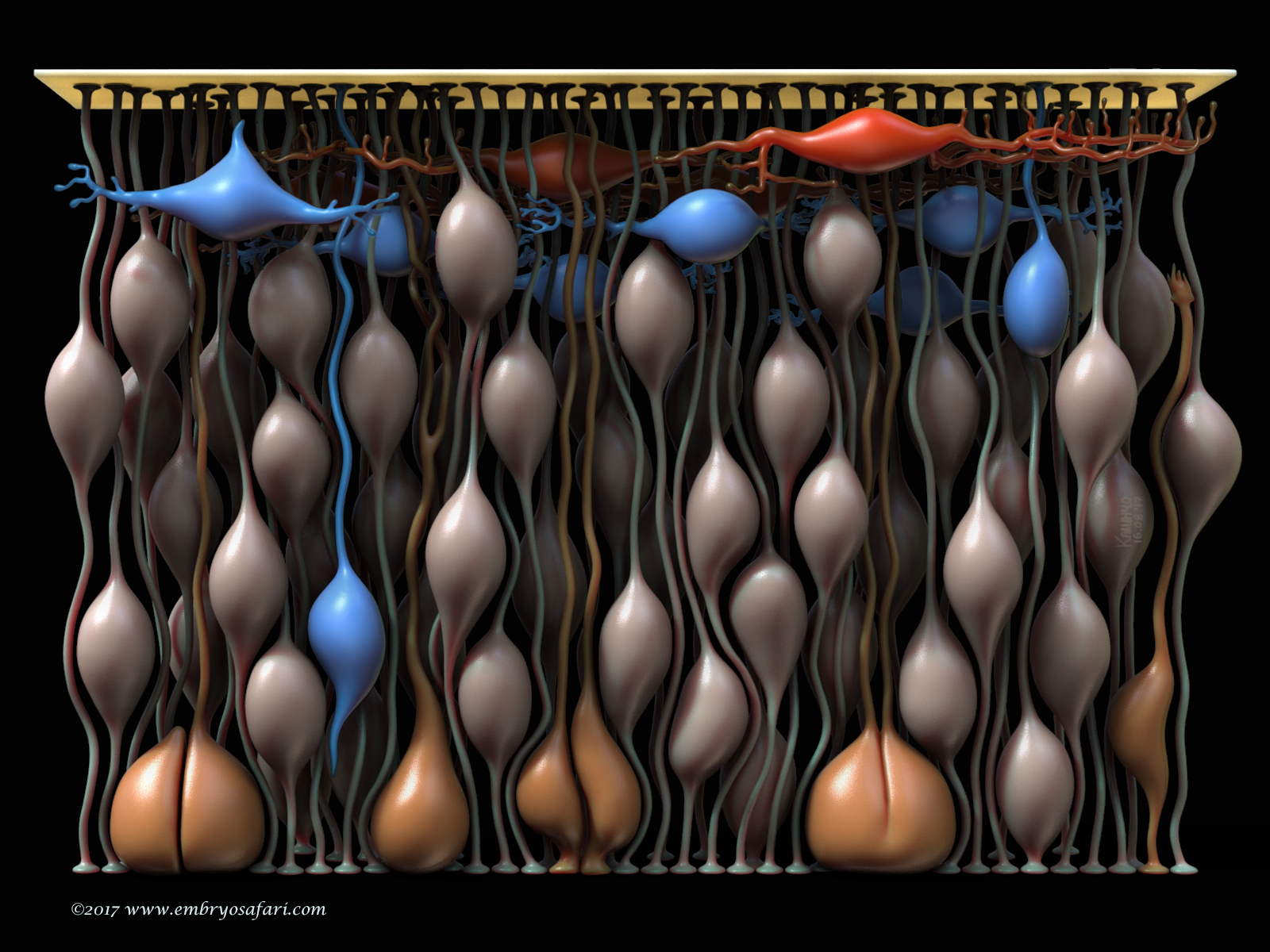
This work represents the cytoarchitecture of the murine telencephalon at E10.5. The goal was to show the cell types at the beginning of neurogenesis. The fleshy (salmon) colour indicates the neuroepithelial cells (NECs) whose nuclei migrate “up and down” the thickness of the epithelium, undergoing INM (interkinetic nuclear migration). Mitoses occur close to the ventricle (the apical side of the epithelium), where the mitotic cells are indicated in orange-ish. What fascinates me is the fact that most NECs, even the mitotic ones, remain anchored to the pial basal lamina (the bright yellow plate on the top) via these long, thread-like basal processes. The first neurons in the telencephalon, those of the preplate, are indicated in red (Cajal-Retzius cells) and blue (prospective subplate neurons). Initially, this model was part of a bigger story which aimed to illustrate gyrification (cortical folding) in mammals, but it was too ambitious (one day I’ll go back to it!).
References:
http://dev.biologists.org/content/141/11/2182
http://emboj.embopress.org/content/23/11/2314
http://onlinelibrary.wiley.com/doi/10.1038/emboj.2008.227/abstract
https://link.springer.com/article/10.1007/BF00391127
“Cardiac Anatomy Of The Human Embryo”
Rarely there is a subject more irresistible than the embryology of our own species. Without any anthropocentric bias, human developmental anatomy is simply beautiful! Of all human embryology subjects, I consider brain, cardiac and craniofacial development most challenging in terms of morphological understanding. And because I enjoy such kind of challenges, I decided to tackle first cardiac anatomy, for which I knew the least. Ideally, I dream of creating illustrated plates for all Carnegie Stages, for both external and internal cardiac morphology, as well as for the major cellular and genetic events in cardiac development. Only time will show how feasible such an endeavour is. For now, I present illustrations on Carnegie Stages 9/10 and 12.
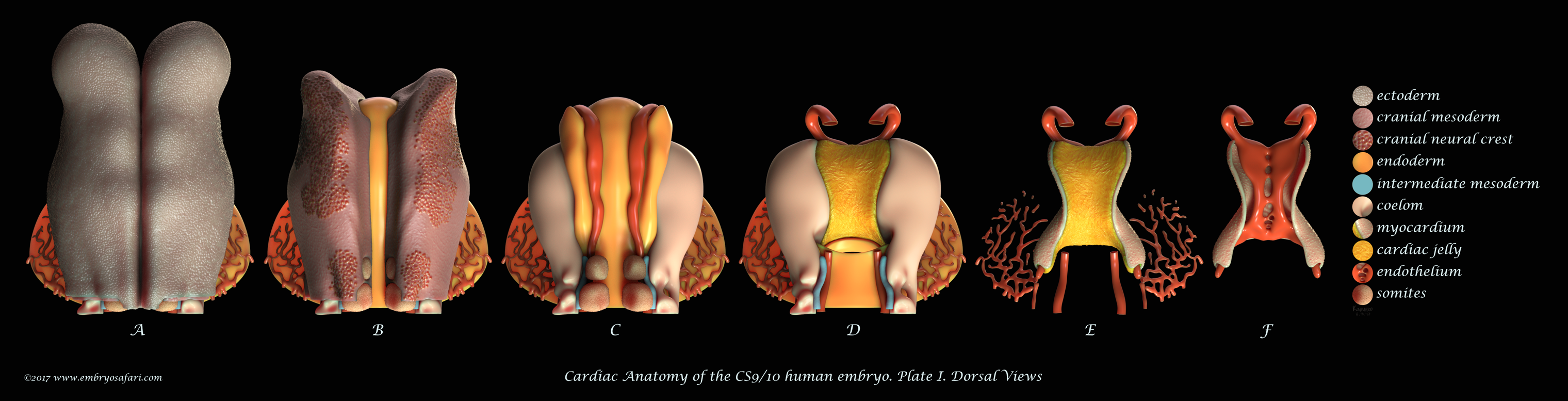
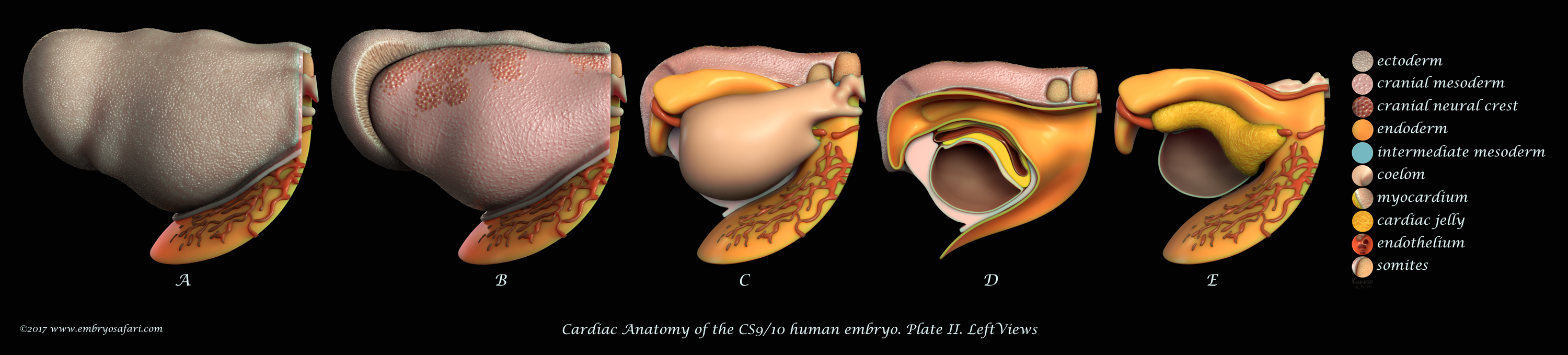
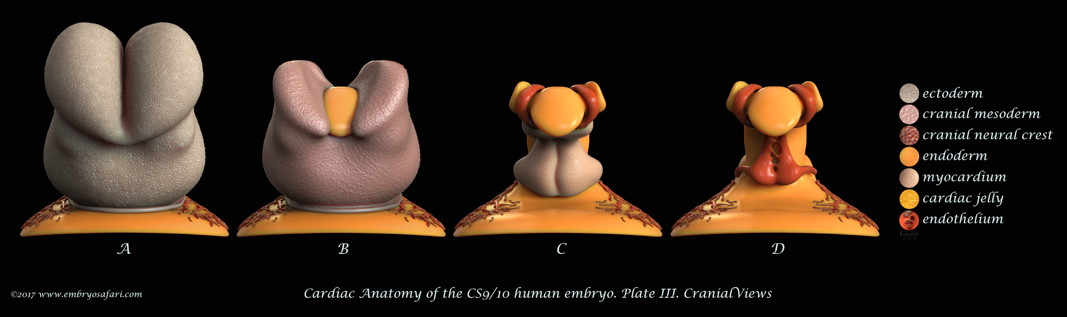
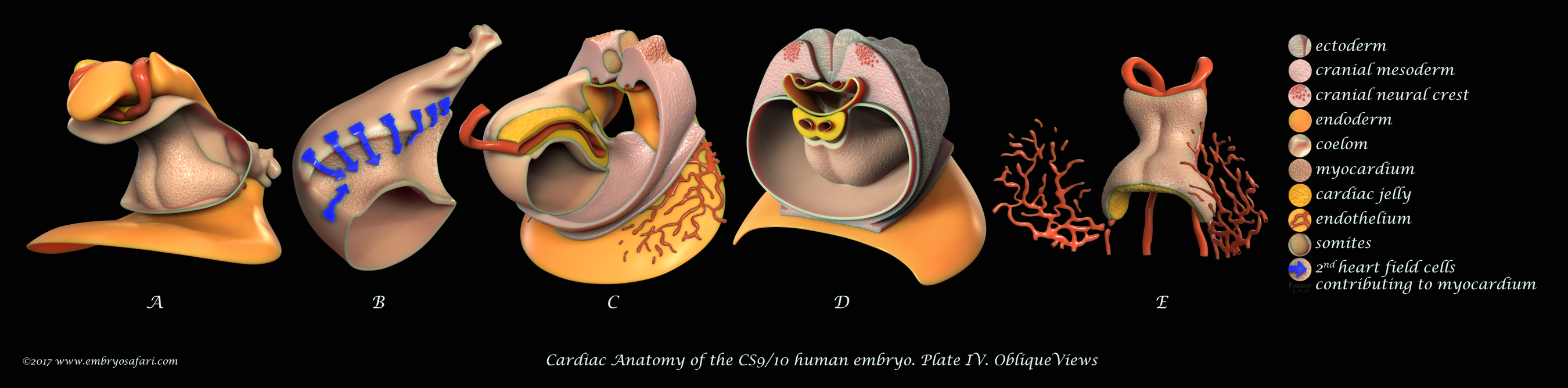
These plates illustrate the anterior half of the human embryo at a morphological stage between CS9 and CS10, viewed from different angles. This stage is interesting for it shows the fusion of the initially bilateral endocardial tubes (visible in Figure J on Plate I), and the presence of a distinct pericardial cavity (anterior coelom), shown in Figures C, D (Plate I) and in A-D (Plate IV). The abundant cardiac jelly, visible in Figure E (Plate I), and in Figures C and D (Plate IV), fills the space between the fusing endocardial tubes and the incipient myocardium. Importantly, myocardial progenitors in the second heart field add up to the growing myocardium (shown as blue arrows in Figure B on Plate IV). In addition to the heart, I spent some time sculpting the closing neural tube, the first two pairs of somites, the septum transversum, and the cranial neural crest (each cell was individually painted, as seen in Figure B on Plate I). For detailed examination, please view the images at full screen mode, or visit https://www.embryosafari.com/gallery.
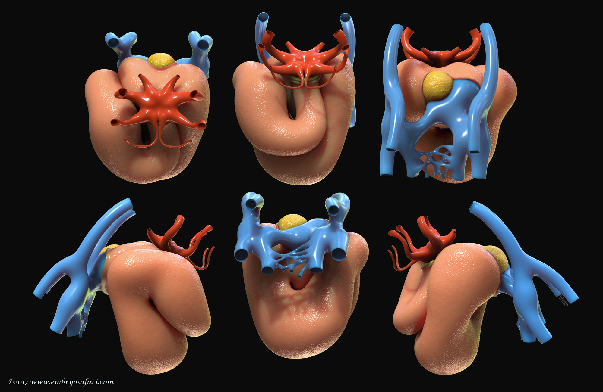
This illustration represents the external morphology of the embryonic human heart at Carnegie Stage 12, viewed from different angles. The pericardial sac has been removed to expose the myocardium. The focus here is on the spatial relationship between the myocardium (salmon colour), the cardiac jelly (in yellow) and the vessels at the venous pole (blue). At this site, the second heart field continues supplying the myocardium with cardiomyocyte progenitors via the dorsal mesocardium (the cut edge in the top 3rd figure, left to right). The red colour indicates the vessels at the arterial pole of the heart, where the highly dynamic pharyngeal arch arteries form and regress in a quick, craniocaudal succession.
References:
http://circ.ahajournals.org/content/123/10/1125
http://circ.ahajournals.org/content/early/2010/08/09/CIRCULATIONAHA.110.953844.full
http://science.sciencemag.org/content/354/6315/aag0053.full
http://perspectivesinmedicine.cshlp.org/content/4/10/a015750.long
http://onlinelibrary.wiley.com/doi/10.1002/ajmg.a.35896/abstract
O’Rahilly R., Müller F., Human Embryology And Teratology, Third Edition, 2001
This is all about my work for now. If you wish to learn more, please jump to https://www.embryosafari.com/gallery
At the end, I would like to thank all curious, luminous and hardworking beings of science, without whom developmental biology wouldn’t have bloomed into its current state.
Thank you.
 (7 votes)
Stunning work, especially for one self-taught in scientific illustration! Thank you for sharing your talent and efforts with the world – and I’ll be keeping an eye out for your continuation of this series!
Thank you for the support, Natalya. I think your Xenopus and natural history illustrations are wonderful!