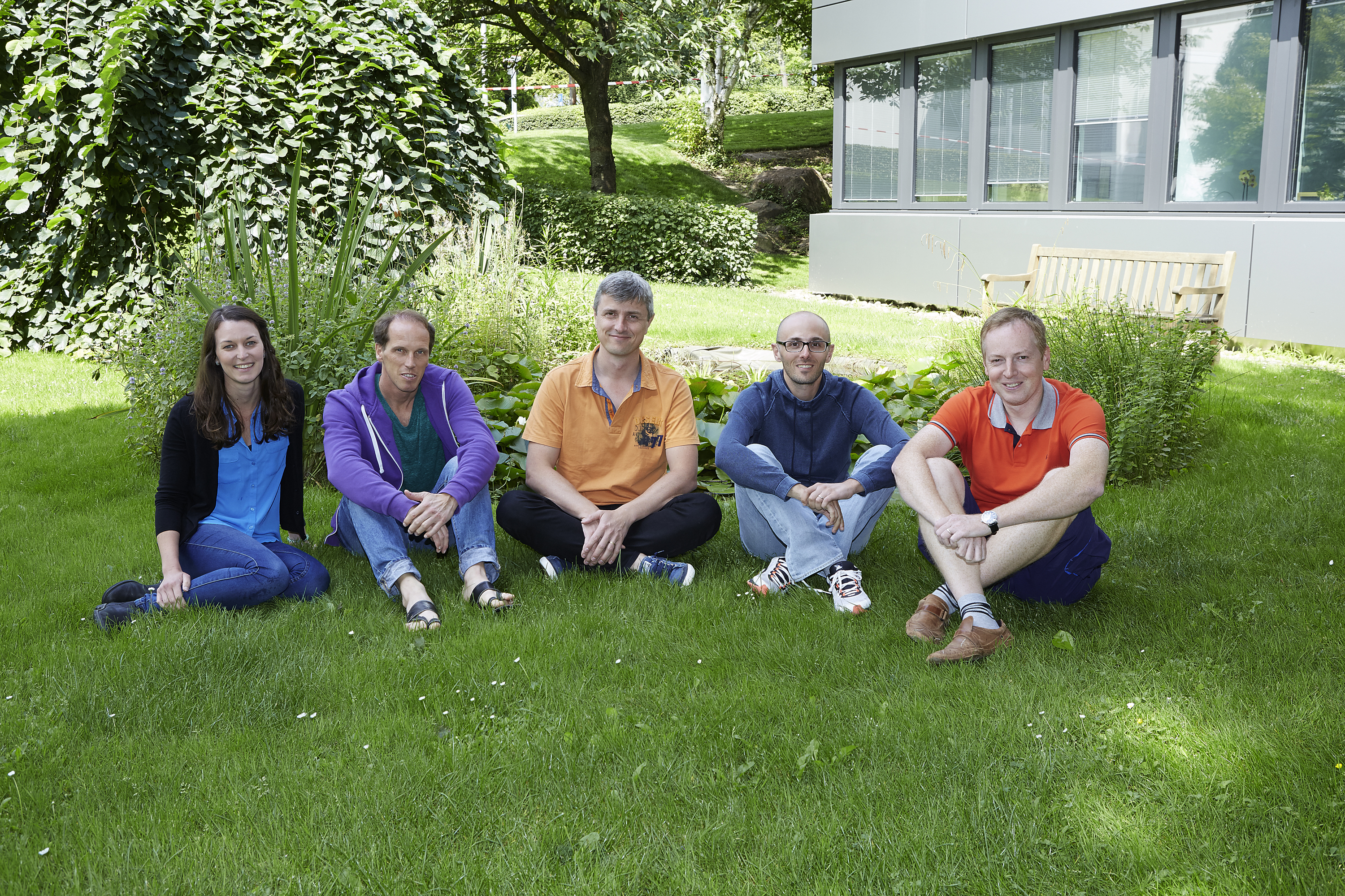
The people behind the papers: Martin Beck, Yannick Schwab, Nicole Schrieber & Paolo Ronchi
Posted by the Node Interviews, on 16 August 2016
In this new series, we interview the people behind some of the most exciting recent papers in developmental biology and related fields, to give context to the work and find out how the story came together.
To inaugurate the series, we start with a paper that came out recently in Cell, and uncovered a mechanism for how nuclear pore complexes are inserted into the nuclear envelope in early Drosophila development.
We hear from five of the people behind the paper, all of whom are based at the EMBL in Heidelberg: lead author Bernhard Hampoelz, his PI Martin Beck, and their EM collaborators Yannick Schwab (PI and head of the EM facility), Nicole Schrieber and Paolo Ronchi.
They gave us their perspectives on this collaborative, multi-disciplinary project.
We’ll start with Martin: can you tell us the brief history of the Beck lab, and what key questions the group is trying to answer?
MB I have a mixed training in structural biology (PhD with Wolfgang Baumeister at the MPI Martinsried) and systems biology (postdoc with Ruedi Aebersold at ETH Zurich). I had already worked on nuclear pores as a student and when I started my own laboratory at EMBL in 2010, I felt that my training positioned me well to attempt to understand nuclear pore complex architecture. We have combined electron microscopic with mass spectrometric approaches to structurally analyse nuclear pores in situ.
I was always intrigued by two biological aspects of this: i) that understanding the assembly pathways of large macromolecular machines can help us to understand their architecture because nature essentially has broken down the problem into smaller pieces for us that are more feasible to approach. And ii), that in order to understand the function of large macromolecular machines in situ, one needs to understand how their structure is spatiotemporally modulated, e.g. across cell types or the cell cycle.
From my perspective, the beauty of Bernhard’s project is that it brings all of this together to elucidate a new phenomenon. It builds on the methodological strength and quantifies a compositional variation of nuclear pores across space and time to discover an unanticipated way to get a nuclear pore complex (NPC) in to the nuclear envelope.
And Bernhard, how did you come to join the Beck lab? Am I right in thinking you brought flies to the lab?
BH Exactly – I think flies were not very popular in Martin’s group before I entered. Also my background was perhaps unusual for the lab, since I came from developmental biology. When working at the IBDM with Thomas Lecuit, my focus was on nuclear morphology and its developmental control. Thomas gave me a lot of freedom in investigating things that popped up and it happened that I observed annulate lamellae (AL; stacked cytoplasmic membranes that are a subset of the ER and decorated with NPCs) insertion into the nuclear envelope by imaging. Aware of the potential of this finding, I knew that in order to nail this down mechanistically I had to convince somebody that works on NPCs at the ultrastructural level to give me the chance to pursue this project. I came to EMBL as a visiting scientist and soon presented my findings to Martin. I am very glad that Martin, although the project was not in the direct focus of his lab at that time, agreed on supporting me to continue this work in his group.
“I knew that in order to nail this down mechanistically I had to convince somebody that works on NPCs at the ultrastructural level to give me the chance to pursue this project.”
So Yannick, you’re neighbours in Heidelberg, but did you know Martin well beforehand? How did you get involved in Bernhard’s project?
YS Martin and I work on the same floor at EMBL. In fact, I head the electron microscopy core facility (EMCF) which is sharing the space where Martin’s Unit (Structural and Computational Biology) has its set of cryo electron microscopes. Therefore, we see each other very often. Even though Martin’s group is focused on structural biology, they do not hesitate to cross the border towards cell biology which is the field of expertise of the facility. Whilst Bernhard had already solid background in EM and was already an advanced user, his project required advanced expertise both in volume EM and in Correlative Light Electron Microscopy (CLEM). For this, he teamed up with some of the EMCF staff (Pedro Machado, Paolo Ronchi and Rachel Mellwig) and with Nicole Schieber a specialist in Focussed Ion Beam Scanning Electron Microscopy (FIB-SEM) from my team. My involvement in this project was mostly at the level of organizing this collaborative work and setting priorities when the last set of experiment had to be done.
Finally, Nicole and Paulo: how were you recruited to this story?
NS I started with the Schwab team when it was just beginning as the research technician. My background has had a strong focus on Electron Microscopy for the past 9 years since my undergraduate degree got me hooked at the University of Queensland, Australia. For the past 3 years here at the EMBL I have shifted more towards the 3D EM techniques, especially FIB-SEM.
My role in the lab means that I try to connect the team to the facility and on some occasions I can step in to help with projects that I either find interesting or see I can add some expertise. For this story, my colleagues Rachel and Pedro from the EMCF were already working on this project together with Bernhard and I had been following its progress in our regular meetings. They had managed to solve some difficulties with the EM sample preparation and we all quickly realised that FIB-SEM would add to the three dimensional picture of the story. Since this would be a demanding task for the FIB-SEM I put my hand up to acquire the data and really enjoyed the challenge as well as being able to team up and work more closely with my colleagues than we normally would.
PR I have been working in the EMCF for 2 years after a postdoc experience at EMBL, working on membrane trafficking. Since I’ve been here, I have always been very keen on pushing CLEM methods in the facility and Bernhard’s project was a great opportunity. But it was a big challenge as well.
I got an email from Yannick asking whether I would be interested in helping Bernhard with a CLEM experiment. I knew his project and I had always found it very interesting, but it was on short notice (it was for the revision of the paper) and I was on holiday! The project required to adapt a high accuracy CLEM method that had been previously developed on yeast cells to the Drosophila embryos. Luckily, I had been setting up the best conditions to treat Drosophila ovaries for another collaboration and thought that Bernhard’s system was similar enough. Therefore I used the same protocol and, for once, everything worked smoothly at the first attempt.
Where did the interest in nuclear pores come from, Bernhard? And what was the key problem you wanted to address with this paper?
BH I have to admit that my interest in NPCs came by accident. In Thomas Lecuit’s group I worked on nuclear morphology and used fluorescently labelled Nucleoporins as means to outline the nucleus in imaging. Naively I realized that these Nups do not only label the nuclear envelope (NE) but also foci in the cytoplasm and I learned about AL, which have been known for decades actually. Curious about their function, I imaged them live and saw that they insert into the NE. Puzzled by the fact that this has not been observed before I started to think how to further develop this project.
What makes the early Drosophila embryo an ideal model for the question of nuclear pore insertion? What are the model’s challenges?
BH The fly embryo offers a couple of advantages. The most intriguing is probably its ease to do live imaging. Basically you just glue an embryo expressing your fluorescently labelled protein of interest onto a coverslip and start to image. Moreover the embryo is susceptible to injection of drugs or genetic manipulation that could interfere with your process of interest. Conceptually, I like that it allows you to put a cell biological question into the context of animal development. A challenge is the short cell cycles, especially for EM – we sectioned numerous embryos that happened to be frozen during mitosis and were thus useless for our purpose.
“Basically you just glue an embryo expressing your fluorescently labelled protein of interest onto a coverslip and start to image.”
Can you briefly describe what you found out about the relationship between annulate lamellae, nuclear pore complexes, and the nuclear envelope?
BH AL are sub-compartments of the endoplasmic reticulum that contain stockpiled NPCs. Because AL are in particular enriched in oocytes and early embryos across species, they were always considered as storage pools of maternally derived NPC material. Whether they could somehow contribute to the NPC pool at the nuclear envelope had remained elusive. Our study proves that AL are indeed inserting into the nuclear envelope when the nucleus expands during interphase in Drosophila embryos at the blastoderm stage. This can work because unlike in differentiated cells NPCs in the early fly embryo are laterally mobile within the NE and thus can redistribute. We reveal that NPCs at AL are pore scaffolds that only mature to the full NPC complement once inserted into the nuclear envelope. And, based on EM, we suggest a topological model how such insertion could happen.
So what makes a nuclear pore at the AL different from one in the nuclear envelope?
MB Its composition. Certain nucleoporin subcomplexes are missing in AL. This includes for example the Nup214 complex that is important for mRNA export out of the nucleus in differentiated cells but also for some import pathways that might play already a role in the early embryo. We believe that it is assembled into AL-originated pores only after they inserted into the nuclear envelope.
And why was EM necessary? I understand three different techniques were used (CLEM, FIB-SEM and tomography): were any of them a particular challenge?
YS EM in general is a challenging set of techniques. From the sample preparation to the image analysis, specific skills and dedicated protocols are required. Fortunately, the EMCF has accumulated a great deal of experience since its creation more than 10 years ago. Benefiting from a good integration within the research scene, it has, among others, developed specific methods for preparing Drosophila embryos (by high pressure freezing and freeze substitution) and for imaging them with 3D EM (tomography and FIBSEM).
NS FIB-SEM is a difficult technique that requires a lot of patience but is extremely rewarding in the results you can obtain. For this project one of the main difficulties came after finding the correct stage of the embryo and an interesting event by TEM thin sections. We then wanted to immediately image this same embryo in the FIB-SEM by looking at the block face itself and use the focussed ion beam (FIB) to ablate very thin slices to allow consecutive images that build the 3D data. During standard preparations for FIB-SEM you have a buffer where you can allow the sample to stabilise during the imaging so that the slice thickness is consistent, and can also test the imaging conditions to get the best possible resolution. In this case we didn’t have such luxuries and this was a challenge especially since the event we were looking for required the best possible resolution. The other challenge came from remounting the sample from how it was sectioned for TEM to the stub for FIB-SEM, here we were dealing with a piece of resin containing the sample that was very small (in the range of several hundred microns) and the orientation of this block was critical.
PR The CLEM method we used was developed by the Briggs and Kaksonen groups at EMBL to study endocytosis on yeast and mammalian cell cultures. Their work has made this technique very popular in the community and therefore our facility in the last 2 years has dealt with a number of projects using this method, adapting it to different organisms and different subcellular structures. Bernhard’s case was more challenging because we had to look for embryos of the right developmental stage and in the right stage of the cell cycle (interphase). After preparing the samples for EM, we therefore sectioned a few embryos and inspect them by conventional EM to identify the ones that displayed interphase nuclei, before proceeding with the CLEM workflow.
What was the key insight given by EM?
YS EM techniques enabled us to visualize for real what a bundle of other techniques could only suggest. For example, live fluorescence imaging strongly suggested the physical connection, continuity, between the AL and the nuclear envelope. Thanks to 3D EM (tomography and FIB-SEM) this continuity appeared obvious. FIB-SEM furthermore demonstrated the organization of the ER sheets relative to the nuclear envelope, an observation that led us to propose the model exposed in the paper. It was very important as well to demonstrate that the very dynamic fluorescent patches observed in vivo were indeed AL. CLEM clearly confirmed this.
“EM techniques enabled us to visualize for real what a bundle of other techniques could only suggest.”
Does the mechanism of AL-NPC insertion change during development, and is this important?
BH Yes, indeed. Insertion of entire AL is a maternal program and declines starting with zygotic induction. One reason might be simply the temporal constraint. AL insertion happens fast and could overcome the slow kinetics of classical interphase NPC assembly, as we know it from tissue culture cells. In the prolonged interphase 14 of Drosophila embryogenesis, AL diminish from the nuclear layer at the embryos’ cortex. The vast abundance of AL in oocytes and early embryos of many animal species also argues that they have a general role in the earliest stages of development.
Did you have a single ‘eureka!’ moment when everything came together or you got a particularly stunning result?
BH I would say two of them: First the photoconversion experiments where I could see that converted Nucleoporins distributed from an extranuclear spot into the nuclear envelope and there dissipated laterally. This really proved to me that AL do insert. And secondly when we could reveal insertion ultrastructurally in the FIB-SEM analysis.
Is there a loose end or surprising result in the paper you would particularly like to get to the bottom of?
MB Various experiments described in our paper show that nuclear envelope organisation before the start of transcription is very different from well-studied conditions in differentiated cells. That overexpression of the lamin B receptor is sufficient to make nuclei in the early embryo look as their counterparts at later stages I personally found very surprising.
And what are you working on now?
BH As always many questions emerge: for example how do the NE openings that seem critical for AL insertion form? How are they stabilised? To get a handle on this on a molecular level is my next goal.
Martin and Yannick, any planned future collaborations?
MB YES!
YS Yes definitely. This collaboration was one of these great and rewarding moments when we witness how the expertise from a service facility helps our colleagues to progress in their science. Martin’s and Bernhard’s enthusiasm and collaborative spirit successfully engaged everyone in this fantastic story. I can tell you they have plenty more, as exciting as this one!
“This collaboration was one of these great and rewarding moments when we witness how the expertise from a service facility helps our colleagues to progress in their science.”
Bernhard Hampoelz, Marie-Therese Mackmull, Pedro Machado, Paolo Ronchi, Khanh Huy Bui5, Nicole Schieber, Rachel Santarella-Mellwig, Aleksandar Necakov, Amparo Andrés-Pons, Jean Marc Philippe, Thomas Lecuit, Yannick Schwab, Martin Beck. Pre-assembled Nuclear Pores Insert into the Nuclear Envelope during Early Development. 2016. Cell. Volume 166, Issue 3, p664–678
You can catch up with the latest People behind the Papers here
 (1 votes)
Get involved
Create an account or log in to post your story on the Node.
Sign up for emails
Subscribe to our mailing lists.