Woods Hole images round 4- choose a movie to be a Development cover!
Posted by the Node, on 5 September 2013
For the last round of Woods Hole images this year we have an exciting development- the last round is a movie round! Below are 4 great movies from last year’s Woods Hole embryology course, and you can vote for your favourite. The most voted movie will be featured in the homepage of Development and a still or collection of stills from the movie will be the cover of a coming issue of the journal. You can see what the cover will look like by clicking on the link below each image.
Voting will close on noon GMT on the 30th of September.
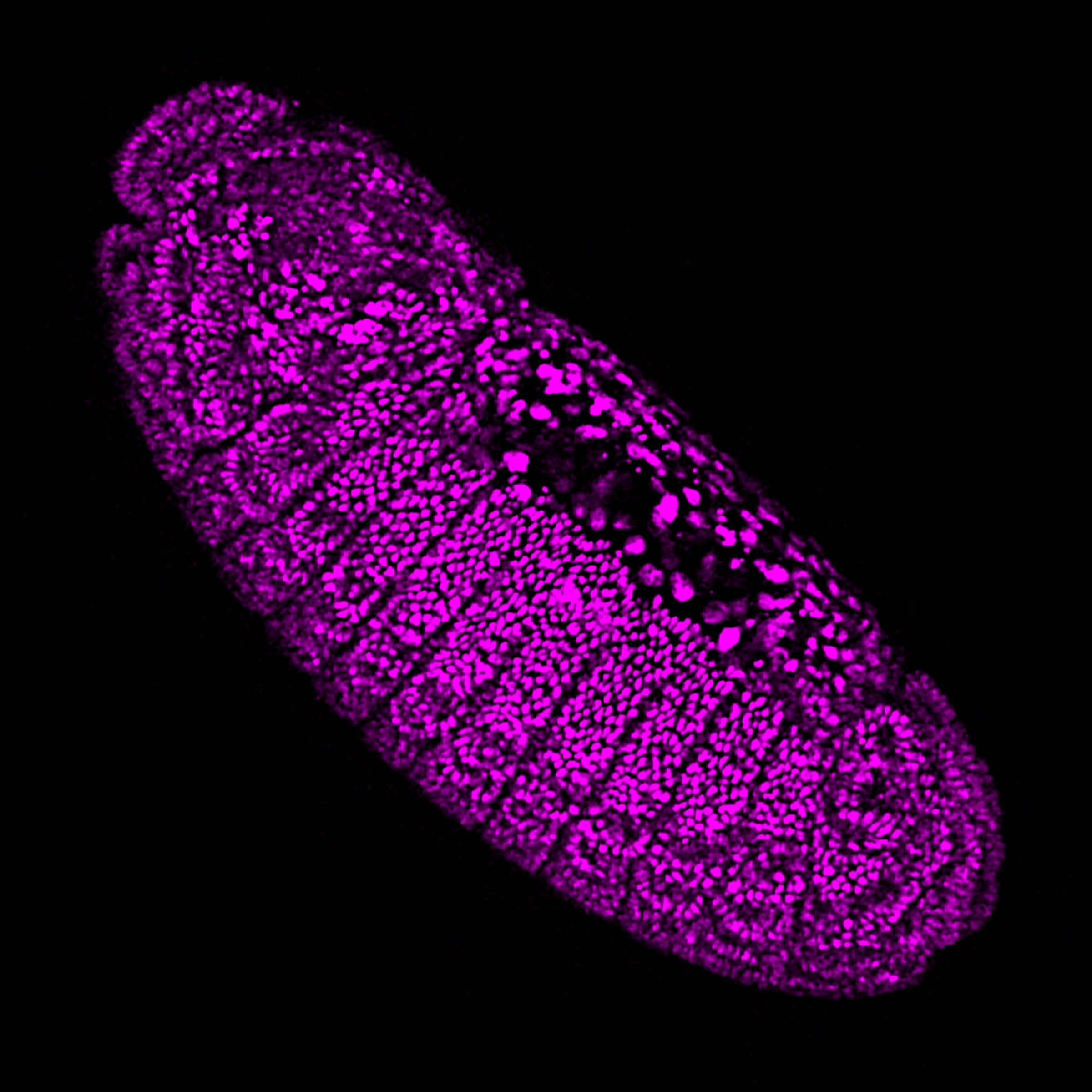
1. Drosophila embryogenesis. Lateral view of a Drosophila melanogaster embryo with anterior to the left and ventral down. The embryo carries a Histone 2A-RFP transgene that allows visualization of all nuclei and was imaged by confocal microscopy (maximum intensity projections created from each timepoint). During the approximately eight hours of development the embryo goes from stage 6 to stage 13, during which time the embryo undergoes gastrulation, germband extension, germband shortening, and the appearance of morphological segmentation. This movie was taken by Marina Venero Galanternik (University of Utah), Rodrigo G. Arzate-Mejía (Universidad Nacional Autonoma de Mexico), Jennifer McKey (Universite Montpellier) and William Munoz (The University of Texas MD Anderson Cancer Center). Cover image.
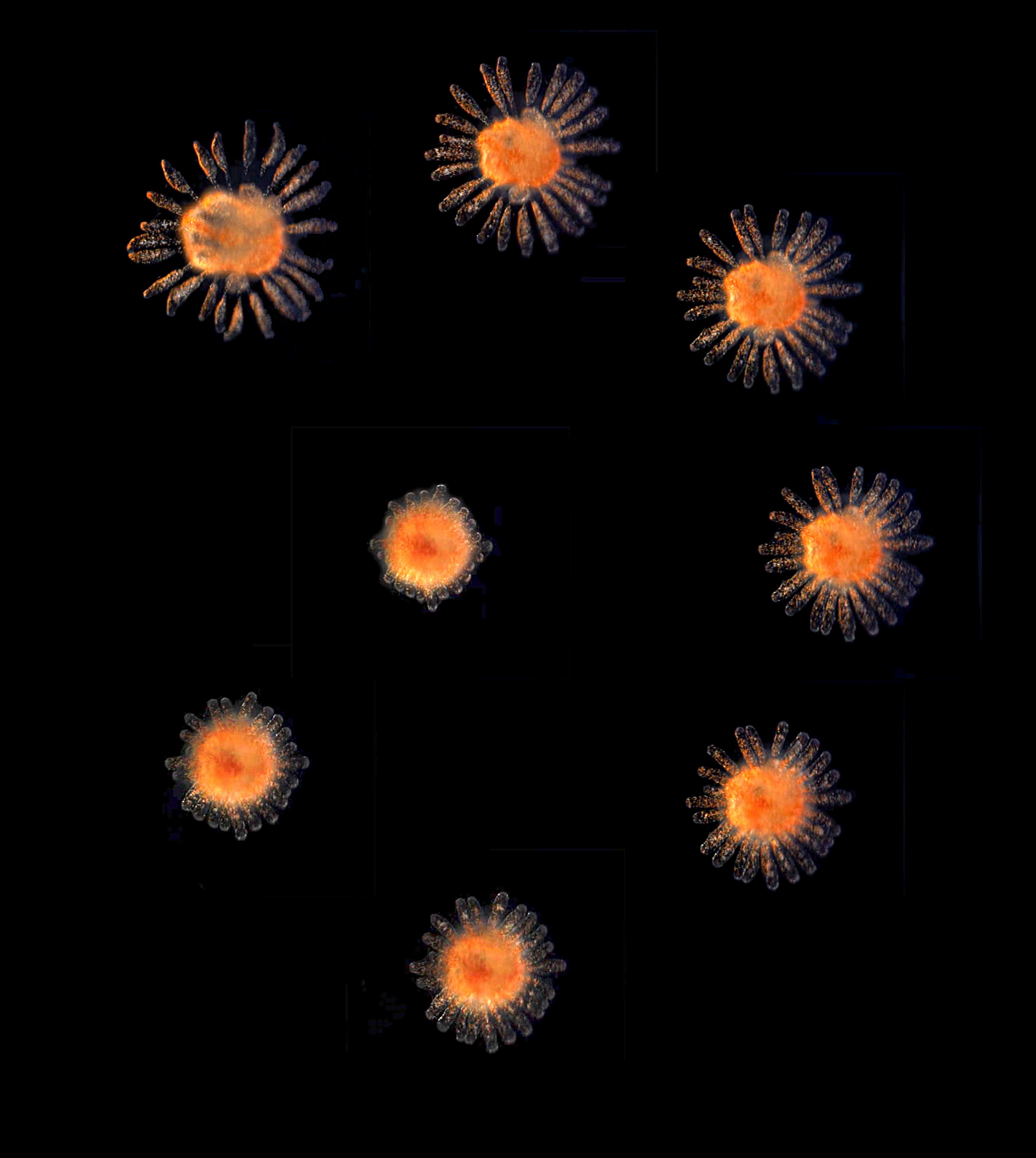
2. Ascidian metamorphosis – extension of ampullae. Metamorphosis of the colonial ascidian, Botrylloides violaceus, imaged by widefield microscopy. During the three-hour period the ampullae extend out over the substrate, and eventually this individual will bud off additional colonies. This movie was taken by Matthew Clark (University of Oregon). Cover image.
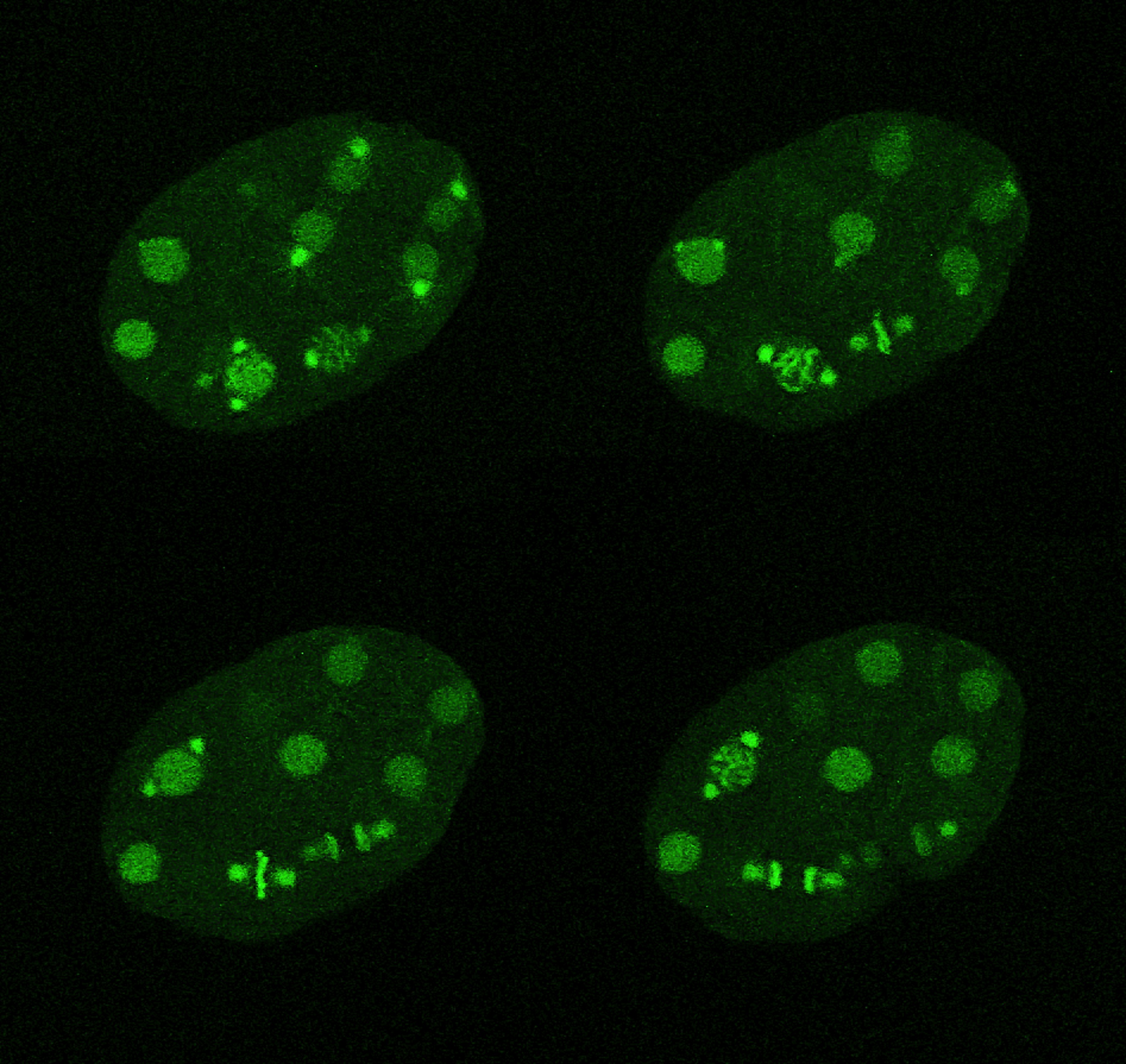
3. C. elegans early cell divisions. The embryo contains both GFP:Histone H2B and GFP:gamma tubulin allowing for visualizaion of both the chromosomes and the centrosomes respectively. At the 8 cells stage, the embryo contains the AB.a1, AB.ar, AB.p1, AB.pr, MS, E, C and P3 cells. The 2 cells that divide at the bottom near the end of the movie are the MS and E cells (with E dividing first). Imaged by confocal microscopy (maximum intensity projections created from each timepoint). Movie covers approximately 17.5 minutes of development. This movie was taken by Daniela Di Bella (Fundacion Instituto Leloir), Joyce Pieretti (University of Chicago), Saori Tani (Kobe University) and Manuela Truebano (Plymouth University). Cover image.
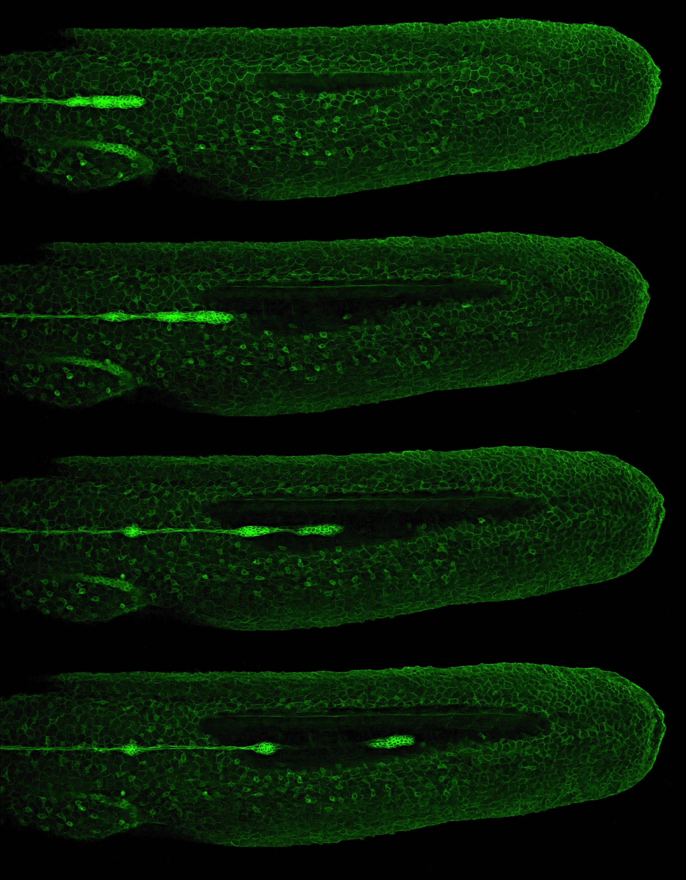
4. Zebrafish lateral line migration. Zebrafish lateral line primordium migration leaves behind clusters of cells that will form neuromasts, which are mechanoreceptive organs that allow the fish to detect water movement. Here the cells of the primordium and neuromast clusters are visualized in the Tg(-8.0cldnb:lynEGFP)zf106 transgenic line in which claudin B-GFP fusion protein highlights the cell membranes. Each frame of the movie is a maximum intensity projection of a confocal Z-stack. Lateral view with anterior to the left and ventral down. Movie covers approximately 8 hrs of development. This movie was taken by Eduardo Zattara (University of Maryland, College Park ). Cover image.
If you are interested in using any of the images or movies in this post, please contact the Node to request permission
 (5 votes)
2 thoughts on “Woods Hole images round 4- choose a movie to be a Development cover!”
Leave a Reply
Get involved
Create an account or log in to post your story on the Node.
Sign up for emails
Subscribe to our mailing lists.
Way to make this hard Node people. They’re all so pretty. There are good things happening at MBL.
Glad you like them! They are really beautiful!