BEE-ST – a technique for monitoring hard tissue development
Posted by Alexandra Bisia, on 3 January 2024
What is this?
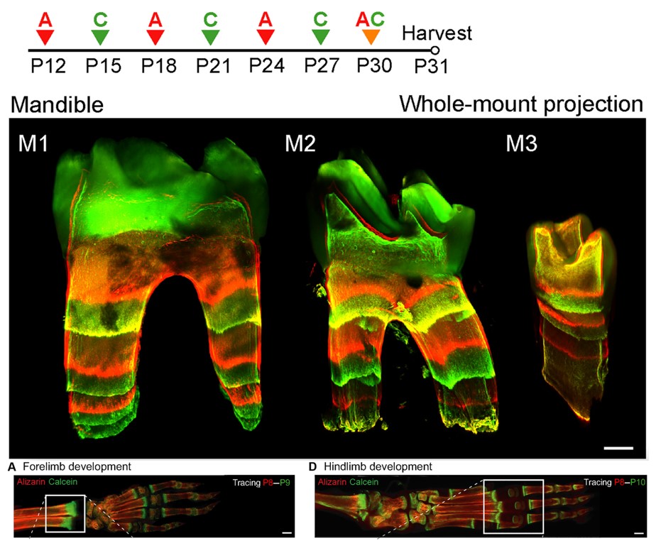
BEE-ST (Bone and tEEth Spatio-Temporal growth monitoring) is a method to monitor mineralization dynamics across species. It is based on the intraperitoneal (abdominal cavity) administration of fluorescent calcium-binding dyes in an animal of interest at the specific time you want to observe newly-forming mineralized tissue. Sequential administration of dyes that fluoresce at different wavelengths (alizarin, red; and calcein, green) enables the observation of bone and tooth mineralization dynamics across time.
Where can this be found?
The technique can be applied to all tissues that undergo mineralization in various species, so the fluorescent dyes will be found in parts of bones and teeth that have developed since the administration of the dyes. The technique was developed by graduate student Marcos González López and his colleagues in Jan Křivánek’s lab at the University of Masaryk in Brno, Czechia.
How was this image taken?
The image was taken by high-resolution confocal laser-scanning microscope. Confocal microscopes use fluorescent lasers to illuminate different depth spots in your sample. By using confocal microscopy, you can obtain several images in different planes that can be put together to recreate a 3D model of your sample, in this case, the mouse molar.
What does it do?
The BEE-ST technique enables researchers to investigate how mineralized tissues (bones and teeth) develop or repair themselves after injury. This is achieved through the binding of dyes to the newly-synthesized or newly-repaired regions of bone or teeth. Once harvested, bones and teeth can be subjected to an optical-clearing protocol, enabling light to pass through them, and a couple of days later the tissue is ready to be imaged by fluorescent laser-scanning microscopy.
Why should people care about this?
The BEE-ST method is a powerful to understand how hard tissues become mineralized.
In the developmental biology and tissue engineering research communities this tool can be utilized to:
- Gain a better understanding about how bone and teeth obtain their shape.
- Evaluate how changes in the expression of genes, proteins or signalling pathways influence bone or tooth regeneration.
- Assess the effectiveness of small molecules or drugs in promoting repair and healing of bones.
How would you explain this to an 8-year-old?
Teeth and bone have a lot of calcium in them. When they grow, or when they break and repair themselves, they add more calcium to themselves. We can use special, coloured dyes that “grab onto” growing teeth and bones to see how fast they grow or repair themselves. This way, when we look at a tooth or bone from an animal that we gave these dyes to, we can see which parts grew recently. Like you can see in the picture, when we switch between red and green dyes, we can find out how long ago each part of the tooth or bone grew, or how quickly. Look at the bones from the front and back leg of the mouse: the red parts grew before the green parts. This way, we can study how teeth and bones grow, and how they repair themselves after they break. We can also test new medicines to see if they can help bones heal more quickly after they break.
Where can people find more about it?
If people want further information on how this method works and its versatile use in all mineralized tissues across species, they can read the recent paper from the Křivánek lab at the Department of Histology and Embryology at Masaryk University in Czechia.
Check out other ‘Show and tell’ posts and how about writing one yourself?
 (No Ratings Yet)
Get involved
Create an account or log in to post your story on the Node.
Sign up for emails
Subscribe to our mailing lists.