Collaboration: All the things we cannot see (alone).
Posted by miriamirosenberg, on 3 June 2019
By Miriam Rosenberg and Suparna Ray

Most of what we know about axial patterning in insects comes from decades of careful, beautiful work done in flies. Thanks to the genetic screens of Christiane Nüsslein-Volhard and Eric Wieschaus in the late 1970’s, we learned that distinct classes of genes, many of them transcription factors, act in an elegant procession to achieve the designation of segment identity along the anterior-posterior and dorsal-ventral axes of the insect embryo. This process is robust when nature gets noisy- buffering against noise in RNA gradients to create precise boundaries, detecting coincidence of different proteins and translating their affinities into procedural specificity. Robustness ensures remarkable reproducibility of developmental processes in the face of novelty, since evolution is restless. And it is restless: two fully developed individuals of the same species with the same life history are full of small genetic differences, due to the persistent insinuation of random mutations. When we take a step backwards to look at two closely related species, we see much larger differences. Recognizing the value of this perspective has led to the field of Evo-Devo: that is, using an evolutionary perspective to better understand a developmental process and, in turn, using the knowledge of development to decipher the mechanisms of evolution.
Strength in numbers.
If you have ever looked at insects, you know that they have tremendous variation in morphologies in nature, and it turns out that this diversity arises in part from variation in their embryonic patterning program. Gerhard Krause described variation in developmental styles from his morphological studies of insect embryos in the 1930’s. Later, pioneers in modern Evo-Devo like Michael Akam, Nipam Patel and Diethard Tautz took this further: molecular comparison of evolutionarily distant insects illuminated developmental programs in a way that looking only at one species could not. Discoveries in beetles especially, but also aphids, crickets and others, showed that flies are not typical among insects and therefore are not the only important game in town: a point that insect evo-devo researchers are always quick to point out. And yet, many of us studying less traditional model systems today, like beetles, wasps, or bugs, nevertheless get siloed in our own organism’s biology. One of the greatest strengths of these insect systems is the ability to put things together by looking at where they drifted apart. It is through the diffracting lens of evolutionary differences that we can understand the origins of robustness and of genetic consensus. The ability to move experimentally in a single project between different organisms permits full expression of this colorful perspective.
Collaboration was essential for synthesis of the discoveries of Nüsslein-Vollhard and Wieschaus into the basis of our understanding of axial patterning. And so too is collaboration between developmental biologists in different (insect) systems to make fundamental discoveries about the origins of robustness and functional variation in developmental programs.
Our recent paper in eLife (Ray* and Rosenberg* et al, eLife 2019) describes such a finding about the ancestral function of a highly conserved peptide, which would not have been possible if the study had been limited to any single insect. In this post, we highlight the importance of the collaboration and how it came about, since this ability to move between insect species, and open data sharing, permitted the most profound insights of our work.
Better together.
Ray and Rosenberg et al., is a work that came together from 4 different groups working in different countries, and carried out over almost 10 years. Our story involves a few main molecular characters: Mille-pattes, a regulatory micropeptide; Shavenbaby, a transcription factor previously known for its key role in epidermis development; and Ubr3, an E3 ubiquitin ligase. Our paper’s story was inspired by the publication by Savard et al. (Cell 2006) showing that mille-pattes (mlpt), which they identified in an expression screen in the flour beetle Tribolium castaneum, is essential in Tribolium for abdominal segment formation. This finding was surprising at first because mlpt doesn’t produce a regular protein, but rather is a an apparently long non-coding RNA encoding only four tiny peptides of 11-32 amino acids in length. The suspense grew later, when studies of the same gene in flies, named polished rice (pri) or tarsal less (tal), revealed functions in leg patterning, trachea and epidermis differentiation, but no role in segmentation of the Drosophila embryo (Kondo et al., Nat Cell Biol 2007; Galindo et al., PLoS Biol 2007).
Our collaboration arose from parallel stories, which made possible the insights from comparison. We have decided to tell each of the stories, that became a powerful synergy and ultimately, enabled the most profound results.
1. Ray and Klingler.

Suparna Ray had joined the lab of Martin Klingler in Erlangen, Germany, towards the end of a collaborative genome-wide screen, iBeetle, whose aim was to uncover all developmental genes in Tribolium. mlpt was the newest kid on the block in the search for a definitive set of gap genes. Of particular interest was its interaction with Notch during Drosophila tarsal leg joint development, since Notch is a missing link long sought between the vertebrate segmentation clock and the segmentation clock of short germ insects, like Tribolium (Dequéant et al., Science 2006; Pueyo and Couso Dev Biol. 2011). Intrigued by the novel regulatory potential of micropeptides encoded from small open reading frames (smORFs) in developmental programs, the Klingler lab began to apply the most cutting edge approaches in Tribolium for functional analysis of gap genes (Schinko et al., BMC Dev Biol 2010 ; Schinko et al., Dev Genes Evol 2012). Shortly after Mlpt was discovered to activate the transcription factor Shavenbaby (Svb) in the fly epidermis (Kondo et al., Science 2010), RNAi knockdown of Tribolium svb in the Klingler lab revealed its role in abdominal segmentation. Furthermore, the mlpt and svb RNAi phenotypes were similar, suggesting a functional interaction of these two genes in Tribolium embryonic segmentation.
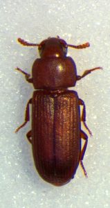
Meanwhile, out of the iBeetle screen (Schmitt-Engel et al., Nature Comm. 2015) came a small group of additional candidate genes showing mlpt-like RNAi phenotype. Intriguingly, these genes did not match any known developmental regulators. Instead, they correspond to enzymes involved in the ubiquitin proteasome system, often seen as a mere degradation factory that removes damaged or misfolded proteins. In particular, two genes located side by side and that give strong mlpt-phenotypes were predicted to encode ubiquitin E3 enzymes. These unexpected results prompted a whole flurry of research questions. Was the proteasome actually involved in Tribolium segmentation? How can a ubiquitous degradation machinery be required for the proper formation of posterior segments? Is the proteasome functionally related to mlpt and svb functions in segmentation, as suggested by strikingly similar phenotypes?
2. Rosenberg and Payre.

Miriam Rosenberg was at New York University in the lab of Claude Desplan, studying Nasonia, a wasp whose embryonic development exhibits intermediate character between Tribolium and Drosophila. The Savard paper opened the possibility of a role of mlpt peptide in abdominal segment specification, a function not yet accounted for in control of Nasonia’s posterior development by known fly genes. She began to investigate Nasonia mlpt and found that it exhibits striking expression in the region of the embryo that gives rise, in a delayed fashion, to the most posterior abdominal segments (Rosenberg et al., eLife 2014). Shortly thereafter, François Payre, who has spent many years elucidating the function and evolution of Svb during epidermal differentiation, came to the Desplan lab for a sabbatical. The Payre lab in Toulouse, France, had just shown that the Svb protein is post-translationally processed, from a transcriptional repressor into an activator, in the response to mlpt (pri/tal) peptides in flies (Kondo et al., Science 2010). During his sabbatical in New York City, he began to characterize the expression of svb and of its target genes during Nasonia embryogenesis.
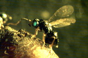
Upon his return to Toulouse, he and Rosenberg continued working together to characterize the expression and function of mlpt and svb in Nasonia. When the Payre lab demonstrated the role of the ubiquitin ligase Ubr3 in fly for Mlpt/Pri-mediated processing of Svb (Zanet et al., Science 2015), ubr3 joined the club in Nasonia, as well. Contrary to what was reported in fly, the long germ embryo of Nasonia showed essential functions for these genes in embryogenesis.
3. Descaras, Toubiana, Khila.

At the same time, in Lyon, France, Abderrahman Khila and his group were studying development of the water strider, Gerris buenoi. Water striders have the remarkable ability to walk on water, enabled by specialized hairs on the legs which allow it to trap air bubbles between leg and water, as well as differential elongation of the T2 leg (Khila et al., PLoS Genetics 2009; Armisén et al., 2018 BMC Genomics). An RNAi screen, searching for genes involved in these features in Gerris was being conducted by Amelie and William. Since Svb is a key regulator of hair formation in flies, they also assayed a putative role of svb in Gerris legs. When svb function was knocked down, they indeed observed a lack of hairs, but a prevalent phenotype in earlier stages was strong embryonic segmentation defects.
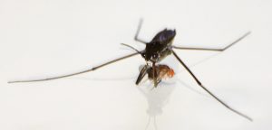
This was the starting point for testing whether the Svb partners were also involved in segment patterning in the water strider.
A collaboration is born!
By this time, Ray and Klingler were deep into the characterization of the E3 ubiquitin ligase when a poster from the Payre lab was presented at the French Fly meeting in Sète in 2014: they reported that the Svb processing triggered by Mlpt/Pri peptides relies on a limited proteasome degradation that critical requires the same E3 ligase, called Ubr3, as deduced from genome-wide functional screening.

Excited by these data, Ray and Klingler made contact, and arranged a visit to the Payre lab in Toulouse to discuss each others’ findings. During this meeting, an important common link was made: the complementarity of expression of mlpt and svb that had been observed in Tribolium was also present in Nasonia, and possibly other, more basal insects.
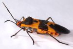

Payre was already in touch with Khila and kept collaborating with Rosenberg, who had moved to Israel and expanded the study of (now) three genes to the milkweed bug, Oncopeltus, working with Tzach Auman, a graduate student in the lab of Ariel Chipman in Jerusalem. Payre suggested to all groups involved that to meet shortly before Christmas of 2017, in Lyon, to discuss interest in joining forces with all insect models into a single story about the functional evolution of this trio of genes.
An eventful meeting.
A consensus was reached, since all data supported the ancient evolutionary origin of mlpt/Svb function in the insect embryo, and in all species with segmentation function, both genes exhibit posterior expression. But in flies, Svb expression is restricted to the head segments. A nice idea was proposed by Amelie Descaras, a student from the Khila lab. Could we use the Drosophila system, with its facile genetic tools, to broadly express Svb in the early fly embryo, to see whether its ability to function in embryonic patterning could be re-awakened? Here, the finesse and fly expertise of Hélène Chanut-Delalande was invaluable in designing and executing experiments that not only established the genetic role for Mlpt in embryo patterning, via Svb, but also highlighted the exquisite specificity of this regulation- using surgical mutations in Svb, required for Ubr3/Mlpt association, that abrogate the segmentation phenotype.
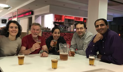
All together now…
Together, our research has traced the story of a multi-protein complex whose conserved functional interaction is critical for epidermal differentiation, leg development, and embryo segmentation. While each component of this complex has been strongly conserved through the evolution of insects, the segmentation function was lost in the most derived species, Drosophila. The dynamic expression patterns of mlpt and svb, which we observe in species that have diverged for hundreds of millions of years, underscore the continued importance of their segmentation function throughout the insect phylum. This ancestral role in segmentation calls out for study in additional, independently evolved, long germ insects. But it was the a-ha’s made possible by studies in divergent species with various modes of segmentation that coalesced into our current understanding of phenotypic plasticity and the evolutionary role of multi protein complexes.
When the Node invited us to highlight our recent advances with a post for young scientists about our work, we decided it was an important opportunity to highlight the significance of one central player in our work who does NOT appear in the published manuscript: the collaboration itself.
The authors would like to thank Martin Klingler, Abdou Khila, François Payre, Ariel Chipman and Galit Ophir for helpful comments on this piece.
Original research article described in this post can be found at: https://elifesciences.org/articles/39748


 (3 votes)