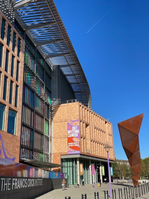
The 2nd Crick-Beddington Developmental Biology Symposium: Meeting Summary
Posted by Alex Eve, on 20 October 2022
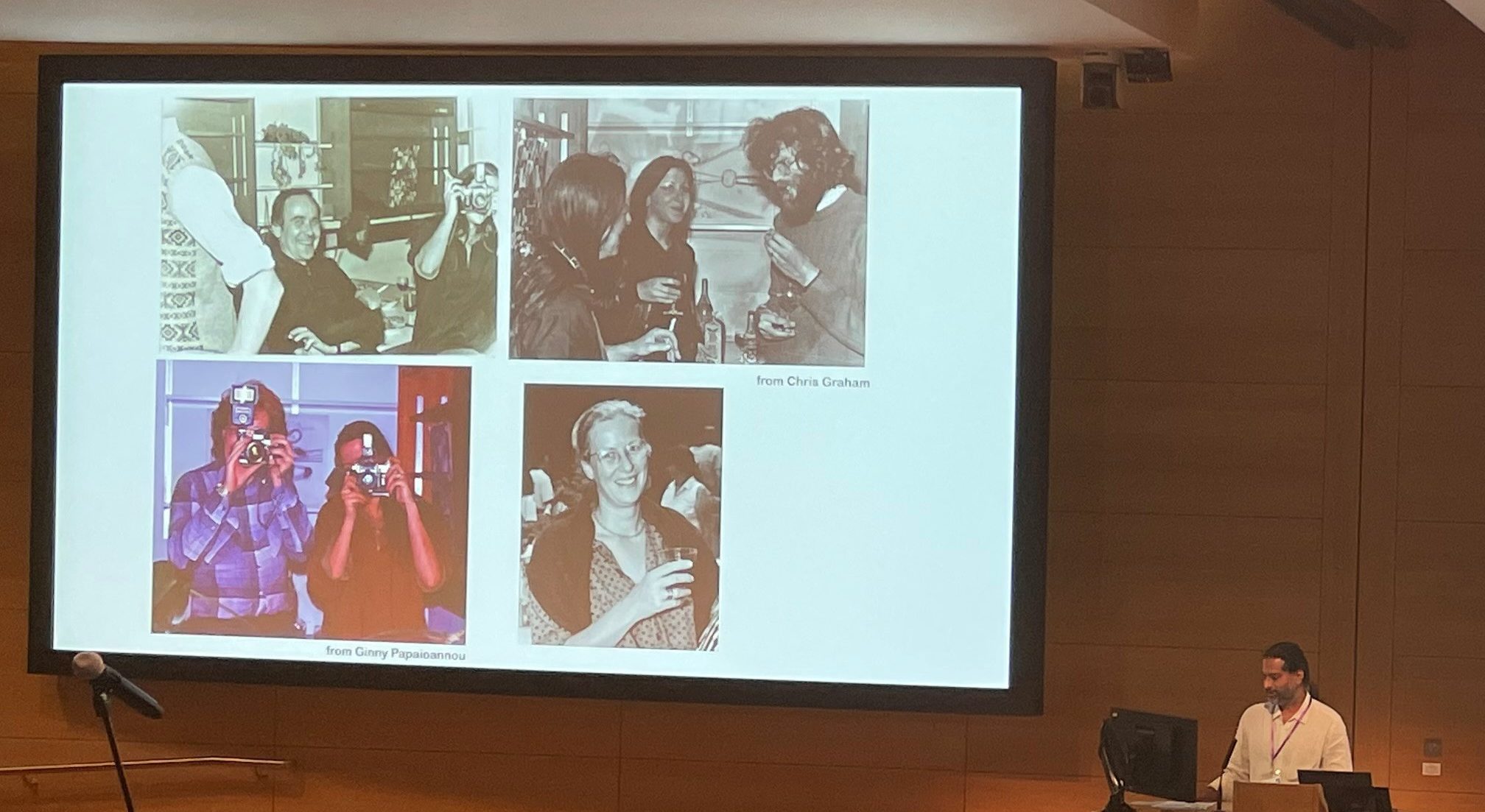
The 2nd Crick-Beddington Developmental Biology Symposium took place at The Francis Crick Institute between 9-10 October 2022. The hybrid symposium was generously funded by the MRC Rosa Beddington fund, which was established to uphold her memory and support curiosity-led science. Although a little later than originally planned, the conference maintained a similar ethos to the 1st symposium in 2019 (see an overview of that meeting from Alex Gould and Vicki Metzis and my previous Meeting Summary); many of the plenary speakers had direct links to Rosa and the talk topics reflected Rosa Beddington’s love of embryos and microscopy, as well as her meticulous description of morphogenesis. Shankar Srivivas, a former postdoc with Rosa and co-organiser of the meeting, opened the symposium with memories and photos of Rosa, mentioning her work on transplanting the mouse node (Beddington, 1994), how she believed in unity between all model organisms and how she often felt more comfortable behind the camera than in front of it.
Patrick Tam (University of Sydney) began the meeting by discussing how he and Rosa had a common interest in mouse gastrulation and were each other’s first collaborators in the late 1970s. Patrick presented a spatial transcriptomic atlas of the gastrula mouse embryo that provides insights into the molecular activity underpinning lineage trajectory during gastrulation. Matthew Stower (Srinivas lab, University of Oxford) also discussed mouse gastrulation, using light-sheet imaging to reveal coordinated, collective migratory behaviours in the dorsal visceral endoderm. In their first talk as an independent group leader, Diana Pinheiro (Research Institute of Molecular Pathology) employs computational modelling to understand the collective internalisation of the mesendoderm in response to morphogen signalling during zebrafish gastrulation. Yanlan Mao (University College London) also uses computational modelling to understand how mechanics generates the three major folds in the Drosophila wing disc and how structures are maintained under stress, such as when larvae are constricted. On this theme of mechanics and morphogenesis, Caren Norden (Gulbenkian Science) discussed optic cup development, revealing the importance of the extracellular matrix for the migration of a population of ‘rim cells’ into the retinal neuroepithelium and Toby Andrews (Priya lab, Crick) zebrafish heart morphogenesis and the role of mechanical stretching for trabecular development. Similarly, Golnar Kolahgar (University of Cambridge) explained how vinculin-dependent mechanosensory activity in Drosophila enterocyte progenitors maintains homeostasis of the intestine. Irene Miguel-Aliaga (MRC London Institute of Medical Sciences) presented a different perspective on the Drosophila gut, discussing how inter-organ communication creates sex differences in intestine geometry and function, revealed using microCT. Simon Bullock (MRC Laboratory of Molecular Cell Biology), a former graduate student with Rosa Beddington, uses Drosophila as a model to understand how dynactin-dynein complexes asymmetrically localise RNA transcripts, including Gap genes. Drosophila segmentation was the focus of Jacques Bothma‘s talk (Hubrecht Institute) as well. Jacques has developed ‘LlamaTags’ for rapid imaging of transcriptional dynamics to reveal how the genome encodes and interprets time. Alain Chédotal (Vision Institute) similarly works on the leading edge of technology, employing virtual reality for the characterisation and 3D analysis of cleared human embryos. Jan Huisken (University of Göttingen) concluded the imaging theme by presenting Flamingo, a low-cost, customisable, modular light-sheet imaging microscopy set-up that can be shipped around the world and operated remotely.
Several of the talks reflected on metabolism. Josh Brickman (reNEW), a former postdoc with Rosa, looked back at their time together with humour, reflecting how Rosa had insisted he work on transcription factors, to which he now credits his career. Josh explained how transcription factor residence on enhancers supports plasticity in foregut stem cells and lipid metabolism alters gene expression via sirtuin-dependent acetylation. Aydan Bulut-Karslioglu (Max Planck Institute for Molecular Genetics) uses mTOR-inhibited embryonic stem cells as a model to show that fatty acid degradation sustains mammalian diapause. Moving from development to cancer, Salvador Aznar-Benitah (Institute for Research in Biomedicine) discussed how metastasis-initiating cells have high fatty acid metabolism and are sensitive to dietary fats, such as palmitic acid. Meritxell Huch also showed how lipid metabolism can reprogram differentiated liver tumour cells and discussed the importance of TET1-mediated demethylation for de-differentiation during liver regeneration. Selin Jessa (Kleinman lab, McGill University) presented the developmental origins of paediatric gliomas, focusing on the links between histone 3 lysine 27 methylation mutations and 3′ Hox genes.
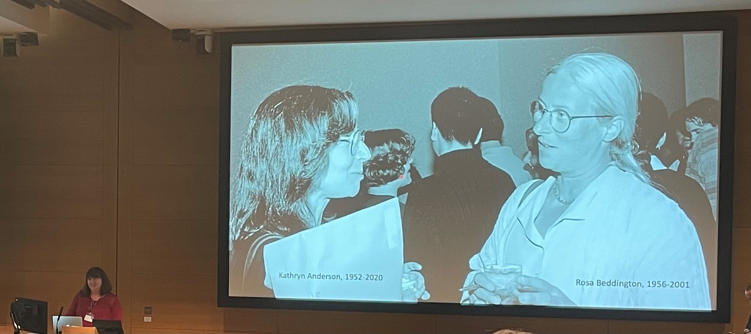
Another emergent theme from the meeting was developmental principles across scales. At the population level, Hugh Ford (Chubb lab, Laboratory for Molecular Cell Biology) uses Dictyostelium to study cAMP signal propagation and cell migration. In tissues, Danelle Davenport (Princeton University) explained how the periodic pattern of skin appendages is established via polarising morphogenesis and Jakub Sumbal (Koledová lab, Masaryk University) presented insights from single-cell RNA-sequencing data of terminal end buds during mammary gland epithelial branching. Val Wilson (University of Edinburgh), a former postdoc in Rosa’s lab, presented research on two populations of axial progenitors in mice (neuromesodermal progenitors and lateral-paraxial mesodermal progenitors) and how signalling controls fate decisions within these populations. At the cellular level, Tamara Caspary (Emory School of Medicine) began her talk by explaining how Kathryn Anderson and Rosa Beddington catalysed her research in mouse ‘hat’ gene mutations, which were later mapped to ciliogenesis and Hedgehog signalling. Tamara showed how these two processes could be uncoupled through the activity of ARL13B in the cilia versus the cytoplasm. Rita Sousa-Nunes (King’s College London), a graduate student with Rosa, similarly aims to uncouple the proteome and transcriptome. Rita began her talk by sharing how Rosa once said to her “if she did it all again she’d [work on] flies and she’d [work with] with Daniel St Johnston”. Using Drosophila neural stem cells as a model, Rita explained how quiescent cells retain untranslated transcripts in their nuclei. Finally, Alex Schier (University of Basal) reminisced about how he once sat next to Rosa before she gave a talk, remarking to him, “I hope I don’t faint”. Alex presented on how the cell develops a specialised form and function by expressing gene ‘modules’, using the differentiation of zebrafish notochord and hatching gland as a model system.
A new addition to this year’s programme was two sessions of flash talks by a selection of the poster presenters. Equipped with just three minutes each, the flash presenters covered a wide range of different topics within developmental biology. They did a fantastic job of enticing the audience to visit their poster during the breaks and poster sessions. Several speakers use live cell imaging to answer their development questions: Sunandan Dhar (Saunders lab, National University of Singapore) studies cell fusion in skeletal muscle; Jean-Francois Derrigand (Spagnoli lab, King’s College London) is exploring exocrine pancreas morphogenesis; Michael Smutny (University of Warwick) is investigating global tissue reshaping zebrafish gastrulation; Markus Schliffka (Maitre lab, Curie Institute) studies the fluid dynamics of microlumens that form in the mammalian blastocyst; and Cerys Manning (University of Manchester) follows dynamic decisions in the development of the eye. Also studying eye development, Jana Sipkova (Franze lab, University of Cambridge) investigates the role of Eph/Ephrin signalling and mechanics in neuronal migration between the eye and the optic tectum. Making the link between mechanics and development, Eirin Maniou (Galea lab, University College London) uses 3D printing to measure mechanical forces during chick neural tube closure in vivo and Sera Weevers (Tsiairis lab, Friedrich Miescher Institute for Biomedical Research) studies how osmosis-driven mechanical stretching underlies the establishment of the Wnt organiser in Hydra. Further understanding the connections between signalling, transcriptional regulation and cell fate, Sergio Menchero (Tuner lab, Crick) aims to uncouple transcriptional and morphological changes by comparing opossum and mouse organogenesis. Similarly, Joaquina Delas (Briscoe lab, Crick) studies how cell fate decisions in the neural tube are regulated at the chromatin level by differential binding and differential element availability and Vicki Metzis (Imperial Collge London) investigates how the posterior neural fate is acquired via CDX2. Furthermore, Lisa Thomann (Lemaire lab, CRBM) uncovers the role of PI3K signalling in ascidian notochord development. Finally, at the subcellular level, Azelle Hawdon (Zenker lab, Australian Regenerative Medicine Institute) studies sub-cellular mRNA asymmetry in mammalian embryogenesis and Chantal Roubinet (Baum lab, University College London) spoke on the asymmetric inheritance of nuclei during Drosophila neuroblast divisions.
As well as the plenaries, selected talks and flash talks, there were also presentations from ThermoFisher and 10X Genomics. For ThermoFisher, Sarawuth Wantha presented the Amira software, suitable for large lattice light-sheet microscopy data, transmission electron microscopy, AI-based segmentation and high-content screening. On behalf of 10X Genomics, Nicola Cahill introduced the Chromium, Visium and Xenium platforms for single-cell sequencing, spatial transcriptome and in situ applications, respectively.
A final variation from the first meeting was the hybrid aspect. I was fortunate to attend most of the meeting in person; however, I didn’t miss out by having to duck out a little early to catch my train. I was able to quickly log in to the live stream on Zoom to watch the concluding remarks. I hope those of attended virtually also found the hybrid format helped accessibility. The switching, too, between in-person and video presenters was seamless and it was great that those who could not attend in person were still able to share their work with us.
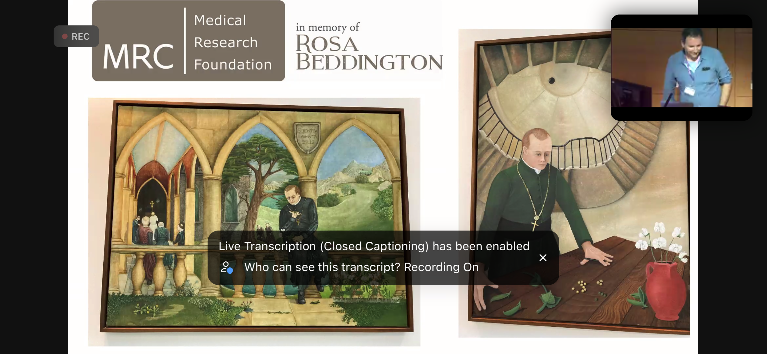
Nic Tapon concluded the symposium by thanking the speakers, the Crick Events team, sponsors and co-organisers; Shankar Srinivas, Alex Gould and Caroline Hill. Nic explained how two of Rosa’s paintings of Mendel that hang in the Crick were especially appropriate this year because it’s the 200th anniversary of Mendel’s birth.
Thank you to the organisers for their effort in assembling the symposium together, chairing sessions and for the smooth execution of the programme. I also thank all the speakers for sharing their stories, both of their science and their memories of Rosa Beddington.
 (1 votes)
Get involved
Create an account or log in to post your story on the Node.
Sign up for emails
Subscribe to our mailing lists.