

Gene expression in the formation of the avian hypoblast
Posted by Rahma Mohamed, on 12 October 2026
This summer, I was able to gain insight into where evolution and development meet during my time working in the laboratory of James DiFrisco. My project focused on understanding the evolution and development of the avian hypoblast and investigating the role of gene expression in its organisation and development.
The hypoblast is an embryonic cell layer that gives rise to extraembryonic tissues. Despite not directly forming the embryo, the hypoblast formation is an essential step before gastrulation – the process which forms the 3 germ layers (endoderm, mesoderm and ectoderm). These germ layers give rise to major tissues and organs in our body. Therefore, understanding the role the hypoblast plays in early development can provide insights into potential causes of developmental failures in the chick and early human development.
A question in evolutionary developmental biology is whether the hypoblast is conserved. This means asking whether there are key characteristics that are maintained during evolution. These characteristics could include signalling pathways or gene expression patterns. We are trying to understand, in this case, whether the hypoblast shares similar molecular signatures across vertebrates.
The early stages of endoderm development are closely assoicated with the hypoblast and can be split into two waves. The first wave begins with cells becoming specified as primitive endoderm prior to gastrulation. This has a role in axis and yolk sac formation. The second wave occurs as a result of gastrulation, allowing cells to form the definitive endoderm.
With R, we used UMAPs to see how cells are organised into clusters based on gene expression similarities. This visualisation was done by condensing single-cell RNA data from chick embryos into a 2D format for easier visualisation. After obtaining UMAPs for several markers, we chose to focus on two well-known endoderm markers Gata6 and Sox17. These markers play key roles in early embryonic development, and their activity as transcription factors allows endodermal identity to be maintained.
Using existing data sets, I first used spatial data to get an idea of how we would expect the expression of our chosen markers to look. My first step was the collection of chicken embryos after incubation at 38°C. HCR-FISH was then carried out, where probes bind to the RNA of a gene of interest during detection. These embryos were then mounted, imaged using spinning disk confocal microscopy to see exactly where the gene expression was and analysed using Fiji to give our final images (Fig. 1).
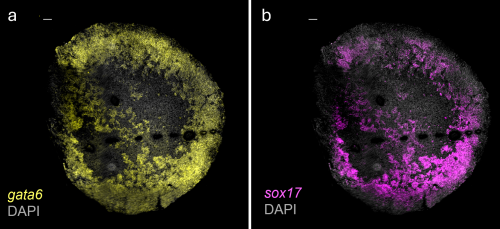
Fig. 1: HCR data images to visualise gene expression at stage EGK XI. Images obtained after completing HCR-FISH on chicken embryos and imaging using spinning disk confocal microscopy (a) Gata6 (b) Sox17
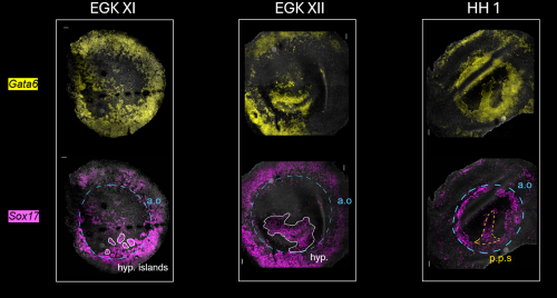
Fig. 2: Expression of Gata6 and Sox17 shifts from the area opaca to embryonic regions in later developmental stages. a.o: area opaca, hyp.: hypoblast, p.p.s: pre-primitive streak. (a) Stage EGK X: area pellucida consists of an upper layer and clumps of cells (hypoblast islands) which indicate a developing lower layer. (b) At stage EGK XI, there are high levels in the area opaca and the hypoblast islands which are moving anteriorly to later merge to form the hypoblast (c) Stage HH 1: Sox17 and Gata6 expression is reduced in the area opaca, shifting to the pre-primitive streak. Timepoints are from post-egg laying, growing in 38 °C incubator.
In the end, I was able to successfully visualise gene expression of markers Gata6 and Sox17 in the chick embryos and to trace the progression of these endodermal signatures to see how it shifts between different regions of the embryo (Fig. 2). In future, this project would benefit greatly by including more replicates and timepoints for validation. Extending this research to other vertebrates such as fish and frog would allow comparative work to begin allowing us to test the hypothesis that the hypoblast is conserved across vertebrates.
I would like to give a huge thank you to the DiFrisco Lab as well as the Briscoe Lab, where I was warmly welcomed and given a space to further enrich my knowledge in developmental research. I am especially grateful to my supervisor, Arsham Nejad Kourki, and to Sumit Garai for their invaluable guidance over the 9 weeks. Furthermore, I would like to thank the Francis Crick Institute for providing this insightful experience, and to the Rosa Beddington Fund for funding my project.
Get involved
Create an account or log in to post your story on the Node.
Sign up for emails
Subscribe to our mailing lists.