The position will provide bioinformatics support, develop new pipelines, and organize practical courses to help our scientists analyze and interpret their next-generation sequencing (NGS) data.
Bioinformatics position in the new informatics cluster supporting the Novo Nordisk Foundation Centers for Stem Cell Biology and Protein Research.
The position will provide bioinformatics support, develop new pipelines, and organize practical courses to help our scientists analyze and interpret their next-generation sequencing (NGS) data.
Bioinformatics position in the new informatics cluster supporting the Novo Nordisk Foundation Centers for Stem Cell Biology and Protein Research
The NNF Centers for Protein Research (CPR) and Stem Cell Biology (DanStem) at Faculty of Health & Medical Sciences at the University of Copenhagen are establishing a new integrated informatics core that will cover a range of technologies. This new core was recently expanded to include bioinformatics as a result of our establishing a new platform for single cell sequencing. We are currently looking for an ambitious bioinformatician who will assist with NGS analysis with in this core and provide new bioinformatics pipelines to individual scientists with in both centers. The individual will be imbedded in a bioinformatics core with expertise that will include NGS, proteomics, and image data analysis. The bioinformatician will join the centers starting from November 2017 or upon specific agreement.
DanStem was established as a result of a series of international recruitments coupled with internationally recognized research groups focused on insulin producing beta cells and cancer research already located at the University of Copenhagen. Current work in DanStem spans a broad range of stem cell and developmental biology and involves state of the art technologies including imaging and single cell NGS technologies. DanStem currently hosts 12 research groups, which are working in wet lab and tissue culture laboratories that are equipped for working with both Embryonic Stem Cells (ESCs) and Induced Pluripotent Stem Cells (iPSC). Together with the Novo Nordisk Foundation Center for Protein Research DanStem is currently operating three scientific platforms: an Imaging facility, a flow cytometry platform and a sequencing facility. The platforms are staffed with experts and provide advanced training along with assistance in using the state-of-the art technologies. DanStem and the CPR recently collaborated to establish a single cell sequencing platform and the new informatics platform will build on the collaborative bridges initiated as a result of this new platform.
CPR has been established at the Faculty of Health and Medical Sciences, University of Copenhagen, to promote basic and applied discovery research on human proteins of medical relevance. The Center comprises a wide range of expertise and resources including proteomics, protein production, bioinformatics and general characterization of disease mechanisms. The Programme for Disease Systems Biology consists of the Big Data Management platform, which handles the data from other technology platforms, as well as two research groups that cover many systems-level aspects of biology and medicine, including the integration of molecular-level data and healthcare data, including biomedical texts.
Job description
The job will sit at the nexus of the two centers and astride several existing joint efforts to bridge computational technologies including image analysis and single cell sequencing. The tasks associated with this position will include providing bioinformatics support to our scientists, constructing pipelines for their analyses, and running courses that will help them acquire bioinformatics literacy. In particular this position will be responsible for developing and executing projects with other center scientists and helping them to make the best use of their own data and that already in the public domain.
Your profile
Candidates must hold a Master and/or PhD degree in computational biology/bioinformatics or have similar relevant educational background and experience
Proficiency in at least one of the scripting languages (e.g. Python) is required
Sound statistic and fluent programming skills in R/Bioconductor is essential
At least two years of experience in the analysis of NGS data, particularly data related to gene expression and regulation
Knowledge of UNIX-like operating system
Experience in interdisciplinary collaborations or bioinformatics services will be an advantage
Scientific understanding of molecular biology and genomics is beneficial
Good English communication skills, both oral and written, are prerequisite for the successful candidate
We offer you
Stimulating, challenging and multifaceted research environment
Possibility for continued education and training
Attractive employment conditions
The employment has an initial duration until the end of 2020 with a possibility of extension. The employment is scheduled to start November 2017 or upon agreement with the chosen candidate. The place of work is at DanStem, University of Copenhagen, Blegdamsvej 3B, Copenhagen. Salary, pension and terms of employment are in accordance with the provisions of the collective agreement between the Danish Government and AC (the Danish Confederation of Professional Associations). In addition to the basic salary a monthly contribution to a pension fund is added (17.1% of the salary). Depending on qualifications, a supplement due to qualifications may be negotiated.
The application must include
Motivation letter
Curriculum vitae incl. education, experience, previous employments, language skills and other relevant skills
Copy of diplomas/degree certificate(s)
Three letters of reference
Questions
For further information about the position, please contact
The University of Copenhagen wishes to reflect the diversity of society and welcomes applications from all qualified candidates regardless of personal background.
Only applications received in time and consisting of the above listed documents will be considered.
Applications and/or any material received after deadline will not be taken into consideration.
Application deadline: October 15th 2017
Founded in 1479, the University of Copenhagen is the oldest university in Denmark. It is among the largest universities in Scandinavia and is one of the highest ranking in Europe. The University´s six faculties include Health and Medical Sciences, Humanities, Law, Science, Social Sciences and Theology www.ku.dk
Our lab studies the early events of craniofacial development using teleost fish, zebrafish (Danio rerio) and Mexican tetra fish (Astyanax mexicanus) as model animals. We are particularly focused on evolution and development of the jaw structures. These include dentitions, sensory systems, and jaw bones. Formation of these structures involve complex interactions and we are interested in finding the molecular signaling pathways which regulate their development. This research addresses the questions related to the human congenital malformations and evolutionary development.
Applications are invited for fully funded studies which lead to the Master degree in Oral Biology in the area of molecular mechanisms of craniofacial development. Looking for a highly motivated student with good communication skills. Fine understanding of any of the following subjects would be highly beneficial: molecular biology, developmental biology and cell biology.
Our lab located at the Department of Oral Biology, College of Dentistry, University of Manitoba. The University is located in Winnipeg, the largest city in the province of Manitoba, Canada. The city has a rich cultural environment and the region provides exciting opportunities for outdoor exploration and recreation in all seasons
Please forward your complete CV with a brief statement of your research interest to Devi.Atukorallaya@umanitoba.ca
This article is recent news from the Babraham Institute in Cambridge, view the original post here and the Nature Communications research paper here.
Credit: Ms Laura Woods. This is a fluorescent microscopy image of the womb of an elderly mouse. The bright green areas show cells which respond to pregnancy hormones. As a mouse ages, the womb becomes less sensitive to hormones, as shown by the uneven, patchy green. This is reflected in the developmental problems we see in the offspring from these older mothers.
Deciding to start a family later in life could be about more than just the age of your eggs. A new study in mice suggests the age of a mother’s womb may also have a part to play. This work, led by Dr Myriam Hemberger at the Babraham Institute and the Centre for Trophoblast Research in Cambridge, UK, is one of the first to look at the effects of age on womb health and it is expected to lead to new research into human pregnancies.
The risks of complications during pregnancy all increase with age. A woman in her late 30s is twice as likely as a younger woman to have a stillbirth, she is also 20% more prone to giving birth prematurely and more likely to experience conditions such as pre-eclampsia. Many of these effects have been linked to the deteriorating quality of ageing egg cells. Yet, this new research, published in Nature Communications, reveals that older wombs also have more trouble adapting to pregnancy.
By examining first pregnancies in aged mice, the team showed that, for mice as for humans, the risk of complications increases with age. Closer examination revealed that the wombs of older mothers are less able to support the growth of a placenta, meaning the developing young have poor blood supply, which slows their growth and can cause birth defects.
The co-first authors were Ms Laura Woods and Dr Vicente Perez-Garcia. Speaking about the findings, Ms Woods said: “We wanted to enhance our understanding of the increased risks of pregnancy in older mothers. When we compared mice who have their first litter in middle age to their younger counterparts, we found that the lining of the uterus does not respond as well to pregnancy hormones and this delays placenta formation. By identifying the key pathways affected by age in mice we have a better idea of what to look for in humans.”
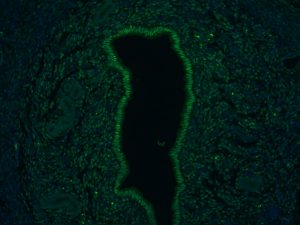
Credit: Ms Laura Woods A fluorescent microscopy image of the womb of a young mouse for comparison. Note that the bright green areas are much more consistent.
Understanding the potential risks of pregnancy with age is an increasingly important issue. In the UK, more and more women are starting families later and in 2015, 53% of UK births were to women aged 30 or over. A 2016 report by the Human Fertilisation and Embryology Authority showed that freezing eggs for later use is also growing in popularity. In 2001, just 29 women opted for the treatment, rising to 816 by 2014.
Lead author, Dr Hemberger, Group Leader in Epigenetics at the Babraham Institute, said: “Overall, our study highlights the importance of the ageing uterine environment as a cause of reproductive decline in female mice. This is one of the first times that the considerable impact of age on pregnancy has been studied in detail beyond the effects of egg fitness. More research will be needed to establish if and how our results translate to humans.”
The shorter lifespan of mice means that they are useful for studying the effects of age on pregnancy but these results cannot always be directly applied to human pregnancies. These new results will help to guide long-term studies in humans but it is not yet clear what the implications of these findings will mean for family planning and human healthcare. It is clear that other factors besides egg quality may need to be considered when planning a family.
As a member of the Royal College of Obstetricians and Gynaecologists, Ashley Moffett, Professor of Reproductive Immunology at the University of Cambridge and expert on placenta formation, said: “We know that the so-called Great Obstetrical Syndromes, in particular pre-eclampsia are more common in older women but it’s still not clear why. Although more work is needed to demonstrate this effect in humans, this study could help advance research into these important questions”.
About the Babraham Institute:
The Babraham Institute receives strategic funding from the Biotechnology and Biological Sciences Research Council (BBSRC) to undertake world-class life sciences research. Its goal is to generate new knowledge of biological mechanisms underpinning ageing, development and the maintenance of health. Research focuses on signalling, gene regulation and the impact of epigenetic regulation at different stages of life. By determining how the body reacts to dietary and environmental stimuli and manages microbial and viral interactions, we aim to improve wellbeing and support healthier ageing.
Publication Reference
Woods L, Perez-Garcia V, Kieckbusch J, Wang X, DeMayo F, Colucci F, Hemberger M, (2017) ‘Decidualisation and placentation defects are a major cause of age-related reproductive decline. Nat Comms 8. http://dx.doi.org/10.1038/s41467-017-00308-x
Research Funding
This work was supported by the Biotechnology and Biological Sciences Research Council (BBSRC; Strategic Programme Grant BB/J004499/1), the Centre for Trophoblast Research, University of Cambridge, UK and a Medical Research Council (MRC) DTP studentship.
Animal Statement:
As a publicly funded research institute, the Babraham Institute is committed to engagement and transparency in all aspects of its research. Animals are only used in Babraham Institute research when their use is essential to address a specific scientific goal, which cannot be studied through other means. The main species used are laboratory strains of rodents, with limited numbers of other species. We do not house cats, dogs, horses or primates at the Babraham Research Campus for research purposes.
The use of animals in this study was performed in full compliance with UK Home Office regulations and with approval of the animal welfare committee (AWERB) at The Babraham Institute, and with the relevant project and personal licences. The study used the wild type C57BL/6 strain of mice housed at the Babraham Institute. Pregnancy was studied in mice between the ages of 8 and 12 weeks or 40 and 58 weeks. Embryo and placenta development were assessed around 11.5 days into pregnancy.
Written By: Margaret Frank, Ora Hazak, Samuel Leiboff, Heike Lindner, Concepcion Manzano, Lena Mueller, Michael Raissig, Annis Richardson, Adam Runions, Sebastian Soyk
A systems biology approach to understanding development
The 2017 FASEB meeting “Mechanisms in Plant Development” launched with a keynote by Philip Benfey (Duke University, USA) about the current understanding of root development. He presented an analysis of the dynamics of the SHORTROOT (SHR) – SCARECROW (SCR) – CyclinD6 (CYCD6) signaling network during root stem cell specification and the effects of specific carotenoid derivatives on lateral root prepatterning. Further, he proposed a minimal gene regulatory network consisting of epidermis-expressed SHR and MYB36 combined with CIF2 peptide treatment, which is sufficient for endodermis differentiation and Casparian Strip formation. Finally, he presented a new root phenotyping system for direct monitoring of maize root architecture in an agricultural field setting.
Next, Neelima Sinha (University of California, Davis, USA) discussed mechanisms of resistance to the parasitic plant dodder (Cuscuta spp.) in tomato. Using transcriptomics and whole genome resequencing in resistant and susceptible cultivars of tomato (S. lycopersicum), she mapped loci conferring resistance to dodder infection. Furthermore, she discussed the roles of lignin deposition in cortex cells for dodder resistance. She outlined a signaling cascade involving NBS-LRR proteins as well as MYB and AP2-type transcription factors that controls the expression of genes involved in lignin biosynthesis in response to dodder infection.
Finishing the first session, Anthony Bishopp (University of Nottingham, UK) presented mathematical approaches to explain vascular patterning in the root. His models suggest that vascular patterning can be established independent of initial asymmetry in auxin concentrations. Instead, a model solely based on spatial constraints and root growth are sufficient to enable vascular pattern in its final form to emerge from a simple genetic network, explaining the vast diversity seen in different flowering plants.
Alternations between generations
The first session on Monday was under the theme Alternations between generations and started off with Thomas Dresselhaus (University of Regensburg, Germany) presenting fascinating work on a number of small signaling peptides that regulate pollen tube attraction and other aspects of fertilization in Arabidopsis and maize. He finished by presenting a recent transcriptome study regarding zygotic genome activation in developing maize embryos showing that 10% of genes are active a few hours after fertilization.
Next, Rita Groß-Hardt (University of Bremen, Germany) explained how the phytohormone ethylene regulates polytubey during pollen tube reception by triggering disintegration of the non-receptive synergid. In addition, she discussed the developmental consequences associated with the attraction of supernumerary pollen tubes.
The next speaker, Stewart Gillmor (CINVESTAV, Mexico), discussed contrasting findings concerning the ratio of maternal and paternal transcripts during early embryo development. He highlighted his recent paper showing delayed paternal rescue of zygotic patterning mutants, and presented a new, comprehensive hybrid embryonic transcriptome suggesting a maternal gene expression bias in young Arabidopsis embryos.
Then, Duarte Figueiredo (Uppsala BioCenter, Sweden) highlighted the recently published work on the role of auxin in seed coat development. Auxin is produced in the endosperm and exported to the integuments in an AGL62-dependent manner, where it seems to repress Polycomb Complex genes and thus allows for seed coat formation.
Sharon Kessler (Purdue University, USA) presented recent work on the importance of the subcellular localization of MLO proteins during pollen tube reception. Additionally, she discussed ongoing work to understand the molecular basis of natural variation in fertility, resistance to powdery mildew, and ovule number.
Moritz Rövekamp (University of Zurich, Switzerland) unraveled the ancient function of the CrRLK1L family in cell elongation during vegetative development by studying the single family member MpFERONIA in the liverwort Marchantia polymorpha. He summarized preliminary results suggesting an additional role for MpFERONIA in sexual reproduction.
Finally, Siobhan Braybrook (The Sainsbury Laboratory, UK) discussed cell wall adaptations of brown algae to different tidal zones. She presented her studies on the re-hydration of artificial cell walls and embryo development in Fucus – and highlighted the importance of charged cell wall polysaccharides for marine plant survival.
Short Range Signaling
Developmental mechanisms are often regulated by release of signaling molecules or mechanical forces and perception of these signals by neighboring cells. The afternoon session of the meeting on Short Range Signaling was opened by Dolf Weijers (Wageningen University, the Netherlands) discussing new players in early embryo development. Weijers showed that polarly-localized SOK proteins influence the direction of cell divisions during early embryo development. In addition, using a whole-genome approach to determine the transcription patterns along the ontogeny axis of the root, his lab found that the main patterns are expressed in two opposing gradients. He proposed the idea that in the root meristem cells gradually switch from undifferentiated to differentiated cells.
Elizabeth Haswell (Washington University in Saint Louis, USA) introduced the role of mechanotransduction and mechanosensitive ion channels in development. In Arabidopsis ten genes encode proteins closely related to the canonical MscS mechanosensitive ion channel from E. coli. Haswell showed that one of these Arabidopsis genes, MSL8, protects pollen grains from the osmotic shocks intrinsic to their development and showed that it serves as a mechanoreceptor.
The next speaker of this session, Joop Vermeer (University of Zurich, Switzerland) focused on cell volume control in endodermal cells overlying the emerging lateral root. He used live cell imaging to observe the cytoskeleton dynamics during lateral root growth through the endodermal cell layer. Vermeer showed that during this process microtubule arrays (visualized with MAP4) rapidly reorganise in the surrounding tissues and that a specific Arabidopsis MAP protein was induced in these cells.
Keiko Torii (University of Washington, USA) took us through the enigmas of receptor-ligand-based mechanisms defining stomata development. Torii discussed the autocrine role of EPF1 peptide in the meristemoid cells as well as ERECTA-LIKE1 receptor distribution in different cell types and its correlation with cell-specific identity.
The next talk by Ora Hazak (University of Lausanne, Switzerland) was dedicated to the role of the first differentiating root vascular tissue called protophloem in sensing of CLE peptides. Protophloem is a dynamic continuously differentiating tissue providing the sugars and hormones necessary for the maintenance of the root meristem. Her findings show that this tissue is responsible for the sensing of high levels of CLE peptides that results in locking of the protophloem in an undifferentiated state and later in the suppression of the root growth.
Nathanaël Prunet (Caltech, USA), presented the results of a collaborative work with Toshiro Ito and Frank Wellmer on the developmental origin of the phenotype of the Arabidopsis superman mutant, whose flowers show an increase in stamen number. Prunet nicely showed that flower patterning associates with auxin depletion and cytokinin accumulation at the boundaries between floral whorls and organs. He could demonstrate that this balance is inverted at the boundary between whorls 3 and 4 in the superman mutant. Prunet and collaborators showed that auxin biosynthetic genes are direct targets of SUPERMAN; they proposed that disinhibition of local auxin biosynthesis at the boundary between stamens and carpels is the cause of the superman phenotype.
Juan-Jose Ripoll (University of California, San Diego, USA) helped us to understand fruit growth processes. After fertilization fruit undergoes a dramatic increase in size that is essential to nourish and protect the growing seeds inside. Ripoll exploited Arabidopsis thaliana as a working platform and combined modeling, live imaging technologies and molecular genetics to follow the mechanisms that regulate fruit size and shape.
Antia Rodrigues-Villalon (ETH Zurich, Switzerland) closed the session with a discussion of the role of phosphoinositide homeostasis in vascular tissues development. During differentiation, vascular conductive cells fully (in xylem) or partially (in phloem) lose their organelles and cytoplasm to become conductive tissues. Rodrigues-Villalon showed that imbalance in phosphoinositide homeostasis at the plasma membrane suppresses differentiation both in xylem and phloem conductive cells by affecting vacuolar biogenesis.
Pluripotency and regeneration
Rüdiger Simon (University of Düsseldorf, Germany) opened the Pluripotency and regeneration session by discussing incoherent feedback loops active during the regulation of shoot apical meristem development in Arabidopsis. From genetic studies and fluorescence microscopy he identified previously uncharacterized members of the CLE peptide family that potentially restrict meristem size from the periphery of the CLAVATA3 domain.
Next, Michael Scanlon (Cornell University, USA) presented genetic and genomic analyses of maize leaf development. He showed the genetic dissection of the narrow sheath (ns) mutant, which results from mutations in two WOX3 transcription factor genes, NS1 and NS2. From RNA-seq and ChIP-seq experiments on laser microdissected shoot apices he concluded that NS1 is expressed in the pre-primordial margins and functions in recruiting the lateral domain of the maize leaf.
Agata Burian (University of Silesia, Poland) then proposed a microRNA-mediated signaling pathway that regulates axillary meristem development in Arabidopsis. Using live imaging she visualized miRNA expression patterns at the position of future axillary meristems that suggest roles of miRNAs in the timing of axillary meristem release.
Akira Iwase (RIKEN, Japan) started the second half of the session, with a discussion on wound-induced cellular reprogramming by the AP2/ERF transcription factor WOUND INDUCED DEDIFFERENTIATION 1 (WIND1). He presented ChIP-seq and RNA-seq experiments that identified direct WIND1 target genes, which may contribute to restoring pluripotency at sites of wounding.
Next, Ken Birnbaum (New York University, USA) talked about mechanisms of tissue reorganization during regeneration, using wounded Arabidopsis root tips as an experimental model. He used single-cell transcriptomics and time-lapse microscopy to show that patterning of the root stem cell niche is re-established across groups of cells rather than from a cryptic stem cell niche, and that this re-establishment of cellular organization is preceded by the rapid expression of MONOPTEROS (a root specification gene) independent of auxin.
Remko Offringa (Leiden University, the Netherlands) presented a molecular switch for rejuvenation and polycarpy in flowering plants that is regulated by members of the AT-HOOK MOTIF CONTAINING NUCLEAR LOCALIZED (AHL) protein family. He showed that enhanced AHL expression rejuvenates axillary meristems in the Arabidopsis inflorescence and allows Arabidopsis plants to flower and set seed multiple times. He concluded his talk with a discussion on the potential to exploit rejuvenation biology for increased crop productivity.
Laura Ragni (University of Tübingen, Germany) closed the session with a presentation about periderm development in the Arabidopsis hypocotyl and root. She discussed roles for Programmed Cell Death (PCD) and abscission during periderm establishment, and further presented data showing that periderm development is not affected when lateral root development is compromised.
Gene Regulatory Networks
From roots to fruits, the gene regulation and regulatory networks session showed how NGS technologies are addressing fundamental plant developmental questions in diverse plant species; including topics such as: (1) How roots cope with a “harsh world”, (2) How fruits decide which tissue develops into the flesh, (3) What defines fruit shape? (4) How the plant hormone, auxin does so much, and (5) Why different mutations in the same gene generate diverse phenotypes.
Siobhan Brady (University of California, Davis, USA) focused on how tomato roots develop the exodermis, a lignified and suberinised layer in the root. Using cell type specific sequencing technologies, they identified an evolutionary shift in cell-type specific expression, resulting in exodermis cells’ monopolizing suberin related genes. During drought these suberin genes are upregulated in commercial tomato varieties (Solanum lycopersicum), but not in the drought-tolerant wild tomato (Solanum pennelli), highlighting exodermis suberinization as a possible target for drought tolerance.
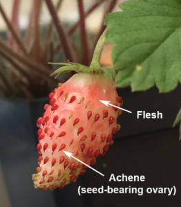
The “inside-out” fruit of strawberry was the focus of the talk by Zhongchi Liu (University of Maryland, USA). Strawberry fruit flesh develops from the inflorescence stem tip, leaving the maturing achenes, seed-containing ovaries, on the outside. Classical experiments demonstrated that strawberry fruit flesh development is regulated by auxin released by the seed-containing ovaries, but the molecular mechanism of communication between the seed and the maternal tissue was unclear. RNAseq and tissue specific auxin measurements, identified bidirectional communication involved in the specification of which tissue becomes the fruit flesh; the endosperm and seed coat produce auxin transported into the stem tip, and there is a novel role for FT in flesh to seed communication.
Next, Lars Ostergaard (John Innes Centre, UK) described how computational modelling and genetics explain the development of heart shaped fruits in Shepherds Purse (Capsella rubella) and the elongated fruits of Arabidopsis thaliana. The FRUITFULL (FUL) gene is required for growth during the post-germination phase and ful mutants have similar phenotypes in both species. They have also identified several additional fruit shape defective mutants by TILLING in Capsella such as heartless, heartbreak and braveheart.
Andrea Gallavotti (Rutgers University, USA) presented research on auxin-dependent transcriptional regulation in maize. Using DAP-seq and ATAC seq, they have identified binding sites for many Auxin Responsive Factors (ARFs), genome-wide. Many known activating and repressing ARFs bind upstream of the same genes (some 60% of ARF target genes), suggesting cooperative or competitive binding may generate auxin’s diverse effects.
Jeff Long (University of California, Los Angeles, USA) explored how different mutations in the same HD ZIP III transcription factors have diverse phenotypic effects in Arabidopsis. Using inducible expression, ChIPseq, and ChIP re-ChIP, they described complex regulatory interactions with transcription factor homo- and heterodimers with both, shared and unique binding sites upstream of important developmental genes, especially in the cytokinin pathway.
Capsella wild type and two fruitfull mutant alleles
A maturing fruit of the wild strawberry studied in the Zhongchi Liu lab
Patterning mechanisms
The session on Patterning mechanisms opened with a talk by Teva Vernoux (ENS de Lyon, France), who discussed the problem of robustly patterning organs at the periphery of the shoot apical meristem (SAM). Combining experiments and computational modeling he showed how spatial-temporal fluctuations in auxin signaling filters out noise to confer robustness on organogenesis, and highlighted a role for the CLAVATA3 domain boundary in the regulation of this process.
Next, Marcus Heisler (University of Sydney, Australia) discussed how phyllotactic patterning in the SAM is limited to the periphery of the meristem. He showed that organs are centered on a small gap between the expression domains of REVOLUTA and KANADI where auxin signaling is maximised, and highlighted how the configuration of this boundary region in the SAM cues both the placement of organs and their dorsoventrality. He also showed that wound-induced auxin depletion in the SAM rearranges the REV and KAN1 expression domains, leading to a new explanation for the influence of wounding on leaf dorsoventrality.
Continuing the theme of patterning boundaries, Annis Richardson (University of California, Berkeley, USA), explained how the understanding of boundary formation in grasses has been impeded by the dearth of mutants with organ boundary defects. She then presented fused leaf 1 (fsl1), a maize mutant with a range of phenotypes related to defective organ boundary formation and maintenance, and summarized work towards identification of the causal mutation in fsl1.
Next, Dominique Bergmann (Stanford University, USA) outlined an integrated picture of stomatal patterning and the establishment of polarities in these lineages. She presented recent work by postdoc Anne Vatén identifying a regulatory circuit involving cytokinin, several cytokinin-regulated CLE peptides and response regulators (A-ARRs), as well as the stomatal transcription factor SPEECHLESS, which provides a means to adaptively regulate stomatal lineages.
Expanding on the theme of tissue polarities Catherine Mansfield (John Innes Centre, UK) presented work investigating coordinated polarities in the leaf, focusing on BREAKING OF ASYMMETRY IN THE STOMATAL LINEAGE (BASL) a protein which polarly localizes to one edge of the cell during stomatal patterning. She showed that broad induction of BASL expression in a developing leaf has coordinated cellular polarities across the entire organ that appear to be independent of the stomatal lineage, consistent with the existence of an intrinsic tissue-wide polarity.
The talk of Aman Husbands (Cold Spring Harbor, USA) followed and returned to the topic of robustly patterning boundaries, focusing on Class III HD-ZIP (HD-ZIPIII) proteins and the role of the StAR-related transfer (START) domain these proteins contain. Using PHABULOSA as a representative protein for the family, he discussed work showing how the START domains – in complex with a so far unidentified ligand – confer switch-like behavior to HD-ZIPIII proteins while simultaneously increasing their transcriptional potency.
Finishing the session, Baoqing Ding (University of Connecticut, USA) discussed the molecular underpinnings of periodic pigment spot patterning in monkeyflowers (Mimulus) by reaction-diffusion. Mutant analysis and transgenic experiments show that these patterns are likely produced by diffusion and reaction of a compound activating anthocyanin formation (a R2R3-MYB activator) and a compound repressing the activator (a R3-MYB repressor), leading to the periodic activation of pigmentation.
Evolution and comparative development
All the investigators in this exciting session generated new molecular, genetic tools in diverse plant model systems to understand the development of complex, ever-evolving plant forms.
Miltos Tsiantis (Max-Planck Institute for Plant Breeding Research, Germany) studies leaf morphological diversity in Cardamine hirsuta, a relative of Arabidopsis thaliana with compound leaves. A computational model of leaf margin development with feedbacks between CUPSHAPED COTELYDON (CUC) transcription factors and the hormone auxin, suggested that changes in transport of auxin by PINFORMED (PIN) efflux carriers and the growth repressive activity of CUCs could generate diverse lobed and compound leaf shapes. Accordingly, the C. hirsutaREDUCED COMPLEXITY (RCO) gene encoding an HD-ZIP-I can reinforce local growth repression by CUCs thus creating compound leaves in C. hirsuta. RCO was lost in A. thaliana and when transformed back into this species it was sufficient to increase leaf complexity but not to cause complete Cardamine-like leaf development— although leaf physiology was altered indicating a potentially adaptive role. The latter was supported by finding positive selection signatures in RCO. Tsiantis suggested that few strong-effect regulators of leaf shape (like RCO) act with additional small effect genes to cause leaf margin diversity. He detailed efforts to isolate those and study their effects on growth
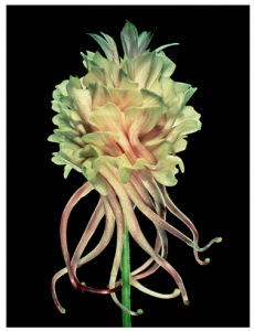
Elena Kramer’s talk (Harvard University, USA) explored the genetic basis of the petal spur, a unique floral structure in columbines. SEM of young petals showed that anisotropic growth contributes to the length of the final petal spur, with sustained cell elongation in Aquilegia species correlating with longer spurs. Transcriptomic profiling of developing petals, followed by inducible gene silencing by VIGS and in situ hybridization, show that division coordination by a TCP transcription factor is necessary for normal spur growth, and the function of STYLISH (SHI/STY) homologs is necessary for nectary development at the spur tip. QTL mapping of spur length in interspecies populations has identified a single mendelian locus that controls the presence and absence of spurs. Nonetheless, the genetic architecture of spur morphology may involve multiple loci, not shared between all columbine species.
Knowing that floral traits such as color, scent, and shape work together to attract pollinators, Cris Kuhlemeier (University of Bern, Switzerland) explored the genetics underlying the complex evolutionary history accompanying changes in pollinator preferences in petunia. QTL mapping of crosses between pollinator types (P. secreta, bee pollinated and P. exerta, bird pollinated) identified alleles of MYB transcription factor ANTHOCYANIN2 (AN2) as key to the evolution of bee pollination in P. secreta. Both species evolved from an ancestor carrying a pseudogene version of AN2 caused by a frame-shift deletion, leading to a white, hawkmoth-pollinated flower. However, in P. secreta, AN2 was ‘resurrected’ by an additional, codon-restoring deletion. Loci for UV floral display mapped to regulatory sequences of flavonol biosynthesis, together suggesting that somewhat simple molecular changes can underlie significant phenotypic and ecological niche changes.
Yoan Coudert (CNRS / Natural History Museum Paris, France) used the leafy shoot axis of the model bryophyte, Physcomitrella patens, to understand gametophytic branching. Unlike flowering plant sporophytes, where polar auxin transport and PIN genes are required to regulate lateral outgrowth, computational models of branch patterning in moss suggest that apolar auxin transport best explains observed architectures. Supporting this hypothesis, mutants in P. patens and manipulation of cell-cell diffusion rates show that the PIN auxin efflux carriers are minor contributors to architecture in P. patens, suggesting that not all plant architectures require polar auxin transport and alternative mechanisms exist in basal land plants.
Devin O’Connor (Sainsbury Laboratory, Cambridge University, UK) examined the function of closely-related PINFORMED (PIN) auxin efflux carriers in Brachypodium distachyon. Using fluorescent fusion proteins, Devin saw that Sister-of-PIN1 (SoPIN1), an efflux protein lost in Arabidopsis but conserved in most flowering plants, is expressed in the epidermis of the meristem and localizes asymmetrically towards auxin maxima. PIN1 paralogs BdPIN1a and BdPIN1b, however, express subepidermally in the developing vascular trace, transporting auxin away from lateral organs. Heterologous expression and complementation in Arabidopsis using the Brachypodium PINs showed that the SoPIN1 and PIN1 clades are not functionally equivalent. Loss of SoPIN1 in Brachypodium is sufficient to give a phenotype like PINFORMED1 in Arabidopsis, whereas PIN1 paralogs BdPIN1a and BdPIN1b together cause an internode elongation defect, suggesting that multiple auxin efflux carriers in Brachypodium may together perform the functions of Arabidopsis PIN1.
The RCO expression domainVirus-induced gene silencing (VIGS) in Cardamine hirsuta in the columbine flower (Aquilegia sp.)
Growth dependent morphogenesis
Adrienne Roeder (Cornell University, USA) launched the session by presenting a fluctuation-based patterning mechanism in Arabidopsis sepals (outermost floral organs). Sepals curvature is influenced by randomly patterned giant cells that form through endoreduplication. Giant cell formation requires the AtML1 transcription factor which has a linear, dosage-dependent role based on mutant and transgenic lines. Time-lapse imaging of YFP-AtML1 revealed that AtML1 expression fluctuates in sepal cells. Mathematical modelling of this, suggests that a specific threshold of AtML1 needs to be surpassed during the G2-phase of the cell cycle to allow endoreduplication and subsequent giant cell formation.
Ari Pekka Mahonen (University of Helsinki, Finland) presented elegant work describing how cambium stem cells are formed post-embryonically in the Arabidopsis root. Cell lineage tracing revealed that only pericycle and procambial cells in contact with xylem precursors contribute to the formation of cambium. Single cell lineage tracing and ablation experiments showed that the central cell layer acts as an “organizer” upon which the division capacity of adjacent cells depends. Conditional mutants also revealed a developmental module consisting of auxin, auxin response factors, HD-ZIP III transcription factors and WOX-like transcription factors required for radial cambium formation, secondary growth and correct patterning in the root.
Carolyn Rasmussen (University of California, Riverside, USA) presented a probabilistic mathematical model to predict 3D division plane orientation in plant cells. Time-lapse imaging of young maize leaves expressing a tubulin-marker line allowed for visualization of division plane (pre-prophase band) establishment and the division itself. The iterative model, seeking minimal division wall area and integration of preprophase band orientation predicts the correct transverse division 97% of the time in-silico (compared to 95% in-vivo), which is much closer to reality than previously published 2D models.
Pilar Cubas (Centro Nacional de Biotecnologia, Spain) presented work on identifying gene regulatory networks that regulate axillary bud dormancy. Both developmental and environmental cues can promote or inhibit bud outgrowth and are integrated by BRANCHED1 (BRC1), an inhibitor of bud growth. Transcriptomics analyses in repressed and activated buds in wild-type and brc1 plants identified three HD-ZIP I transcription factors, which together with BRC1 induce ABA biosynthesis in dormant buds triggering outgrowth. The triple mutant has a bushy phenotype when grown under shade conditions, which usually inhibit lateral growth and promote apical dominance.
Sebastian Soyk (CSHL, USA) presented recent work on the negative epistasis effects of MADS-box transcription factors on tomato yield. When stacking a trait that was selected during domestication and is linked to bigger fruit size with the “jointless” locus that facilitates mechanical harvesting, tomato inflorescences became highly branched and semi-sterile. Using a combination of natural alleles and CRISPR engineering, this can be overcome, resulting in a continuum of inflorescence branching, where some hybrid combinations produced single-branched inflorescences and significantly higher yields.
Hernan Lopez-Marin (Max-Planck Institute for Plant Breeding Research, Germany) reported on the identification of the super determinant (sde) mutant that fails to properly initiate axillary meristems in tomato. The SDE gene shows similarities to PRC1-like components but lacks the canonical RING domain. He proposes that a non-canonical PRC1 complex establishes competence for axillary meristem initiation.
Finally, Katy Guthrie (University of Missouri – Columbia, USA) presented work on the Suppressor of sessile spikelet 2 (Sos2) mutant, which suppresses the second spikelet formation specific to the maize clade of grasses. She showed that the Sos2 mutation seems to have roles in inflorescence, spikelet and flower meristems, and that the Sos2 mutation could partially suppress the indeterminate meristem mutation ramosa2.
Environmental Adaptations
The final session of the meeting on Environmental Adaptations kicked off with a talk by Stacy Harmer (University of California, Davis, USA), who introduced us to Sunflower as a model system for studying circadian rhythms. Before dawn, sunflower inflorescences re-orient themselves to face east in preparation for the rising sun. Harmer demonstrated that this ability to anticipate sunrise is dependent upon a spatio-temporal separation of auxin responses between day and night across the East and West halves of the sunflower stem. Notably, she was able to show that inflorescence orientation impacts temperature-dependent pollen shedding, which has a significant influence on the frequency of pollinator visits and yield.
The next talk was by Daniel Chitwood (Independent Researcher), who talked about a new method for measuring topology, called Persistent Homology, can be used to compare diverse 2-dimensional and 3-dimensional shapes. He presented several studies using persistent homology, including comparisons between roots and shoots, grape rachises from the core grape diversity collection in Geneva, NY, and a massive “leaf morphospace” that was generated from 182,707 leaves from different seed plant families. Surprisingly, he was able to show that there is predictive value to these measurements; 30% of the leaves in this massive leaf morphospace could be accurately assigned back to their respective plant families.
Next, Jessica Guseman (USDA-ARS Appalachian Fruit Research Station, USA) shed light on how environmental signals are integrated into the molecular control of shoot architecture in Arabidopsis and Prunus species. TAC1 and LAZY1 (members of the IGT gene family members) work together to influence branch angles. Guseman demonstrated that each of the IGT genes responds differently to light treatments and mutant combinations with core components of the light signaling pathway. Further work investigating how light influences the IGT family will help us to understand complex architecture problems in trees, for example – how understory branches respond to shading from the top of the tree.
Next, Ben Holt (Associate Professor at the University of Oklahoma, USA) took us through a rigorous series of experiments looking into the functional specificity for how the CONSTANS (CO) DNA binding complex regulates expression of the floral promoting factor FT. Using three-dimensional protein structures, chromatin modeling, and amino acid sequence alignments between the CO DNA binding complex and its functional analog, the Nuclear Factor-Y complex, Holt was able to pinpoint specific residues of the CO DNA binding domain that specify the enhancer element binding capacity of this complex.
Salehin Mohammad (University of California, San Diego, USA) spoke next on the connection between AUX/IAA signaling and glucosinolate biosynthesis. Mohammad used RNA-sequencing, glucosinolate profiling (with LC-MS), and genetic stacking, to delineate a signaling pathway that connects IAA5, IAA6, and IAA19 with the regulation of aliphatic glucosinolate biosynthesis. Surprisingly, he was able to demonstrate that mutants in this pathway are drought sensitive, suggesting a new role for aliphatic glucosinolates in abiotic stress tolerance.
The meeting concluded with an illuminating talk by Heike Lindner (The Carnegie Institute of Washington, USA) on the phenotyping capacity of GLO-Roots, a robotic system that uses a luciferase reporter system to track root architecture over time. Underground environmental pressures (for example water availability and nutrient composition) have the ability to shape root architecture over evolutionary time. Lindner is leveraging the geographic and genetic diversity of Arabidopsis to perform a GWAS for environmentally selected root architectural traits across a collection of ecotypes.
Conference attendance summary:
The 15th FASEB meeting “Mechanisms in Plant Development” 2017 took place once again at the Vermont Academy in Saxons River, VT. The organizers Prof. Doris Wagner (University of Pennsylvania), and Prof. Marja Timmermans (University of Tübingen) invited 26 speakers (including the keynote) representing the international “who is who” in plant developmental biology. With an almost equal ratio of 46% to 54% female and male speakers, respectively, this meeting represents an outstanding example for gender equality in science. One interesting fact, however, was that the vast majority of the invited female speakers were from US Universities (8 vs. 3 from Europe, 1 from Japan), whereas the vast majority of European invited speakers were male (9 vs. 3 from US, 1 from China/substituted by Australian). Unfortunately, 3 invited speakers could not attend the meeting and were substituted by equally qualified scientists. To promote visibility of early career researchers, 30 short talks were selected from submitted abstracts. 15 of those talks were given by PIs in pre-tenure stages of their career (7 female, 8 male), 3 by grad students (2 female, 1 male), and 12 by postdocs (4 female, 8 male, representing roughly the ratios of submitted abstracts). The outstanding program and the unrivaled reputation of this bi-annual meeting attracted 145 international attendees (54.5% male and 45.5% female) with 57.2% attendees coming from US Universities and research institutions, 32.6% from Europe, and the rest from other countries all over the world. Of the 119 attendees that disclosed their ethnic background, 12% were from underrepresented minorities (hispanic/latino, black/african american, native american/pacific island), 27% were asian, and 61% were caucasian.
One special feature of this conference is and has always been that speakers present a good amount of unpublished data to receive input and opinions from other experts in the field. In addition, all 145 attendees were invited to bring posters, and evening poster sessions in a relaxed environment gave the opportunity for very fruitful discussions and great interactions between young researchers and established PIs. An additional poster session was dedicated to grad students to promote visibility and guarantee interaction with more established scientists. Two “meet the speakers” lunches and the newly established “career development” lunch with topics as diverse as “how to publish your science” to “science and social responsibilities” encouraged interactions across different career stages. Overall, the friendly, enthusiastic, encouraging and inclusive environment of this conference is a shining example of how conferences should be and offers diverse role models for young scientists.
During the election of the organizers for the 2019 meeting, the community of plant developmental biologists once again proved how progressive it is, and voted Kenneth Birnbaum (New York University) and Jill Harrison (University of Bristol) as the next organizers. We are excited for the next FASEB meeting “Mechanisms in Plant Development” 2019 at St. Bonaventure University and we thank the Vermont Academy for being a great host of the past FASEB meetings.
A postdoctoral position is available in the laboratory of Dr. Sophie Astrof at Thomas Jefferson University to study roles of cell-extracellular matrix (ECM) interactions in cardiovascular development and congenital heart disease. We have recently discovered that progenitors within the second heart field (SHF) give rise to endothelial cells composing pharyngeal arch arteries. Projects in the lab focus on the role of ECM in regulating cell fate and migration of SHF-derived cells, and in the regulation of signaling between endoderm-, mesoderm-, and ectoderm-derived tissues during the morphogenesis of the pharyngeal vasculature. The project will combine genetic manipulation, embryology, cell biology, and confocal imaging. My laboratory is a part of a modern and well-equipped Center for Translational Medicine at Jefferson Medical College (http://www.jefferson.edu/university/research/researcher/researcher-faculty/astrof-laboratory.html) located in the heart of Philadelphia. For further information on our work, please see the following publications: Wang et al., Development, 143:88-100, 2016 and Wang et al, Developmental Biology 421:108-217, 2017. To apply, please send a letter of interest detailing your expertise, CV and names and contact information of three references to sophie.astrof@gmail.com
This post, funded by The Leverhulme Trust (grant RPG-2017-249) “Dynamic Self-Organisation of Stochastic Intracellular Transport”, is available to study the transport of E-cadherin on microtubule cytoskeleton in the group led by Dr Natalia Bulgakova: www.sheffield.ac.uk/bms/research/bulgakova.
Cell-cell adhesion plays a vital role during the development and adult life of multicellular organisms. The focus of the Bulgakova group is on discovering roles and regulation of cell-cell adhesion using Drosophila as a model organism. We have recently discovered that microtubule organization in epithelial cells is driven by cell shape, and is required for distribution of E-cadherin, a key cell-cell adhesion molecule, around cell periphery. The successful applicant will study the mechanisms of how E-cadherin is transported along a dynamic network of microtubules to achieve correct and reproducible E-cadherin distribution. The project will employ a combination of molecular genetics, immunohistochemistry and live imaging. The work will be a part of collaboration with the group of Dr Lyubov Chumakova, University of Edinburgh, who will complement and support experimental findings by creating a mathematical model of E-cadherin transport.
The successful applicant will have a PhD or equivalent experience relevant to studying cell-cell adhesion or intracellular trafficking, knowledge of working with genetic model systems and be highly motivated. Experience in using Drosophila, molecular biology, confocal microscopy, and live imaging, and knowledge of cell-cell adhesion, cell polarity and intracellular trafficking will be an advantage. The successful candidate will be expected to have excellent interpersonal and communication skills, be highly independent, and committed to research in a fast-moving and competitive field.
For more information and to apply, please see http://www.jobs.ac.uk/job/BEA999/research-associate-in-cell-and-developmental-biology/
As a medical doctor specialising in respiratory diseases, with limited experience in real lab-based research, I was disappointed how little could be done for patients with end-stage lung disease (ESLD). Although respiratory diseases account for the third highest mortality of non-infectious disease deaths worldwide and lung cancer is the most common cancer (World Health Organization, 2011), treatment is largely supportive and lung tissue destruction is irreversible.
I started my PhD with Emma Rawlins at the Gurdon Institute, University of Cambridge, as part of the Wellcome PhD Programme for Clinicians by looking at human induced pluripotent stem cells (hiPSCs). These seemed to provide an opportunity to apply the discoveries of developmental biology to treat disease (Takahashi and Yamanaka, 2006). Differentiating patient-specific iPSCs into any relevant cell type could potentially be used for disease modelling and drug screening, and also bring us a step closer to cell-based therapies including personalised organogenesis. Research on murine lung development as a model for human lung development has been crucial to current protocols for differentiating human iPSCs into lung (Firth et al., 2014; Firth et al., 2015; Ghaedi et al., 2013; Huang et al., 2015; Huang et al., 2013; Longmire et al., 2012; Mou et al., 2012; Wong et al., 2012; Wong et al., 2015). I initially started work on iPSC directed differentiation to lung and wanted to use human embryonic lungs simply for validation purposes. However, it became evident very quickly that little developmental biology had been done on human embryonic lungs and it was not clear how closely the published step-wise iPSC differentiation protocols actually reflect human embryonic and foetal lung development.
Characterising the different progenitor populations in both the developing mouse and human embryonic lung seemed to me an essential part of regenerative medicine. The molecular understanding of developing human lungs was inadequate, as human lung development had not been characterised using cells derived from the human embryonic lung itself, but only using iPS-derived lung cells.
Mouse lung development
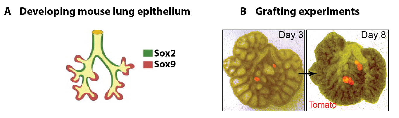
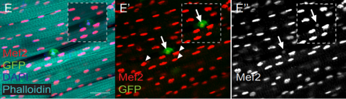
In parallel to my human work, I also worked on mouse lung development since it has been so extensively used as a model for human lung development. Considering the various anatomical and molecular differences already known between mouse and human lungs, I felt it was important to understand both mouse and human lung development more fully in order to ascertain to what extent the mouse findings could indeed be applied to human lungs. In both species, the lung develops as a blind-ending tube which branches multiple times, a process which results in the production of gas-delivering bronchioles and gas-exchanging alveoli. We know from lineage tracing experiments that the most important epithelial progenitor population in the developing mouse lung is found in the branching tips, giving rise to all the various epithelial cell types in the adult lung, both bronchiolar and alveolar cells (Rawlins et al., 2009). In mice, these epithelial tip cells are classically Sox9+ and initially give rise to the neighbouring Sox2+ stalk cells which are bronchiolar progenitors and give rise to the future airway (Fig.1 A). Using technically challenging grafting experiments I showed, among other things, that extrinsic signalling is the key in determining the tip and stalk progenitor cell fate, but that a significant degree of plasticity is present and that this gradually decreases throughout development (Fig.1 B) (Laresgoiti et al., 2016). I got a lot of practice in dissecting the epithelium free from any mesenchyme, which was very useful later on for the human work (Fig. 3B).
The finding that the environment is crucial in determining distal tip progenitor fate suggested that it would be possible to define in vitro culture conditions for human embryonic lung epithelium, both for the self-renewal of tip cells and their differentiation towards alveolar and bronchiolar lineages. This was an exciting realization as successful identification of culture conditions for developing human lungs would make molecular genetics possible.
Figure 1 A. Branching mouse lung is characterised by Sox9+ tips and Sox2+ stalks (reproduced from (Wang et al., 2013). B. Epithelial tip progenitors were microdissected from donor E12.5 (A) or E16.5 (B) Tomato (Rosa26RmTmG/+) lungs and grafted into the mesenchyme of unlabelled E12.5 hosts. Hosts/grafts were cultured for 8 days. Tip grafts grew (shown), integrated into the host lung and formed a lumen (not shown).
Main challenges of working on human embryonic lungs
One of the main limitations of working with human tissue is the lack of continuous tissue supply. This changed quickly and unexpectedly when I went to one of the Cambridge pubs to discuss my research plans and get some advice from a post-doctoral friend. He mentioned the “TransEuro” trial going on at the University of Cambridge – a clinical trial in which brain tissue from terminations of pregnancies is used for neurosurgical transplantation into treatment-resistant Parkinson’s patients. This meant that developing lung tissue was regularly discarded without any scientific benefit. This seemed to be the ideal source of human lung tissue for validation of my attempts at iPS-derived directed differentiation, but it quickly became clear what a huge potential an in-depth molecular characterisation of human embryonic lungs would have for the entire lung developmental and stem cell biology community. Additionally, the Joint MRC/Wellcome Human Developmental Biology Resource (www.HDBR.org) coordinated tissue harvesting from all over the UK via London and Newcastle. This meant that we had lung tissue provided to us multiple times per month from all over the country. The developmental age ranged from 5 to 20 weeks’ developmental age (7-22 weeks’ gestational age) and the samples used had no known genetic abnormalities.
Another challenge was that tissue used for sequencing and in vitro culture had to be as fresh as possible. Initially, we did not want to take any chances and dissection usually occurred as soon as possible after tissue was obtained, which was often late in the day. Experiments frequently continued until the early hours of the morning. I must say I was grateful once it became clear that overnight storage in specifically designed embryonic tissue storage medium did not affect gene expression, reproducibility and viability!
Since this was an exciting new area with huge potential, I was very grateful for the help I received from two amazing undergraduate/masters students, Oriol Caritg and Dawei Sun, who assisted me with my human work, as well as another PhD student, Jo Johnson.
Main achievements
Molecular comparison of human and mouse embryonic lungs
Molecular characterisation of human embryonic and foetal lungs using immunostaining, qRT-PCR and RNAseq transcriptome analysis revealed both major similarities and differences between mouse and human lung epithelial progenitors (Nikolić et al., 2017). Interestingly, 96% of orthologous genes that were expressed in human tips were also present in mouse.
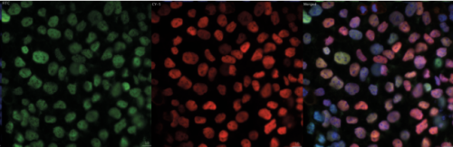
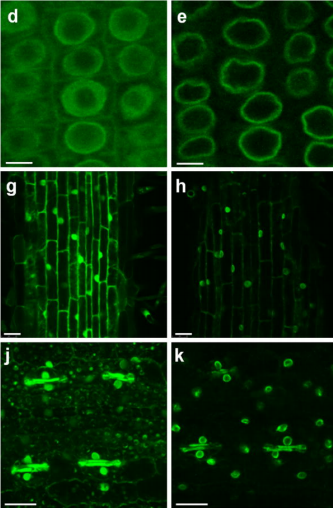
The most important difference was the discovery that the classical stalk marker SOX2 is expressed in the human SOX9+ distal tip progenitors (Fig. 2), in stark contrast to the expression pattern in mice. We showed that this is a true interspecies difference and cannot be explained by relative age. This is an exciting finding, since Sox9 has been used as a classical mouse tip marker and Sox2 as a classical mouse stalk marker, with a clear demarcation between them (Chang et al., 2013; Liu and Hogan, 2002). My work suggests that SOX2 cannot be used as a unique stalk marker in human embryonic lungs and for validation during iPS-derived directed differentiation. Furthermore, there were more subtle differences in tip signalling pathways between mouse and human (Figure 3). The same signalling pathways were present, but these were wired in fundamentally different ways. For example, BMP2 and BMP7 were highly enriched in human tips, whereas Bmp4 was enriched in the mouse (Bellusci et al., 1996); IHH was expressed in human lungs whereas mouse lungs express Shh (Bellusci et al., 1997).
Figure 2. In the early stages of lung development, Sox2 is only expressed in the stalk in mouse (E11-15), whereas in human SOX2 expression extends into the tip (weeks 5-16).
On the one hand these data provide support for the use of mouse as a model for human lung development, but on the other hand they question its applicability in view of the many genetic differences, the functional consequences of which are not yet known. Therefore, to study the functional consequences of manipulating these genes, a genetically modifiable system for studying human lung development is required.
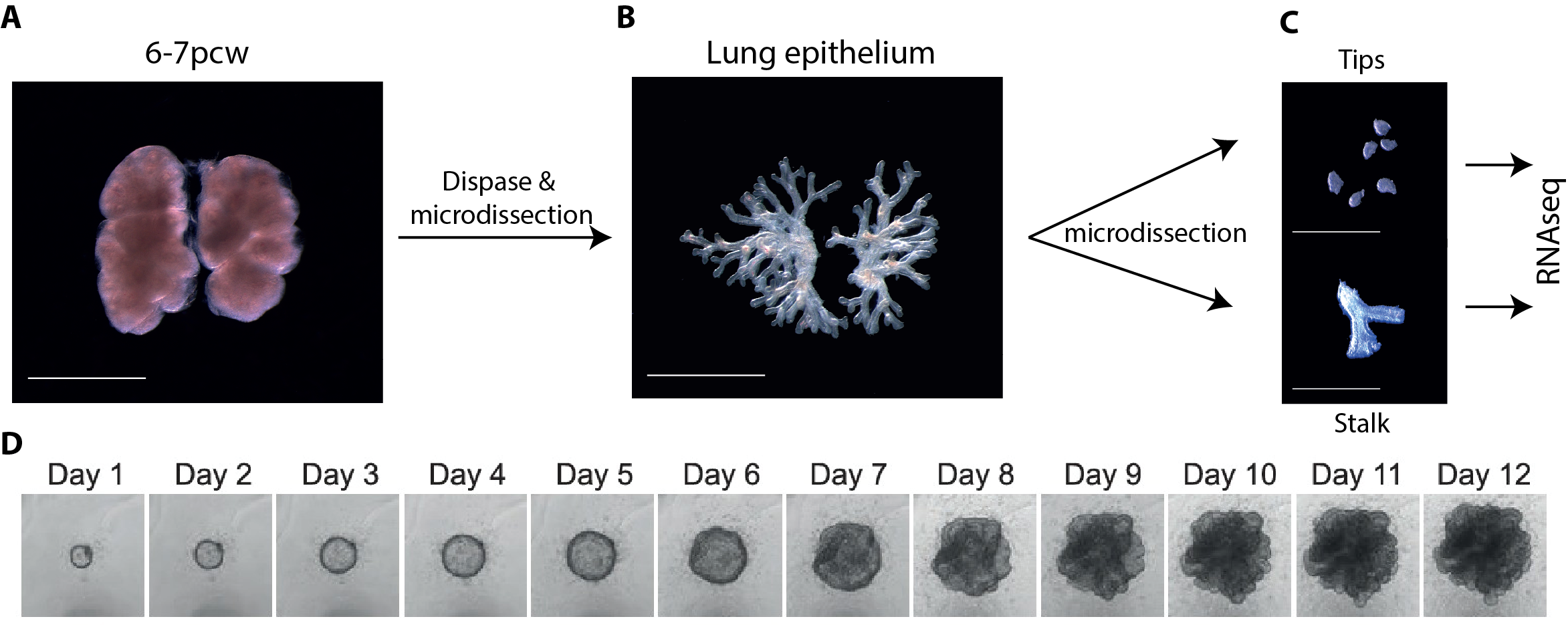
Figure 3 A-C. 6-7 post-conception week (pcw) human embryonic lung was incubated in dispase for 2 minutes, upon which the epithelium was microdissected free from any macroscopically obvious mesenchyme. The epithelium was further microdissected into tips and stalk, and then processed for RNAseq. D. Bright field images of one tip taken every 24 hours for 12 days cultured in self-renewing media (R-SPONDIN1, EGF, Noggin, FGF10, FGF7, CHIR99021, SB431542).
Self-renewing 3D organoid culture of human tip progenitors
We therefore developed culture conditions which have the ability to expand human embryonic lung epithelial stem cells as an indefinitely self-renewing, expanding population of undifferentiated cells with a organoid forming efficiency of 100% (Figure 3). The concept is similar to capturing the inner cells mass of a blastocyst as a self-renewing embryonic stem cell line. Our lung organoids could also be frozen and thawed, and the technical advice obtained from Meritxell Huch’s lab within the Gurdon Institute was immensely helpful. This now allows the in vitro study of human embryonic lung development without the need to obtain fresh tissue for every experiment.
However, in mouse, the same culture conditions were not sufficient to maintain mouse lung epithelial progenitor cells after several passages. This suggests that acquiring a self-renewing state is possible with further optimisation of the culture conditions, but the required signalling mechanisms are likely to be different. This was not surprising based on the transcriptome comparison between mouse and human tip progenitors.
RNAseq of cultured tips and stalks further validated my self-renewing culture conditions, since key genes are shared by cultured tip, cultured stalk and fresh tip cells without expression of differentiation markers. This suggests that the in vitro conditions mimic the in vivo conditions very closely.
Furthermore, single cell efficiency was high at about 35% and unpublished results showed that we were able to genetically manipulate human epithelial tip progenitors using CRISPR-Cas9. Knocking out SOX9 led to loss-of-self-renewing state phenotype, showing that SOX9 expression is essential for organoid maintenance. What this means is that we can now study human lung progenitor self-renewal in vitro in a genetically modifiable system which is extremely efficient without introducing chromosomal abnormalities, and which reproduces what is happening in vivo. This opens up a whole new research field of studying the mechanism of self-renewal and differentiation.
In vivo and in vitro differentiation as proof for a self-renewing state
One of the most important proofs that we had actually achieved a self-renewing state was the ability to differentiate our expanded organoid cells towards both bronchiolar and alveolar lineages. If real stem cells do it, then our expanded ones should do it do. Indeed, human embryonic epithelial lung progenitors, which had been expanded as 3D organoids, could be differentiated in vitro towards both alveolar and bronchiolar lineages. Similarly, xenotransplantation of organoid cells mixed with E13.5 dissociated mouse lungs, into the mouse kidney capsule led to efficient bronchiolar differentiation and the appearance of ciliated cells, mucus cells and basal cells, although alveolar differentiation was much less efficient. This strongly supported our hypothesis that a self-renewing state had been achieved.
We have also performed xenotransplantation into bleomycin-injured adult mouse lungs which showed highly efficient integration and proliferation of intratracheally administered single organoid cells. It also showed that the organoid cells can differentiate into bronchiolar cells, suggesting that lung identity is retained within the organoids. Such efficient engraftment has not been achieved previously. This is an exciting new model with many future possibilities.
Impact and future directions
In conclusion, the platform that we have developed for in vitro analysis of the embryonic human lung will be the first system available for functional experiments on human embryonic lungs. It will allow human embryonic lung development to be studied using modern molecular genetic techniques. The fact that we can genetically modify our organoid system also means that we can use CRISPR-Cas9 to identify novel genes required for alveolar and bronchiolar differentiation. This has been done in pancreatic development using iPS-derived cells (McGrath et al., 2015). However, our approach would be more reliable, as we would be doing this with real lung progenitors, albeit expanded ones, rather than iPS-derived ones. This research has the long-term potential to be transformative for lung regeneration by disease modelling, specifically by determining the role of genetic variants associated with lung diseases, by developing improved therapies for premature neonatal lung maturation, and also for in vitro differentiation of iPS cells. It has been claimed that iPSCs offer unparalleled opportunities to model lung development and disease using human cells. I would argue that our culture system using real human embryonic lung cells provides a more authentic and reliable platform to model lung development and disease.
From a clinical point of view, I hope that the work will make a difference to at least three groups of patients:
Patients with end-stage lung disease: our work will guide iPS-derived directed differentiation, which could then be used to recellularise a decellularised lung or lobar scaffold to be used for lung transplantation without the need for immunosuppression.
Premature neonates with lung failure due to immaturity: the 3D organoid culture system we developed will allow the study of late foetal development using real lung progenitors rather than iPS-derived ones. Understanding how the lung matures in these final stages of lung development will be crucial to improve survival for babies born prematurely.
Patients with rare congenital lung conditions. Gene editing in our 3D culture system will enable disease modelling of conditions such as surfactant protein B and C deficiencies.
Publishing first and fast versus publishing “big”
Every scientist would like their work to be recognised as important and groundbreaking by those in their field. However, as far as career progression is concerned, the journal chosen often appears to be far more important than the actual quality of the work. It seems to me that journal impact factor is used as a substitute for quality by those who are not experts in a particular field, or who do not understand the impact of the work. In my experience, important factors in choosing a journal include the speed of the review process, the availability of person-power to do the revision work, the awareness of competitors, and related guesswork of what stage those competitors have reached and whether their work is yet of a standard suitable for publication. During our paper revision period, I was in full-time clinical training and I was reliant on the goodwill of my colleagues for the revision experiments, especially Oriol Caritg and Quitz Jeng. On reviewing all these aspects, we chose a journal known for its efficient and fair review process rather than focusing simply on journal impact factor. Put differently, we chose to publish first and fast, rather than attempting to publish “big”. I remain hopeful that our work (as well as that of others) will be judged purely by its contents and the impact it has on the field, rather than by the impact factor of the journal in which it was published.
Marko Nikolić
Wellcome Trust/CRUK Gurdon Institute & Wellcome Trust/MRC Stem Cell Institute,
Liu, Y. and Hogan, B. L. M. (2002). Differential gene expression in the distal tip endoderm of the embryonic mouse lung. Gene Expr. Patterns2, 229–233.
Takahashi, K. and Yamanaka, S. (2006). Induction of pluripotent stem cells from mouse embryonic and adult fibroblast cultures by defined factors. Cell126, 663–676.
Our latest monthly trawl for developmental biology (and other cool) preprints. See last year’s introductory post for background, and let us know if we missed anything
It all went very meta this month with Matthew Cobb’s PeerJ preprint about a forgotten experiment in preprints from the 1960s. The story was highlighted in Science, a journal, Cobb explains, whose editor disparaged the initial preprint experiment in the 60s. Elsewhere, doubts were raised about last month’s headline-making human embryo CRISPR paper, as highlighted on the Niche (which also highlighted Shoukhrat Mitalipov’s response to the prepint).
“T47D_rep2 and b1913e6c1_51720e9cf were two Hi-C samples. They were born and processed at the same time, yet their fates were very different. The life of b1913e6c1_51720e9cf was simple and fruitful, while that of T47D_rep2 was full of accidents and sorrow…”
The preprints were hosted on bioRxiv, PeerJ and arXiv. Use these links to get to the section you want:
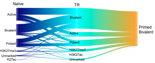
Resolution of Reprogramming Transition States by Single Cell RNA-Sequencing. Lin Guo, Xiaoshan Wang, Mingwei Gao, Lihui Lin, Junqi Kuang, Yuanbang Mai, Fang Wu, He Liu, Jiaqi Yang, Shilong Chu, Hong Song, Yujian Liu, Jiadong Liu, Jinyong Wang, Guangjin Pan, Andrew P. Hutchins, Jing Liu, Jiekai Chen, Duanqing Pei
Inhibition of granulocyte ROS production by opioids prevents regeneration. Elodie Labit, Lise Rabiller, Christophe Guissard, Mireille Andre, Christine Rampon, Corinne Barreau, Beatrice Cousin, Audrey Carriere, Margaux Raffin, Gilles Mithieux, Mohamad Ala Eddine, Bernard Pipy, Anne Lorsignol, Sophie Vriz, Cecile Dromard, Louis Casteilla
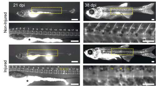
Tracking ectopic vertebra formation in injured zebrafish, from Lopez-Baez, et al.’s preprint
Hemimetabolous genomes reveal molecular basis of termite eusociality. Mark C. Harrison, Evelien Jongepier, Hugh M. Robertson, Nicolas Arning, Tristan Bitard-Feildel, Hsu Chao, Christopher P. Childers, Huyen Dinh, Harshavardhan Doddapaneni, Shannon Dugan, Johannes Gowin, Carolin Greiner, Yi Han, Haofu Hu, Daniel S. T. Hughes, Ann-Kathrin Huylmans, Carsten Kemena, Lukas P. M. Kremer, Sandra L. Lee, Alberto Lopez-Ezquerra, Ludovic Mallet, Jose M. Monroy-Kuhn, Annabell Moser, Shwetha C. Murali, Donna M. Muzny, Saria Otani, Maria-Dolors Piulachs, Monica Poelchau, Jiaxin Qu, Florentine Schaub, Ayako Wada-Katsumata, Kim C. Worley, Qiaolin Xie, Guillem Ylla, Michael Poulsen, Richard A. Gibbs, Coby Schal, Stephen Richards, Xavier Belles, Judith Korb, Erich Bornberg-Bauer
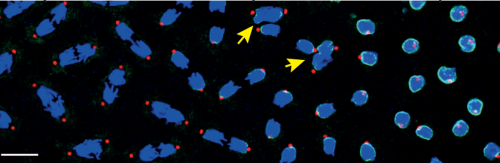
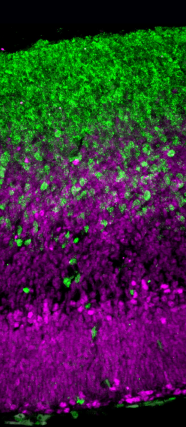
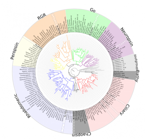
Structural and functional diversity of a dense sample of retinal ganglion cells. J. Alexander Bae, Shang Mu, Jinseop S. Kim, Nicholas L. Turner, Ignacio Tartavull, Nico Kemnitz, Chris S. Jordan, Alex D. Norton, William M. Silversmith, Rachel Prentki, Marissa Sorek, Celia David, Devon L. Jones, Doug Bland, Amy L. R. Sterling, Jungman Park, Kevin L. Briggman, H. Sebastian Seung, the EyeWirers
An improved ATAC-seq protocol reduces background and enables interrogation of frozen tissues. M. Ryan Corces, Alexandro E. Trevino, Emily G. Hamilton, Peyton G. Greenside, Nicholas A. Sinnott-Armstrong, Sam Vesuna, Ansuman T. Satpathy, Adam J. Rubin, Kathleen S. Montine, Beijing Wu, Arwa Kathiria, Seung Woo Cho, Maxwell R. Mumbach, Ava C. Carter, Maya Kasowski, Lisa A. Orloff, Viviana I. Risca, Anshul Kundaje, Paul A. Khavari, Thomas J. Montine, William J. Greenleaf, Howard Y. Chang
Active ribosome profiling with RiboLace. Massimiliano Clamer, Toma Tebaldi, Fabio Lauria, Paola Bernabo, Rodolfo F. Gomez-Biagi, Elena Perenthaler, Daniele Gubert, Laura Pasquardini, Graziano Guella, Ewout J. N. Groen, Thomas H. Gillingwater, Alessandro Quattrone, Gabriella Viero
A Large-Scale Binding and Functional Map of Human RNA Binding Proteins. Eric L Van Nostrand, Peter Freese, Gabriel A Pratt, Xiaofeng Wang, Xintao Wei, Steven M Blue, Daniel Dominguez, Neal A L Cody, Sara Olson, Balaji Sundararaman, Rui Xiao, Lijun Zhan, Cassandra Bazile, Louis Philip Benoit Bouvrette, Jiayu Chen, Michael O Duff, Keri Garcia, Chelsea Gelboin-Burkhart, Abigail Hochman, Nicole J Lambert, Hairi Li, Thai B Nguyen, Tsultrim Palden, Ines Rabano, Shashank Sathe, Rebecca Stanton, Ashley L Louie, Stefan Aigner, Julie Bergalet, Bing Zhou, Amanda Su, Ruth Wang, Brian A Yee, Xiang-Dong Fu, Eric Lecuyer, Christopher B Burge, Brenton Graveley, Gene W Yeo
SEA: The Small RNA Expression Atlas. Raza-Ur Rahman, Abdul Sattar, Maksims Fiosins, Abhivyakti Gautam, Daniel Sumner Magruder, Joern Bethune, Sumit Madan, Juliane Fluck, Stefan Bonn
Enabling cross-study analysis of RNA-Sequencing data. Qingguo Wang, Joshua Armenia, Chao Zhang, Alexander V Penson, Ed Reznik, Liguo Zhang, Thais Minet, Angelica Ochoa, Benjamin E Gross, Christine A Iacobuzio-Donahue, Doron Betel, Barry S Taylor, Jianjiong Gao, Nikolaus Schultz
A Data Citation Roadmap for Scientific Publishers. Helena Cousijn, Amye Kenall, Emma Ganley, Melissa Harrison,David Kernohan, Thomas Lemberger, Fiona Murphy, Patrick Polischuk, Simone Taylor, Maryann Martone, Timothy Clark
Bioinformatics Core Competencies for Undergraduate Life Sciences Education. Melissa A. Wilson Sayres, Charles Hauser, Michael Sierk, Srebrenka Robic, Anne G. Rosenwald, Todd M. Smith, Eric W. Triplett, Jason J. Williams, Elizabeth Dinsdale, William Morgan, James M. Burnette III, Sam S. Donovan, Jennifer C. Drew, Sarah C. R. Elgin, Edison R. Fowlks, Sebastian Galindo-Gonzalez, Anya L. Goodman, Neal F. Grandgenett, Carlos C. Goller, John Jungck, Jeffrey D. Newman, William R. Pearson, Elizabeth Ryder, Rafael Tosado-Acevedo, William Tapprich, Tammy C. Tobin, Arlín Toro-Martínez, Lonnie R. Welch, Robin Wright, David Ebenbach, Kimberly C. Olney, Mindy McWilliams, Mark A. Pauley
A position for a Postdoctoral Fellow is available in the Department of Development and Stem Cells, in the lab of Bill Keyes, IGBMC, Strasbourg, France (www.igbmc.fr/keyes). Previously, the group discovered roles for cellular senescence during embryonic development, and instructing cell plasticity and stemness in tissue regeneration and cancer. We are seeking a postdoctoral candidate to continue and develop these projects. As such, preference will be given to candidates with a proven record in developmental biology or with animal models of regeneration. These projects will involve the use of tissue culture and animal models coupled with high-throughput genomic analysis and molecular biology approaches.
For further information on the project and the work from the lab in general, see recent publications: Keyes et al, Cell Stem Cell, 2011, 8(2) 164-176; Doles et al, Genes & Development, 2012, 26(19): 2144-53; Storer et al, Cell, 2013, 155(5), 1119–1130; Ritschka et al, Genes & Development, 2017, 31(2):172-183
Candidate requirements
Ph.D or M.D./Ph.D. (already obtained or soon-to-be), with at least one first author publication.
Highly motivated person with strong interest in science research.
Interactive person, with ability to work independently.
Good communication skills, and fluency in English is required.
Previous experience in developmental biology will be heavily favored. Experience in molecular biology is desirable.
Work Environment
The candidate will join an international group of scientists, working on highly competitive topics. The candidate will benefit from access to a modern well-equipped laboratory, as well as access to the IGBMC’s renowned Core Facilities and support.
Conditions
Starting date: January 15th, 2018
Salary: salary is available for one year, but additional funding is sought, and the candidate will be supported to apply for competitive external fellowships.
Application procedure
Deadline: October 1st, 2017
All applications must include a CV with a letter describing your motivation and research interests (past and present), and the contact information for 2-3 referees.
Please submit your application to the following email addresses: bill.keyes@igbmc.fr
 (No Ratings Yet)
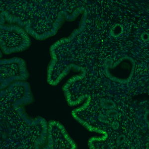
 (1 votes)
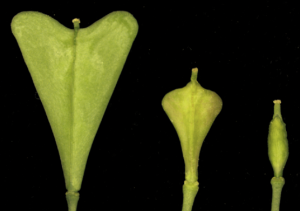

 (10 votes)