Department/Location: Wellcome Trust – Medical Research Council Cambridge Stem Cell Institute, University of Cambridge
Salary: £28,982-£37,768
Reference: PS10005
Closing date: 04 October 2016
Post-doctoral fellow: healthy ageing of human haematopoietic stem cells.
Fixed-term: The funds for this post are available until 31 October 2019 in the first instance.
The Wellcome Trust-Medical Research Council Cambridge Stem Cell Institute is an international centre of excellence for stem cell research and regenerative medicine. Scientists in the Institute collaborate to advance our knowledge of various stem cell types and to perform pioneering work in translational research areas, providing the foundation for new medical treatments. The Institute currently comprises 29 research groups based across 6 sites in Cambridge. In 2018 all researchers will move to a new building on the Cambridge Biomedical Campus.
Applications are invited for a research associate to join Dr Laurenti’s group. We combine state-of-the-art experimental and computational methods to study the unique biological and molecular properties of human Haematopoietic Stem Cells (HSCs).
The post holder will join a new multidisciplinary project funded by the BBSRC. This collaboration between Dr Laurenti and Prof. Göttgens laboratories has as principal aim the investigation of the functional and molecular heterogeneity of HSCs throughout a human lifetime, with a particular focus on the effects of healthy ageing. The project will combine single cell, transcriptomics, epigenomics, flow cytometry, single cell functional assays in vitro and in vivo.
The successful candidate is expected to creatively and independently carry out their own research project, while collaborating on a regular basis with a team of experimentalists and computational biologists. They will also effectively communicate their work in writing and oral presentations at internal meetings and international conferences.
Candidates should hold a PhD in a relevant field. In particular, they will have a strong background either in stem cell biology, haematology, immunology or ageing. Extensive experience with flow cytometry, mouse models and tissue culture is required. The candidate should also possess molecular biology skills. Additional expertise with high-throughput sequencing data and the R programming language would be desirable. Finally, they will have demonstrated scientific achievement with an excellent publication record.
The start date is flexible but can be as early as November 2016.
Once an offer of employment has been accepted, the successful candidate will be required to undergo a health assessment and a security check.
To apply online for this vacancy and to view further information about the role, please visit: http://www.jobs.cam.ac.uk/job/11327. This will take you to the role on the University’s Job Opportunities pages. There you will need to click on the ‘Apply online’ button and register an account with the University’s Web Recruitment System (if you have not already) and log in before completing the online application form.
The closing date for all applications is the Tuesday 04 October 2016.
Here we highlight some developmental biology related content from other journals published by The Company of Biologists.
Meritxell Huch, Wellcome Trust Sir Henry Dale Fellow at the Gurdon Institute in Cambridge, was interviewed as a Cell Scientist to Watch(she’s certainly one for developmental biologists to watch too!).
Kang-Yell Choi and colleagues found that retinoic acid stabilisation of HRas is required for neuronal differentiation during brain development
The Gönczy lab identified Aurora-A kinases as crucial regulators of spindle positioning from worms to humans.
Paul Lasko and colleagues identified the centrosomal protein Bsg25 as a crucial regulator of mitosis and embryonic development.
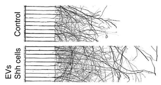
Pamela Yao and colleagues investigateda pool of Sonic Hedgehog contained in extracellular vesicles around neurons.
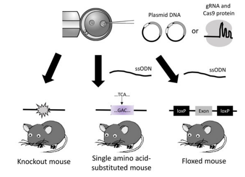
Takashi Yamamoto and colleagues presenteda super-ovulation method paired with genome engineering in mice.
Judith West-Mays and colleagues showed that loss of the transcription factor AP-2β in neural crest cells leads to multiple eye defects, providing a model for early-onset glaucoma
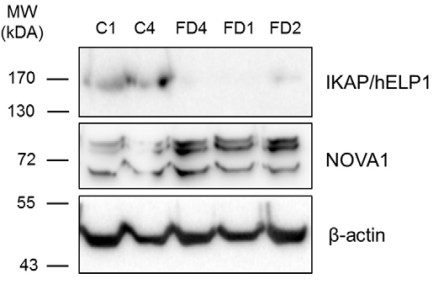
Using patient-derived stem cells, Mylène Hervé and El Chérif Ibrahim implicated miRNAs and the splicing factor NOVA-1 in familial dysautonomia
Kathryn Knight reportedfrom ‘Improving Experimental Approaches in Animal Biology: Implementing the 3Rs’, a conference held in June that showcased the various ways researchers can change their habits to ensure the ethical use of animals in research.
Glen Watson and colleagues found that repair of adult murine hair cells – which is normally only minimal – is enhanced by sea anemone repair proteins.
Adriana Briscoe and colleagues identifiedan expanded number of photoreceptor classes and sexual dimorphism in their expression in a species of nymphalid butterflies
We are seeking a postdoctoral fellow interested in studying social behavior using zebrafish models at the Novartis Institutes for Biomedical Research (NIBR), Cambridge, USA. The goal of the study is to identify genetic and neural basis of social behavior in zebrafish.
Education and preferred qualifications: Ph.D. (completed or near completion) in behavioral neuroscience or related fields; experience in quantitative, large-scale analysis of behavior is a plus; prior experience in zebrafish is not required.
Interested candidates may send their CV, and contact address of three potential referees to ajeet.singh[at]novartis.com. For general information regarding postdoctoral fellowships at the NIBR, please look at http://postdoc.nibr.com/.
Our latest monthly trawl for developmental biology (and other cool) preprints. See June’s post for background, and let us know if we missed anything
It was another bumper month for preprints, as bioRxiv’s Richard Sever celebrated:
This month we found three pieces on the development of the spinal cord, three investigations into gene expression networks in the early fly embryo, some in vivo cell biology featuring centrosomes and kinesins, as well as a lot of evolutionary content and some very cool sounding tools, perhaps the most striking of which is a hand-powered centrifuge modelled on a whirligig! bioRxivprovided the bulk of the preprints, with one coming from PeerJ.
Assembly of Radically Recoded E. coli Genome Segments. Julie E. Norville, Cameron L. Gardner, Eduardo Aponte, Conor K. Camplisson, Alexandra Gonzales, David K. Barclay, Katerina A. Turner, Victoria Longe, Maria Mincheva, Jun Teramoto, Kento Tominaga, Ryota Sugimoto, James E. DiCarlo, Marc Guell, Eriona Hysolli, John Aach, Christopher J. Gregg, Barry L. Wanner, George M. Church
Highly parallel direct RNA sequencing on an array of nanopores. Daniel R Garalde, Elizabeth A Snell, Daniel Jachimowicz, Andrew J Heron, Mark Bruce, Joseph Lloyd, Anthony Warland, Nadia Pantic, Tigist Admassu, Jonah Ciccone, Sabrina Serra, Jemma Keenan, Samuel Martin, Luke McNeill, Jayne Wallace, Lakmal Jayasinghe, Chris Wright, Javier Blasco, Botond Sipos, Stephen Young, Sissel Juul, James Clarke, Daniel J Turner
August was meant to be a quiet month, with researchers (in Europe, at least) leaving the lab for a well earned break and winding down before the start of the academic year…but here on the Node we’ve had another bumper month full of diverse content. Happy reading!
Meetings
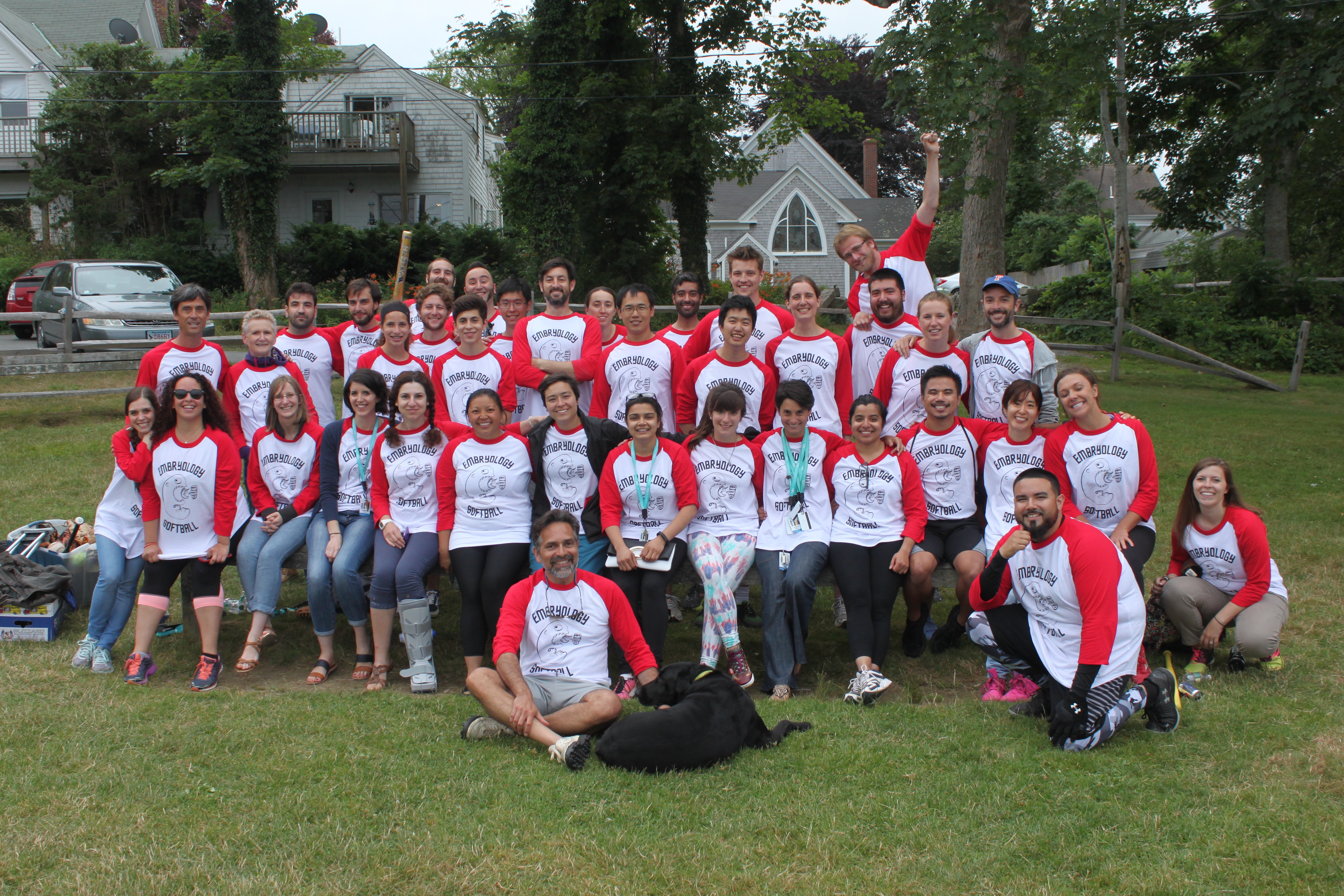
We had a wonderful report from Joaquín Navajas Acedo, Aleisha Symon and Tsai-Ming Lu from the Woods Hole Embryology Course. They really managed to capture the intense excitement and slight mania of the course, and I’m sure made a lot of readers eager to sign up for next year. I reported from the BSD-ISD in Boston: a fantastic conference, and a wonderful city. I also got to meet Yusuff and Mathew who carried on the SDB-BSDB interview chain (long may it continue!).
We started a new series to complement our usual research posts: The People Behind the Papers, highlighting the faces behind recent exciting developmental biology papers. We started with an interdisciplinary team from Heidelberg who uncovered how nuclear pore complexes get into the nuclear envelope, and then featuredThomas Lozito, author of a recent Development paper on lizard tail regeneration.
Applications are invited for a tenure track Assistant Professor faculty position in the Division of Developmental Biology (DB) at Cincinnati Children’s Hospital Medical Center (CCHMC).
DB is home to a large community of developmental biologists (please visit our web site at: http://www.cincinnatichildrens.org/research/divisions/d/dev-biology/default/). We are a highly collaborative interdisciplinary group of basic scientists and clinicians studying fundamental questions in developmental biology, the genetic basis of pediatric disease and regenerative medicine. Being embedded in CCHMC, one of the top ranking pediatric research centers in the world, creates a unique environment in which clinical translation of basic science is greatly enhanced. CCHMC is making a major long-term investment in basic and translational research and developmental biology is one of the pillars in this effort.
DB faculty have access to state of the art subsidized cores, to high-quality graduate and MD/PhD programs, and to training programs for postdoctoral fellows, clinical fellows, residents and clinical faculty. Successful candidates must hold the PhD, MD, or MD/PhD degrees, have recently completed postdoctoral training, have published significant original work and have a mature research plan.
Applicants should submit their curriculum vitae, two to three page research statement focused on future plans, and contact information for three people who will provide letters of recommendation to DB_devbiologists@cchmc.org. Applications must be submitted by December 2, 2016.
The Cincinnati Children’s Hospital Medical Center, and the University of Cincinnati are Affirmative Action/Equal Opportunity Employers. Qualified women and minority candidates are especially encouraged to apply.
So Jeffrey, can you give us the brief history of the Gross lab, and what key questions your group is trying to answer?
JG I started the lab 11 years ago at the University of Texas at Austin, and we moved about a year ago to the University of Pittsburgh Medical School. We’re broadly interested in eye development and diseases. Major questions in the lab focus on optic cup morphogenesis, the epigenetic regulation of retinal and retinal pigment epithelium development and regeneration.
To be honest, we’re rather unfocused, which I think is great! I encourage the students and postdocs in the lab to come up with their own questions and interests and then to go for them; this is the way I was trained as a Ph.D. student with Dave McClay, and I think it is the best way for Ph.D. students and postdocs to learn how to do science. As a result, we’ve worked on a fairly diverse set of questions over the time I’ve had the lab and it really makes this is a fun job.
“I encourage the students and postdocs in the lab to come up with their own questions and interests and then to go for them; this is the way I was trained as a Ph.D. student with Dave McClay, and I think it is the best way for Ph.D. students and postdocs to learn how to do science.”
And Kristen: how did you come to join the Gross lab?
KK I decided to go to the University of Texas for graduate school because it seemed like an exciting research environment that had all the scientific resources one could want but was not a competitive or negative place to work. When choosing the Gross lab, I knew I wanted to study evolutionary questions but I was also concerned with generating a positive relationship with my advisor. The eye is an excellent system to study the evolution and development of complexity and Jeff was amazingly supportive of crazy ideas so it was a good fit. Also, the developmental biology community at UT was and still is a really exciting and supportive group of researchers.
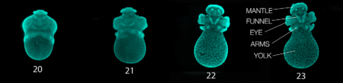
Stages 20-23 embryos stained with Sytox Green, from Fig. 1 in the paper.
While your lab usually works on zebrafish, your Development paper focuses on eye development and photoreceptor differentiation in the longfin inshore squid (Doryteuthis paeleii). So why cephalopods, and this squid in particular, for this project?
JG I’ve always been interested in evo-devo work and wanted us to do some work on eye evolution, but this project really didn’t start until Kristen Koenig joined the lab as a Ph.D. student. The squid project was her idea and I just provided some advice on getting it started – she’s really the brains of the operation!
There is a long history of research using Doryteuthis pealeii at the Marine Biological Laboratory (MBL) in Woods Hole, MA and there is terrific infrastructure for collecting adults and obtaining gametes at the Marine Resources Center at the MBL. Moreover, Kristen and I have spent a significant amount of time at the MBL, which is truly a magical place, so it made a lot of sense to use the resources there and our connections to build Doryteuthis pealeii as a model.There are also worse places in the world than Woods Hole to have to spend a summer working in the lab!
“They are definitely charismatic embryos. It is a pleasure to look at them for as long as I do.”
What’s it like working with squid? They look kind of cute…
KK Doryteuthis pealeii are great to work with. They are a good size, develop at a reasonable pace, they are abundant and resilient to manipulations. Their eyes are quite large during development so for someone interested in visual systems they are quite exciting organisms. As you mentioned, they are definitely charismatic embryos. It is a pleasure to look at them for as long as I do.
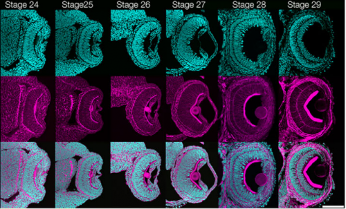
Stages 24-29 of Doryteuthis eye development, from Fig. 2 in the paper
And you get the embryos from Woods Hole?
KK Yes. During most of my dissertation work I spent every summer at the Marine Biological Labs in Woods Hole. As Jeff mentioned, the Marine Resources Center there has an excellent capacity to provide access to adult squid and their eggs. Without the MRC, none of this work would have been possible. I only have embryos during the summer months so I have to plan my experiments for the year during that time.
Could you give us the paper’s key results in a short paragraph?
JG, KK Our research interest is to better understand visual system evolution across the Bilateria from a developmental perspective. We established the squid, Doryteuthis pealeii, as a lophotrochozoan model for complex eye development. Utilizing histological, transcriptomic and molecular assays we characterized eye formation in Doryteuthis pealeii. Through lineage tracing and gene expression analyses, we demonstrated that cells expressing Pax and Six genes incorporate into the lens, cornea and iris tissue, suggesting a convergent involvement in lens formation. We identified the sole source of retinal tissue in the squid and functional assays demonstrated that Notch signaling is required for photoreceptor cell differentiation and retina organization. These assays support a conserved role for notch signaling in neurogenesis in the cephalopod eye.
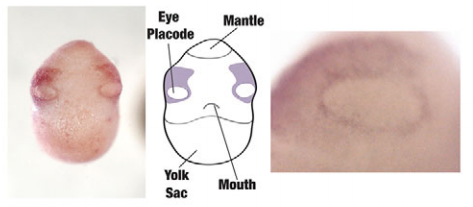
Pax6 expression in the placode stage, from Fig. 11 in the paper
Were there any particularly surprising results that came out of the lineage tracing?
JG, KK Our lineage tracing showed that the cells generating the optic lobe originated from a different place than originally thought. This ultimately has consequences on how we interpret gene expression at these early stages and also suggests that these cells might be migrating to incorporate into the developing optic lobes.
I’m wondering about the question of conservation or convergence (of photoreceptor cell types and the molecular pathways that generate them, in metazoa). What does your work bring to the debate?
JG, KK The photoreceptor cells in the squid retina are rhabdomeric and express r-opsins. There is still a lot to understand about the molecular pathways that generate these cells to enable gene regulatory network comparisons across organisms. Our work supports a role for notch signalling during neurogenesis within the squid retina. This may illuminate a conserved function for notch within pseudostratified neuroepithelia.
“Just seeing the basics of eye morphogenesis continues to blow my mind.”
Was there any part of the work you were most proud of?
KK That’s a tough question because each part of the project presented its own set of unique challenges. The part I still think about regularly is the staging series of eye formation. Just seeing the basics of eye morphogenesis continues to blow my mind. There is so much information in those images and when I look at them it only generates more curiosity and questions.
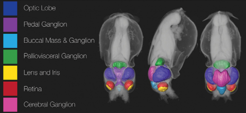
Squid fate map, mapped onto a microCT reconstruction, from Fig. 12 in the paper
And any part of the work that was particularly frustrating or intractable?
KK Some parts of the lineage tracing were challenging. The experiment consisted of hundreds of embryos and had many steps and it was important to keeping track of every individual embryo at each step. Also, half way through the experiment I had to trust the mail delivery system that my specimens wouldn’t get stalled in transit back to Texas and melt into a sad pool of defeat. In the end I knew the experiment was possible but it was just very important to stay consistently focused and organized.
And you’ve left the Gross lab now? What’s next for you?
KK Yes, I just recently left Texas. I am now at the FAS Center for Systems Biology at Harvard University. I am a John Harvard Distinguished Science Fellow and am starting my own lab continuing to study squid eye formation and the evolution and development of visual systems more broadly.
Now you have this squid developmental resource, any future plans to carry on with Doryteuthis? What do you want to know now?
JG I hope to, if Kristen doesn’t mind letting me squat in her lab at Harvard from time to time! The way the lens develops in the squid is fascinating, and I think the cell biology underlying this will be fun to study. I hope that we’ll be able to take a look at this in more detail over the next few summers, and use some of the molecular and imaging tools that Kristen has developed to drill down to the mechanism.
The story of this paper is also the story of my PhD. It begins as most papers and PhDs do: with a distinct and often unrelated starting project or plan. It is great to have a plan. But time and luck and data bend and twist the plan; until it finally breaks and you end up ditching said plan.
Then the real fun can begin.
I – Enter the plan
Vertebrates are an incredibly diverse group of organisms, as demonstrated by the wide range of body shapes and sizes animals belonging to this group can exhibit. This remarkable flexibility was likely a decisive factor allowing for the amazing adaptation of vertebrates to almost all habitats throughout the globe. The general body plan of a vertebrate is usually determined in embryonic development during the process of axial extension, in which the embryo elongates in a rostral to caudal progression by the activity of a very particular population of cells located in the posterior embryonic end. These are the axial progenitors, and their job is to proliferate and give rise to all post-cranial structures of the embryo (Wilson et al., 2009). Yet, an in-depth study of this important cell population was – and still is – hindered by the lack of specific molecular markers.
My initial project was then, to do a molecular characterization of axial progenitors in the mouse embryo; and my supervisor, Moisés Mallo, and I had a nice, neat strategy designed to fish these markers out. At the same time – like a responsible and efficient (and somewhat neurotic) PhD student – I had compiled quite a list of candidate genes that might be expressed and/or could potentially be important to axial progenitors just by searching for truncation phenotypes in existing databases. This way I could screen those genes while waiting for the other experiment’s results. One of these candidate genes was Oct4.
II – How I met Oct4
Oct4 is the gene equivalent of a rock star. Besides being an integral part of the core pluripotency network, Oct4 is also famously known for being an indispensable part of the reprogramming cocktail able to turn somatic cells back into pluripotent stem cells (Takahashi and Yamanaka, 2006). All vertebrate species studied so far seem to have a functional Oct4 counterpart (Frankenberg et al., 2014). In the mouse embryo, Oct4 is expressed as early as the two-cell stage, becoming gradually down regulated as development progresses until its complete silencing in somatic tissues by mid-gestation stages. However, in 2013, a paper came out by DeVeale and colleagues (DeVeale et al., 2013), which showed that, if Oct4 was inactivated during a very particular window of developmental time, some of the resulting mouse embryos could exhibit significant axial truncations, while others showed dramatically shortened trunks, yet perfectly specified tails. The latter were remarkably similar to a variety of transgenic embryos we had described just a few months before, in which the type I TGF-β receptor Alk5 was constitutively active in axial progenitor-containing areas (Jurberg et al., 2013). This showed that not only Oct4 seemed to be important for trunk development, but also suggested the existence of some sort of inverse functional link between Oct4 expression and TGF-β signalling through Alk5 during mouse axis elongation.
But was that really true? At that point, I and my good friend and former labmate Arnon D. Jurberg had been working with this pathway for quite some time, specifically with the Gdf11 mutant. Gdf11 is the main ligand for the Alk5 receptor in vivo, and mutants for both these proteins have characteristically longer trunks due to a delayed trunk to tail transition (Andersson et al., 2006; Jurberg et al., 2013; McPherron et al., 1999). So we definitely knew that these molecules were important for axial progenitors during the extension process. Thus, we decided to put this putative functional connection to the test using our faithful Gdf11 mutant. We reasoned that if premature activation of Alk5 activity was equivalent to the absence of Oct4 during axis formation, and this excess signalling was able to generate shorter trunks, then perhaps the lack of signalling through Alk5 – by absence of Gdf11, for example – could result in an increased expression of Oct4 and that could maybe account for the longer trunks in these mutants. Admittedly, it was a bit of a stretch. But to our vast surprise and amazement, our preliminary results showed us that we might actually be right.
III – Oct4 and the “snakefied” mice
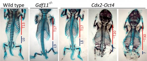
Proving beyond any doubt that Oct4 was really and truly ectopically expressed in Gdf11 mutants took sensibly two years, three different methodologies and a healthy dose of stubbornness. However, in the meantime, we still wanted to know if Oct4 missexpression was responsible for the longer trunks these mutants showcased, which required a way to overcome its progressive downregulation throughout development. Therefore, we chose to take advantage of the Cdx2 promoter, which we had previously demonstrated to be active in axial progenitors, and use it to drive Oct4 expression in transgenic mouse embryos. So it was a dark December day – and a national holiday to boot – when Moisés and I were lucky to dissect together a particularly good batch of E18.5 transgenic embryos courtesy of Ana Nóvoa, transgenic wizard extraordinaire. And the results surpassed even our wildest expectations. Not only were we able to obtain embryos that mimicked the Gdf11 mutant axial phenotype, we also got fetuses that yielded up to 20 or 30 trunk segments (Fig.1). Which meant two things: first, that Oct4 missexpression was most likely the main factor responsible by the longer trunks in Gdf11 mutant embryos; and second, that the trunk to tail transition could be delayed almost indefinitely if you just maintained the right level of Oct4 activity during axial extension.
Fig.1:Sustained Oct4 expression in axial progenitors extends the trunks in mouse embryos. Skeletal analysis of a wild type, a Gdf11-/- and three Cdx2-Oct4 transgenic embryos at E18.5 with increasing strengths in their phenotype. The number of thoracic (T) and lumbar (L) vertebrae is shown.
IV – How the snake got its curves
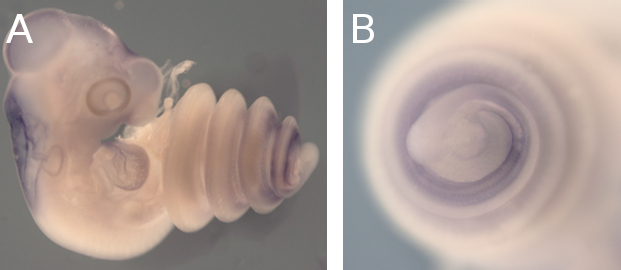
Needless to say, if something has long, rib-packed and organ-filled trunks… well, it’s snakes. Could Oct4 have a role in the making of their long trunks too? For that we needed to check its expression during snake embryonic development, which was most definitely not our area of expertise. We decided to ask for help from Francisca Leal and Martin J. Cohn from the University of Florida, proud owners of a lovely snake facility and source of infinite wisdom in all snake-related matters. We hit the jackpot: expression studies revealed that Oct4 was still present in snake embryos long after it was gone in equivalently staged mice embryos (Fig.2). That made for a classic case of gene expression heterochrony, in which the timing of a gene’s activation or silencing changes from one species to another.
Fig.2:Oct4 expression snake embryos. A. Oct4 expression in a corn snake embryo shortly after it underwent trunk to tail transition. B. Close up of the lower trunk/tail region.
What could have possibly happened during evolution for Oct4 to be so differently regulated in these two groups of organisms? Thanks to Francisca and her sequence analysis skills, we found out that the genomic landscape upstream of the snake Oct4 locus had diverged dramatically from mice and even lizards. Yet, this entire region was extremely well conserved among snakes. As sequence conservation generally means conservation in function, we wondered if these conserved non-coding sequences 5’ of snake Oct4 had any regulatory potential by testing them in mice. We found that a tiny, little 250bp region – the only region conserved in both lizard and snake species – was extremely active on its own, whereas when embedded within larger snake-specific sequences it was a lot less able to drive reporter gene expression. But these were only scattered, isolated pieces of a bigger puzzle. When we used a snake BAC containing the whole genomic context around Oct4 – a kind gift from Isabel Guerreiro and Denis Duboule – and got no expression in transgenic mice, we realized that this entire region most likely operated as a complex regulatory module, composed by a 250bp core regulatory enhancer under the additional control of neighbouring sequences. In the end, we think that the remarkable conservation in snake non-coding sequences probably resulted from strong selective pressures over Oct4 expression due to the adaptation to a fossorial lifestyle, which favoured long trunks for burrowing and prey constriction.
V – Epilogue
So there you have it: the story of how ditching the initial plan can be a good thing, if you just pay attention and let it happen. Especially when results start acquiring a life, to tell a story of their own. Go with the flow. Get really curious and excited. Talk to people, ask for help, and help out even more. And maybe, just maybe, you might stumble across something that, ultimately, turns out to be your dream Evo-Devo project.
Andersson, O., Reissmann, E. and Ibáñez, C. F. (2006). Growth differentiation factor 11 signals through the transforming growth factor-beta receptor ALK5 to regionalize the anterior-posterior axis. EMBO Rep.7, 831–7.
DeVeale, B., Brokhman, I., Mohseni, P., Babak, T., Yoon, C., Lin, A., Onishi, K., Tomilin, A., Pevny, L., Zandstra, P. W., et al. (2013). Oct4 Is Required ˜E7.5 for Proliferation in the Primitive Streak. PLoS Genet.9, e1003957.
Frankenberg, S. R., Frank, D., Harland, R., Johnson, A. D., Nichols, J., Niwa, H., Schöler, H. R., Tanaka, E., Wylie, C. and Brickman, J. M. (2014). The POU-er of gene nomenclature. Development141, 2921–3.
Jurberg, A. D., Aires, R., Varela-Lasheras, I., Nóvoa, A. and Mallo, M. (2013). Switching Axial Progenitors from Producing Trunk to Tail Tissues in Vertebrate Embryos. Dev. Cell25, 451–462.
McPherron, A. C., Lawler, A. M. and Lee, S.-J. J. (1999). Regulation of anterior/posterior patterning of the axial skeleton by growth/differentiation factor 11. Nat. Genet.22, 260–264.
Takahashi, K. and Yamanaka, S. (2006). Induction of pluripotent stem cells from mouse embryonic and adult fibroblast cultures by defined factors. Cell126, 663–676.
Wilson, V., Olivera-Martínez, I. and Storey, K. G. (2009). Stem cells, signals and vertebrate body axis extension. Development136, 2133–2133.
Who are we?
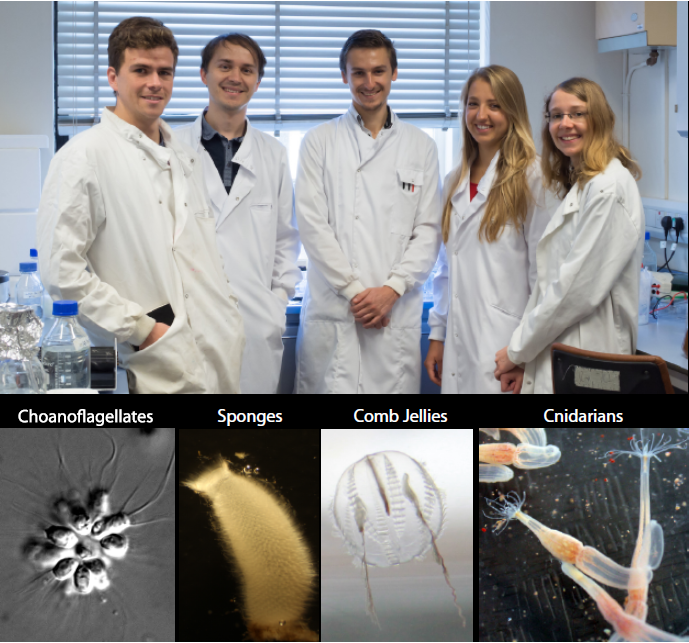
Hi, my name is Ruairi Kavanagh and I’m a Master’s student at Plymouth University. For my dissertation I am based in The Marine Biological Association (MBA). I am carrying out my research in the recently established Burkhardt Lab. Our lab’s research is focused on tracing the origin and evolution of synaptic proteins, to better understand how synapses and neurons evolved in animals. Strikingly, many of the building blocks of animal synapses originated before the first synapses and neurons evolved. We work on a number of model organisms, including choanoflagellates, sponges, cnidarians, and of course ctenophores (Figure 1). We use choanoflagellates, the closest unicellular relatives of animals, to better understand which synaptic signalling machineries for neuronal functions have been co-opted. Sponges, basal animals with no synapses and neurons, are used to elucidate the origin of synapses and neurons. Ctenophores and cnidarians, basal animals with synapses and neurons, are helping us to answer the question, did synapses and neurons originate once or multiple times independently? Typical analyses are centred on identifying the function of key “neuronal” proteins in these organisms. I work on ctenophores and have identified homologs of synaptic proteins from a transcriptome of the ctenophore Pleurobrachia pileus, a local species that is found in Plymouth sound.
Fig. 1: The 2016 “Origin and evolution of synaptic proteins” team. From left: Ruairi Kavanagh, Pawel Burkhardt, Davis Laundon, Florentina Winkelmann and Tarja Hoffmeyer. The organisms we work with in the lab are shown below. Team photo taken by A. Harvey (MBA), photos of organisms by P. Burkhardt (MBA).
Ctenophore characteristics
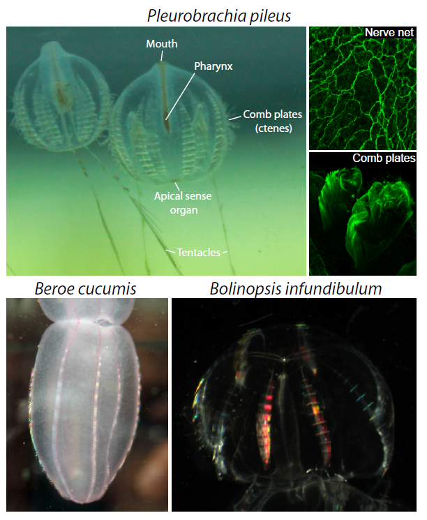
Ctenophores or “comb jellies” are fantastic creatures who have recently come into the evolutionary limelight. There are about 150 known species, almost all of which are pelagic marine predators. Comb jellies contain eight longitudinal rows of ciliated “combs” (or ctenes – ctenophore means “comb-bearing”), which are a distinct apomorphy of the phylum (Figure 2). A nerve net envelops the animal in a kind of polygonal mesh, which is most densely concentrated at the apical sense organ (also called the statocyst). The apical sense organ is thought to detect stimuli such as light, gravity and pressure, to coordinate beating of the ciliated combs, and to control the animal’s orientation. At the oral end, the gastro-vascular tract is connected to the mouth, via a pharynx. At the aboral end there are usually two anal pores, on either side of the apical organ. Interior canals distribute nutrients around the body. Ctenophores are generally found with two tentacles, but some species have secondarily lost the need for them. If tentacles are present, they are lined with sticky cells known as colloblasts, which are used in prey capture (and are another apomorphy). The tentacles of the sea gooseberry, Pleurobrachia (pileus), can be 10-15 times the length of its body. Watching them fish for prey really is an amazing sight. They will extend out their tentacles like big nets and sit in the water column waiting for something to swim into their trap. If they feel the slightest contact, they instantly retract the tentacles towards the mouth, which also triggers them to do a sort of death roll, as they suck down whatever it is they have caught. The two other species that occur here in Plymouth are Beroe cucumis and Bolinopsis infundibulum (Figure 2).
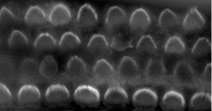
Figure 2: Three ctenophore species, Pleurobrachia pileus, Bolinopsis infundibulum and Beroe cucumis are common all around Britain and Ireland. The nerve net and combs of P. pileus visualized by alpha-Tubulin staining. Photos of P. pileus and B. infundibulum taken by P. Burkhardt (MBA), photo of B. cucumis by A. Harvey (MBA).
Why study ctenophores?
For a long time, ctenophores were considered to be closely related to cnidarians, since they look superficially quite similar, are both gelatinous, contain comparable tissue-level organization, and perform similar ecological roles. However on closer inspection it is clear that there are significant differences between the two phyla. The most obvious difference between them has to do with locomotion. If we look at jellyfish, they move by contractions of the “bell”, and can only move in one direction. Whereas I have already mentioned that ctenophores use their ciliated combs. The coordinated beating of these combs enables the animal to swim forwards or backwards, and allows for a very efficient way of rapidly changing their orientation.
The coordinated beating of combs from the ctenophore Beroe cucumis. Video taken by Ruairi Kavanagh and Pawel Burkhardt (MBA).
In addition, recent phylogenetic studies proposed that ctenophores might after all not be as closely related to cnidarians as previously thought. And here is why: the genomes of two different ctenophores have recently been sequenced. The first ctenophore genome sequenced was the one from Mnemiopsis leidyi, and the second from Pleurobrachia bachei. These studies, using whole genome sequences, showed that ctenophores and cnidarians are actually not closely related. But even more strikingly, these studies showed that ctenophores, and not sponges, are the sister-group to the rest of animals. If true, this would have important implications: for example, as all ctenophores possess synapses & neurons, in contrast to sponges, an independent origin of synapses and neurons in this phylum is possible. Indeed, neuronal components that are found widely in other animal groups appear to be missing from the genomes of P. pileus and M. leidyi, and many neuronal components do in fact not localize to neurons, but to other, non-neuronal cells. Other researchers have called for more detailed phylogenetic analyses and the need to sequence more ctenophore genomes before such contrary conclusions can be drawn. Ctenophores have, after all, been in the spotlight for only a short period of time. Our work will provide valuable insights into ctenophore biology, and will hopefully help to resolve the current dispute on the origin of synapses and neurons.
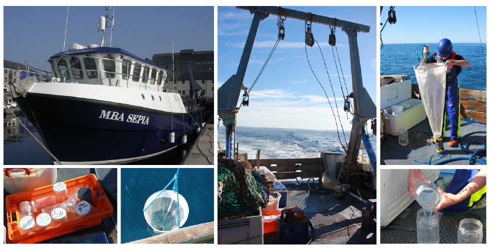
Figure 3: Our research vessel MBA Sepia is used for ctenophore sampling. We use standard plankton nets and do both vertical and horizontal trawls. Photos: P. Burkhardt (MBA).
A day in the lab
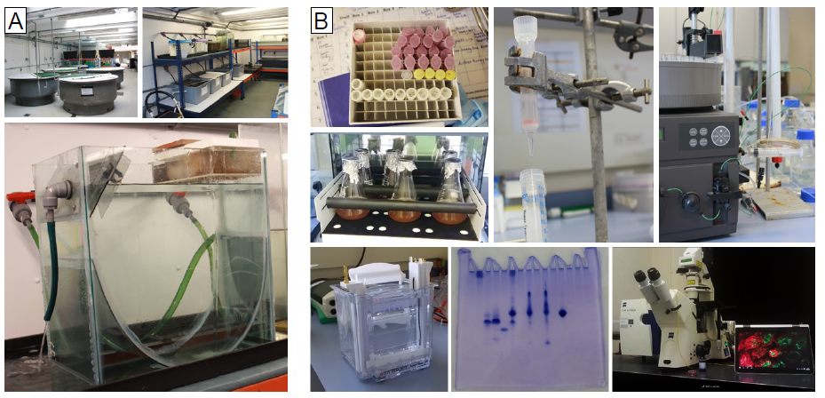
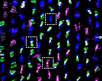
A typical day for us, as you might have guessed, begins at the crack of dawn. The first job of the day is to go ctenophore hunting. For this we use our research vessel named MBA Sepia (Figure 3). The MBA Sepia can accommodate 12 passengers and offers a spacious laboratory. Ctenophore sampling is done using a standard plankton net, and we do both vertical and horizontal trawls (Figure 3). Ctenophores are made up of 99% water and 0.4% salt so one can imagine how difficult it is to take them from the sea, and get them into culture intact back at the lab. In fact, it is difficult to do anything with these buggers! But we persevere. The MBA houses a state-of-the-art seawater facility for marine biological research with a wide range of tanks and we culture ctenophores in so called pseudo-kreisels (Figure 4A). Basically this is a tank that has fresh sea water pumped in from one end and a filter at the other. A cyclical current naturally occurs, which the ctenophores love. One important lesson (that has been learned the hard way) is that you don’t put Pleurobrachia and Beroe together in the same tank, as Beroe is one of Pleurobrachia’s main predators. Ideally I like to leave Pleurobrachia in the tank for 24 hours or so, as this allows them time to recover from the trauma of being handled. They are then ready to use for my experiments, for example for live imaging microscopy and immunostaining assays (Figure 4B). We have generated a couple of antibodies against ctenophore synaptic proteins and are in the process of studying their intracellular localization. In addition, we are using many different biochemical methods, for example immunoprecipitation techniques, to isolate synaptic proteins from ctenophores to determine their in vivo binding partners (Figure 4B).
Figure 4: (A) Sea water hall and ctenophore culture system. (B) Variety of techniques to study ctenophore synaptic proteins, ranging from gel-filtration & ion-exchange chromatography, immunoprecipitation, immunohistochemistry to live cell imaging. Photos: P. Burkhardt (MBA).
Working in the Burkhardt Lab has been a brilliant experience. Probably the most valuable thing I have learned from my time here has been how to properly manage my time in the lab. The MBA molecular lab corridor can be a very busy place. Different lab groups are constantly bustling about, and access to resources has to be negotiated amongst the various research groups. If I want to get everything finished by the end of the day then I have to be very efficient, which means I will usually have three or four things going on at any one time. This takes a lot of concentration, and by the end of the day I am absolutely shattered! My favourite thing about the MBA is that it’s right on the coast, so I like to finish the day with a swim to clear my head. My other favourite thing about the MBA is that there are loads of Italians working here. Tip of the day: in my experience Italian people equals food. After my swim I usually head to one of their houses for some home-made pasta, a glass of vino, and a chat. By the time I get home I am asleep before my head hits the pillow.
The bigger picture
What really attracted me to this project was one simple question. How could such a complex and specialised cell as a neuron possibly have evolved twice in animals? Until very recently, most of us took it for granted that synapses and neurons emerged only once during animal evolution. The two newly available ctenophore genomes have caused us to question our current understanding of the course of neuron evolution. It is exciting to know that the questions surrounding ctenophores and the origin of neurons are only a few years old. As such, these strange and beautiful creatures are likely to remain an important source of information, as we strive to unravel the mysteries of past evolutionary events. For me it will be rewarding to trace the evolutionary origin of synapses and neurons, and find out more about our own evolutionary history. Furthermore, I get to study one of the coolest animals I have ever seen. If this does not excite a biologist, then I don’t know what does!
References
Achim, K. & Arendt, D. Structural evolution of cell types by step-wise assembly of cellular modules. Curr. Opin. Genet. Dev. 27, 102–108 (2014).
Brusca, R. C., Wendy, M. & Shuster, S. M. Invertebrates. Sinauer Associates, (2016).
Jager, M. & Manuel, M. Ctenophores: an evolutionary-developmental perspective Curr. Opin. Genet. Dev. 39, 85–92 (2016).
Jekely, G., Paps, J. & Nielsen, C. The phylogenetic position of ctenophores and the origin(s) of nervous systems. Evodevo 6:1 (2015).
Moroz, L. L. et al. The ctenophore genome and the evolutionary origins of neural systems. Nature 510, 109–14 (2014).
Moroz, L. L. & Kohn, A. B. Unbiased View of Synaptic and Neuronal Gene Complement in Ctenophores: Are There Pan-neuronal and Pan-synaptic Genes across Metazoa? Integrative and Comparative Biology 55, 1028–1049 (2015).
Pang, K. & Martindale, M. Q. Comb jellies (Ctenophora): A model for basal metazoan evolution and development. Cold Spring Harb. Protoc. 3, (2008).
Philippe, H. et al. Phylogenomics Revives Traditional Views on Deep Animal Relationships. Curr. Biol. 19, 706–712 (2009).
Pisani, D. et al. Genomic data do not support comb jellies as the sister group to all other animals. Proc. Natl. Acad. Sci. 112, 201518127 (2015).
Ryan, J. F. et al. The genome of the ctenophore Mnemiopsis leidyi and its implications for cell type evolution. Science 342, 1336–1344 (2013).
Ryan, J. F. Did the ctenophore nervous system evolve independently? Zoology 117, 225–226 (2014).
Whelan, N. V, Kocot, K. M., Moroz, L. L. & Halanych, K. M. Error, signal, and the placement of Ctenophora sister to all other animals. Proc. Natl. Acad. Sci. U. S. A. 112, 5773–8 (2015).
This Editorial originally appeared in Development, Vol 143, Issue 17.
Olivier Pourquié andKatherine Brown
At Development, we are always trying to improve our processes and service – for authors and readers. In April 2015, we made some changes to our peer review process, which aimed at encouraging a more constructive approach to peer review. As we wrote in the Editorial announcing these changes, ‘there can be a tendency for a review to read like a “shopping list” of potential experiments … Instead, we believe that referees should focus on two key questions: how important is the work for the community, and how well do the data support the conclusions? … In other words, what are the necessary revisions, not the “nice-to-have”s?’ (Pourquié and Brown, 2015). Our new system has been in place for over a year now, and we have seen a change in the tone and nature of referee reports: in general, referees have embraced our new guidelines and we feel that this has had a positive effect – both in helping editors to take a decision, and in providing more streamlined feedback for authors. However, and as we recognised at the time, ‘…these changes are conservative compared with some of the more radical approaches in peer review that have been implemented and trialled elsewhere’ (Pourquié and Brown, 2015). At the beginning of 2016, and while reviewing various aspects of the Company’s journals’ peer review processes and online functionalities, we therefore conducted a community survey to gauge your opinions on how we should further improve. Over 300 of you completed the survey (over 800 across all our journals), and we are very grateful for your detailed responses.
In the first part of the survey, we asked you to rank possible improvements or changes to the way we do peer review in order of importance (see Box 1). Perhaps unsurprisingly, you ranked the two statements that relate to speed of publication as the top two priorities. While our own research and author surveys suggest that our speeds are reasonably competitive compared with other journals, there are always improvements to be made and we are actively looking at ways in which we can accelerate the process. We also recognise that multiple rounds of review and revision can be frustrating for the author, so we do try to avoid these wherever possible – while still ensuring the highest standards of peer review. Moreover, such efforts are rarely made in vain: we make a strong commitment to papers at first decision, accepting over 95% of manuscripts where we invite a revision.
Box 1. Peer review
Our January survey asked:
‘Unbiased, independent peer review is at the heart of our publishing decisions but there is always room for improvement and we are open to experimentation and change based on the needs of our community. Please rank the following in order of importance to you.’
Your responses ranked the statements in the order below – from most to least important.
Improve speed from submission to first decision
Reduce number of rounds of review and revision
Collaborative peer review trial (inter-referee discussion before a decision is made)
Name of the Editor who handled the paper published with the article
Double-blind peer review trial (author identity withheld from referees)
Network for transferring referee reports between journals in related fields
Post the peer review reports alongside the published articles
‘Open’ peer review trial (referee identity known to authors but not made public)
Post-publication commenting
Speed aside, you ranked ‘collaborative peer review trial’ as the most important potential innovation – in fact, you rated this almost as important as reducing the number of rounds of review. This chimed with regular feedback we receive from the community, who, as authors, have found such models of peer review helpful. The idea is that, once all the reports on a paper have been returned, the editor shares these among the referees, asking for further feedback that might clarify the decision. This can help to resolve differences between referees, identify unnecessary or unreasonable requests, or – conversely – highlight valid concerns raised by one referee but overlooked by the others. Many journals, Development included, have been doing this informally with a subset of difficult cases for many years and it can prove invaluable in helping editors to make the right decision on a paper.
Given your enthusiasm for such a model, we have carefully assessed a number of potential models, including those already in place at other journals. These range from ‘cross-referee commenting’ as implemented by The EMBO Journal (Pulverer, 2010) and others, to the more active discussions embraced by eLife (Schekman et al., 2013). Bearing in mind the need to balance gathering additional feedback against the consequent effects on speed to decision, as well as the fact that some papers will benefit from further input more than others, we believe that a cross-referee commenting model is most appropriate for Development.
For all research papers submitted after mid-September, the full set of referee reports (minus any confidential comments) will be shared among all the referees. These may be accompanied by specific requests for feedback from the Editor. Referees will then be given two working days to respond before the Editor takes a decision – thus minimising any impact on speed (although the Editor may choose to wait for input in cases where the decision is particularly difficult or borderline). We anticipate that in many cases, feedback will either not be received or will not alter the Editor’s decision. For a minority of papers, however, we believe that this process will significantly aid decision-making and help the authors to move forwards with the paper – whether the decision is positive or negative. And, because it is not always possible to predict what will come out of such a process, we think it important to implement this as standard across all papers. We know that our referees already do a great job in helping authors to improve the papers we review, and we are hugely grateful for their efforts. We hope they (you!) will engage in this new development, and that authors will find it helpful. We will also continue to review other possible improvements to how we handle papers – bearing in mind your feedback from the survey.
The second part of the survey focused on how we present the work we publish (see Box 2). Once again, it seems that what matters most to you is not how the paper looks when it comes out, but how quickly it appears. Since mid 2015, we have been posting the author-accepted versions of manuscripts on our Advance Articles page before issue publication, minimising the time between acceptance and appearance of the work online. While there is still some delay (1-2 weeks) before online posting, this is primarily to allow us time to run our standard ethics checks on all papers – helping to ensure the integrity of the work we publish. Articles then typically appear in an issue around 6 weeks after acceptance: this reflects the time required to copy-edit, typeset and prepare the issue, although again we are working on ways of accelerating these processes.
Box 2. Online developments
Our January survey asked:
‘We recently rolled out a new-look website to make it easier for you to find and read content, but new features and functionality are being developed all the time. Which areas do you think should be our focus for 2016 and beyond? Please rank the following in order of importance to you.’
Your responses ranked the statements in the order below – from most to least important.
Improve speed from acceptance to online publication
Easier viewing of figures alongside the relevant text
Easier viewing of supplementary material including movies
Graphical abstracts (diagrammatical summaries of papers)
Publish final versions of articles one by one, gradually building an issue, rather than waiting for an issue to be complete before publication
Easier access to related articles, special issues and subject collections
Better text and datamining services
Annotation of article PDFs e.g. ReadCube
More community web content such as feeds from third-party bloggers
In terms of potential innovations to our online display, you ranked easier viewing of figures and better display of Supplementary Information, including movies, as the most important. This feedback fitted precisely with the top priorities on our own development wish-list. Improving online functionalities is a long-haul project, particularly given the need to work with external partners, to ensure that any new tools integrate seamlessly with our platform and to be confident that they will continue to serve their purpose long-term. However, we are making progress, particularly with Supplementary Information. We are now able to make the Supplementary Information available with the Advance Article version of the manuscript, meaning that readers do not have to wait until issue publication to view the Supplementary Information. We can also announce that we have partnered with Glencoe Software (glencoesoftware.com) for better display of movies. Once in place, this will mean that movies can be viewed directly from within the HTML version of the article. Given that movies are often an integral part of a developmental biology paper, we are delighted that we will finally be able to give them the prominence they deserve. Other innovations to our online display, aimed at better integration of text, figures, movies and data are in the pipeline.
As a journal, we are always looking for ways to improve the way we both handle and present your work. Along with other recent innovations – including co-submission to bioRxiv, integrated data deposition with Dryad, adoption of ORCiD for unambiguous author identification and promotion of the CRediT taxonomy for author contributions – we hope that these announcements will help authors to better disseminate and gain recognition for their research, and help readers to better access and utilise it. As always, we welcome your feedback and suggestions for future improvements
 (No Ratings Yet)
 The Tanentzapf lab


 (3 votes)
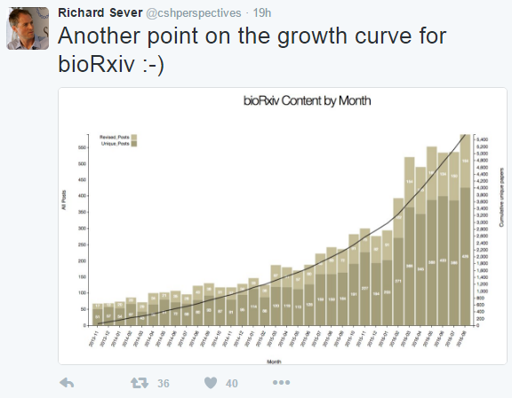


 (8 votes)