The last two days of the EMBO meeting were quite busy, with lots of interesting plenary talks, concurrent sessions, and special symposia.
On Monday evening two interesting non-biology talks were happening at the same time: one about Women in Science, and one about Science Policy & Publishing. I attended the Women in Science talk.
The Women in Science talk was given by Brian Nosek – an associate professor of psychology at the University of Virginia. He studies stereotypes and social judgment, and he let the audience take part in some common tests to identify gender bias. Even though the audience (of predominantly female scientists) knew what they were being tested on, the bias was still apparent when it came to quickly assigning groups of words to work, family, male or female. It’s a bit difficult to explain the test, but you can see it in this video (starting at the 1:17 minute mark) and similar tests are also on Nosek’s site Project Impact.
If the prejudice that “science is for men” is so engrained, as Nosek showed with examples from several studies, is there anything that can be done to change it? The prejudice comes from two sides: those making decisions (e.g. hiring people in academic positions), and those on the receiving end of the prejudice. For the first group, education, blinding (selecting interview candidates from CV’s without names) and comparative assessment were all listed as ways to combat prejudice. For the second group – women themselves – Nosek suggested: role models, confidence building, and removing identity threats. He gave a great example of how confidence building helped female college students perform better on science tests.
Of course there were also many talks about biology these last few days. Linda Partridge’s Monday morning lecture on the effect of diet on lifespan had several people regret their large servings of French cheese the evening before. I interviewed her later in the day, so will summarize that in a separate post.
It was once again difficult to choose between concurrent sessions these last two days, but I settled on “Genes to Shape”, “Genetic Diversity” and “Animal Germline”. I won’t be able to cover all those talks, but here are a few of the talks that Node readers might be interested in:
 Enrico Coen talked about cell polarity in the “Genes to Shape” session. He compared cells to people waiting in a queue at an airport gate. (I took a similar picture of people waiting to board the plane to Nice from Heathrow – see left). These people are all facing the same direction, determined by the location of the gate and airplane, but they’re already polarized (with a front and back) before they join the queue. Likewise, Coen suggested, cells can be polarized before they group together, and cell-cell interaction or signals just coordinate (not cause) the polarisation. He showed a few simulations that demonstrated that such behaviour could then shape a leaf in plants. In the same session, Floris Bosveld showed how detailed image analysis of fly embryos revealed how the Fat/Dachsous/Four-jointed PCP pathway mechanically controls morphogenesis and Markus Affolter demonstrated how blood vessels are formed from unicellular tubes through cell rearrangement.
The germline session on the last day of the meeting was very interesting. Ruth Lehmann, who was chairing this session, used her talk to outline the three main threats to the germline (somatic differentiation, transposable elements, and pathogens) and how germ cells are protected from them. Saadi Kochbin talked about epigenetic guidance of male genome programming. Specifically, he showed how the bromodomain-dependent factor Brdt drives meiosis and haploid cell differentiation.
Some other talks in this session were about unpublished research that I can’t mention yet, but keep an eye out for Mitinori Saitou’s upcoming Science paper – it will be out soon!
The meeting closed with a plenary session on “oxygen sensing, vasculogenesis, and disease”. I noticed that all three speakers in this session – Peter Carmeliet, Anne Eichmann, and Kari Alitalo – who each covered one of the three main topics in the session, had either collaborated with, or directly worked with each other at some point. I suppose that shows that the field of vascular research is as interconnected as the vascular system itself.
All in all, this was an interesting and very varied conference. It was impossible to be at all the talks I wanted to be, but the organisers had done a great job at planning the sessions. Thanks to everyone who dropped by the Company of Biologists stand (and to the people at the career table for promoting my Leaving the Lab article!)
 (2 votes)
 Loading...

 (No Ratings Yet)
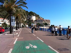 EMBO meeting
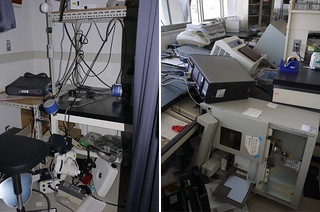 Research – the Tamura lab’s earthquake experience


