
The community site for and by
developmental and stem cell biologists

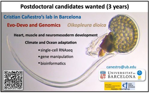
Postdoctoral candidates wanted – Juan de la Cierva call 2026 (3 years) – (deadline contact November 12th)

Posted by , on 31 October 2026

Closing Date: 31 December 2026


Job offer: Postdoc candidate (3 years) (deadline contact November 12th)

Cristian Cañestro’s lab at the University of Barcelona is seeking motivated postdoctoral candidates to apply for the Juan de la Cierva 2026 fellowship, funded by the Spanish Ministry of Science, Innovation and Universities, to join our newly granted project PID2025-170547NB-I00, “Gene loss impact: evolution of cardioparaxial-neuromesodermal development and genome scrambling in Oikopleura dioica as a case study” (OikoLoss).

REQUIREMENTS AND PROFILE OF CANDIDATES:

According to the Juan de la Cierva 2026 call:

  • Applicants must have obtained their PhD between 1 January 2024 and 31 December 2026, with specific extensions or exceptions under the official regulations.
  • Applicants of any nationality are welcome, provided they meet the eligibility requirements of the programme.

We particularly welcome candidates with research interests in evolutionary developmental biology, comparative genomics, genomic structural variation, bioinformatics, and population genetics. Expertise in single-cell omics will be considered an additional asset.

THE PROJECT:

OikoLoss investigates how gene loss can drive evolutionary innovation. Using the chordate Oikopleura dioica as a natural “evolutionary knockout” model, the project integrates EvoDevo, functional genomics, single-cell transcriptomics, population genomics and ecology through two complementary aims:

  • Aim 1. To determine how massive gene loss has reshaped heart, axial muscle and neuromesodermal development in chordates, using single-cell omics, gene-expression analyses and functional experiments.
  • Aim 2. To uncover how genome scrambling and chromosomal breakpoints generate gene-loss alleles, and to assess their potential adaptive impact under different ocean environmental conditions.

The successful candidate will contribute to the project according to their expertise and research interests, with opportunities to develop independent research directions within its scientific framework.

RESEARCH TECHNIQUES:

The project combines experimental and computational approaches, including embryo culture and manipulation, microinjection, CRISPR knockouts, interference-based knockdowns, HCR whole-mount in situ hybridization, confocal microscopy, single-cell RNA sequencing, PacBio HiFi long-read genomic sequencing, comparative and population genomics, and bioinformatics.

TRAINING AND INTERNATIONAL COLLABORATIONS:

The successful candidate will join a multidisciplinary research environment at the University of Barcelona, with opportunities to interact with international collaborators and undertake research stays, particularly at OIST in (Okinawa), IGFL (Lyon), SARS (Bergen), Imperial College (London) and CRG (Barcelona).

FUNDING:

The Juan de la Cierva 2026 programme offers:

  • Duration: 3-year postdoctoral employment contract.
  • Funding: €35,700 per year to co-finance gross salary and employer Social Security contributions. The final salary and ent conditions will follow the applicable University of Barcelona regulations.
  • Additional research funding: €15,000 for research-related expenses over the fellowship.
  • Selection: Competitive national evaluation based on the candidate’s scientific track record and the quality of the host team.

DEADLINES AND APPLICATION TIMELINE

Expressions of interest and pre-selection are NOW OPEN. Early contact is strongly encouraged.

  • 12 November 2026: Official application period opens.
  • 26 November 2026, 14:00 CET: Deadline for candidates to complete their application forms.

Because of the short application window, interested candidates are strongly encouraged to contact us as soon as possible (before November 12th) to allow sufficient time for evaluation and preparation of the application.

CONTACT and PRE-SELECTION:

Interested candidates should email Cristian Cañestro at canestro@ub.edu, attaching a single PDF containing:

  • A motivation letter describing their research interests, relevant experience and reasons for joining our laboratory.
  • An updated academic CV, including date of their PhD award or expected PhD defense, publication record, research experience and technical expertise.

Shortlisted candidates will be invited to an online interview. Pre-selection process will follow principles of equal opportunity and non-discrimination, with particular attention to promoting gender balance.

Further information, related publications and updates:

https://evodevogenomics-unibarcelona.weebly.com/join-us.html

Official Juan de la Cierva 2026 call:

https://www.aei.gob.es/convocatorias/buscador-convocatorias/ayudas-contratos-juan-cierva-2026
Screenshot

(No Ratings Yet)

Tags: , , , , , , , , , , ,
Categories: Jobs

Navigate the archive

Use our Advanced Search tool to search and filter posts by date, category, tags and authors.

How do sea anemones make babies?

Posted by , on 24 October 2026

Inspiration for scientific research projects can come from many unexpected places. With the publication of my second PhD research paper [1], I am looking back at my sources of inspiration as well as some hurdles I had to overcome to see this work come to fruition.

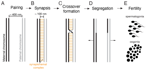
As a PhD student at the Stowers Institute for Medical Research in Kansas City, I was going through the first semester trying to absorb as much information as possible while deciding on my rotation labs. I had decided on my first rotation in the lab of Dr. Scott Hawley very quickly after joining the program. The esteemed Drosophila geneticist was particularly enthralled by the synaptonemal complex, a multi-protein structure that assembles between the maternal and paternal copy of each chromosome in early meiosis (Fig. 1). This structure is crucial for the formation of crossovers between these homologous chromosomes and thereby ensures proper chromosome segregation and fertiltiy (Fig. 1). Apart from this fundamental function, I was fascinated by the fact that this massive proteinaceous structure is able to assemble across the entire length of chromosomes.

As is common at many research institutes, we regularly heard seminar talks from researchers coming to us from all over the world. One of these presentations was given by Dr. Catriona Munro, a scientist who studies meiosis in the jellyfish Clytia hemisphaerica [2]. Previously, I hadn’t really considered the possibility of doing research with unusual organisms like jellyfish. For my bachelor’s thesis in Germany, I did strictly in vitro work. For my year-long research stint at Purdue University in the US, I worked with Drosophila melanogaster. All fairly standard research systems. Seeing that someone performed systematic studies using a jellyifsh was truly fascinating and unexpectdely paved the way for my own future research.

Shortly after this talk, I heard from Dr. Matt Gibson at the Stowers Institute about his lab’s research using the sea anemone Nematostella vectensis. His research team investigates various kinds of fundamental biological questions ranging from how stinging cells in cnidarian species function [3] to neuroscience [4] and Hox-mediated segmentation during embryogenesis [5]. What particularly caught my attention was the fact the Nematostella is relatively large and that each adult has eight gonads that took up the majority of the animal’s body mass. Immediately, I thought that this would make a pretty good meiosis model organism. Scott had previously told me that most meiosis models come with the difficulty that meiotic material is fairly limited. This certainly wouldn’t be a problem with Nematostella.

After I went home for the day, I googled whether anyone was doing research on Nematostella meiosis and its synaptonemal complex. I was surprised to find that research on cnidarian meiosis in general was quite sparse. When it came to the synaptonemal complex, almost 10 years ago a research team in Germany had identified specific protein homologs in the related cnidarian species Hydra vulgaris [6]. In addition, Catriona had looked at the synaptonemal complex in Clytia briefly as part of her research [2]. Other than that, there was no systematic research being done in this area. Considering the importance of the synaptonemal complex for the maintenance of an organisms’ fertility, I was convinced that we needed to understand how it functions in cnidarians. Particularly, as cnidarians like corals are facing tremendous challenges to their fertility. I gathered my scientific curiosity and courage to email Scott and Matt, proposing a research project on studying the conservation of synaptonemal complex proteins in Nematostella. The answers came quickly: Scott absolutely loved the idea. Considering his lab’s newest research with the silk moth Bombyx mori [7] which had quite an unusual synaptonemal complex, he practically couldn’t wait to learn about what novelties the Nematostella equivalent could reveal. Matt was similarly intrigued by this new research direction and my enthusiasm for cnidarian research.

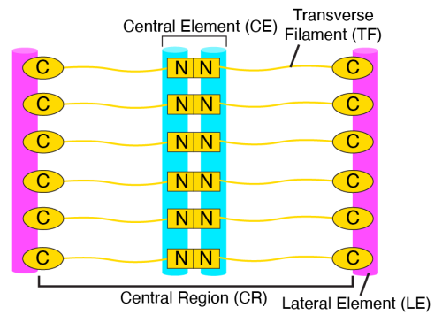
Once I completed my three lab rotations, I decided to pursue my PhD thesis research co-mentored by Scott and Matt. My particular focus was to study whether the architectural functions of Nematostella synaptonemal complex proteins were conserved. Curiously, synaptonemal complex proteins exhibit rapid sequence divergence, despite their crucial function in ensuring meiotic success throughout eukaryotes [8]. As a result of this divergence, it was actually surprising how easy the identification of protein homologs of vertebrate synaptonemal complex proteins in Nematostella was. Most protein homologs were already identified by other researchers but I was able to expand these findings [1]. Now we needed to try and figure out whether these proteins actually function in the same way as they did in other organisms. Considering that the synaptonemal complex consists of three distinct structural elements (Fig. 2), I wanted to generate three individual mutants each disrupting one of these elements. This would allow us to understand the functional importance of each element as well.

Fig. 2: The synaptonemal complex consists of distinct structural compartments. The lateral elements are connected to the chromosomes respectively. In the central region, a central element with distinct structural components is found. The transverse filaments span the entire width of the synaptonemal complex. Adapted from [1]

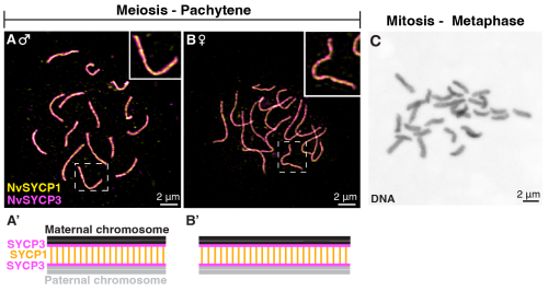
While Nematostella has quickly become my favorite laboratory model, one of the major downsides of working with it is the relatively long generation time of roughly 3 months. This meant that I was raising animals and making crosses for over a year with mutants I had no idea would show any interesting phenotypes. In the meantime, I established necessary tools to study meiosis and the synaptonemal complex in this model, allowing me to investigate the wild-type structure. Some techniques that I had to adapt was preparing chromosome spreads of meiotic and mitotic stages (Fig. 3). During this time, I also worked on a second project investigating the synaptonemal complex in Drosophila species, which you may learn more about here.

Fig. 3: Chromosome spreads enable to visualization of chromosomes and synaptonemal complexes in meiosis (A) – (B) and mitosis (C). (A) Chromosome spread from a male Nematostella individual at pachytene of meiosis I. The stained synaptonemal complexes are shown with SYCP3 (lateral element) in magenta and SYCP1 (transverse filament) in yellow. (B) Chromosome spread from a female Nematostella individual. (A’) and (B’) are schematic representations of what can be seen in the above image. (C) Mitotic chromosome spread at metaphase of a 64-/128-cell stage embryo with DNA visualized in grey. Adapted from [1].

Unfortunately, in my case, this long waiting period also meant that my co-supervisor Scott passed away only one month before I was able to start working with the homozygous mutant adults I had established and raised. His death was not only felt deeply because of the sudden loss of scientific support but also because he was so much more than just a scientific supervisor to so many people – including me.

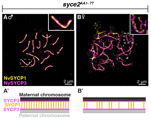
Once I was able to exit the sudden stupor that Scott’s death led me into, I began investigating my homozygous mutants. The three Nematostella proteins I mutated shared approximately 30% of sequence identity with their vertebrate orthologs. While this may seem very low, it is worth emphasizing that orthologous proteins between vertebrates and Drosophila, for example, share no identifiable sequence homology. Thus, I hypothesized that the Nematostella synaptonemal complex proteins and their architectural functions are fully conserved in comparison to vertebrates. First and foremost, this meant that each individual protein component is essential for the successfull assembly of the synaptonemal complex. Thus, I definitely thought I must have made a huge mistake when my first microscopy session on one of my mutants’ chromosome spreads showed no significant assembly defects (Fig. 4). As it turns out, it was not a mistake but the fact that this particular protein is not essential for synaptonemal complex assembly in Nematostella males. Looking at all three of my synaptonemal complex mutants, I was able to reveal that some architectural functions of synaptonemal complex proteins in Nematostella are distinct from those found in vertebrates and, additionally, exhibit dramatic sexual dimorphisms.

Fig. 4: Example image of meiotic pachytene-like chromosome spreads from syce2 mutant Nematostella individuals. (A) synaptonemal complexes from a syce2 mutant male. SYCP3 (lateral element) is shown in magenta, SYCP1 (transverse filament) is shown in yellow. (B) synaptonemal complexes from a syce2 mutant female. (A’) and (B’) are schematic representations of what can be seen in the microscopy images. Adapted from [1].

While these findings by themselves were deeply fascinating, I also wanted to understand the functional relevance of the synaptonemal complex and its protein components on overall fertility and chromosome segregation. While a TUNEL assay was supposed to provide me with a quick answer to the question whether spermatocytes were undergoing increased rates of cell death (hinting at meiotic checkpoint activation), this experiment turned into a 6-months long ordeal of troubleshooting and protocol optimizations. Nonetheless, I persevered and was able to confirm that even those proteins that appeared to be non-essential for synaptonemal complex assembly were necessary to ensure proper meiotic progression. Taken together, it became clear that the synaptonemal complex in Nematostella was quite distinct in its architectural composition when compared to vertebrates. The sexual dimorphisms I observed further complicated this picture but made it all the more intriguing.

When I envisioned this research project, I could have never foreseen where this work would go. Practically all my hypotheses were proven wrong. In addition, I discovered a research interest that follows me into my current postdoc and likely beyond. By establishing Nematostella vectensis as a model for investigating meiosis, this work now opens the door to exploring how the mechanisms that ensure reproductive success have evolved across animals. As this work is the first systematic investigation into how cnidarian synaptonemal complexes funciton mechanistically, this also paves the road to understand how fertiltiy is ensured in these basal metazoans. Considering that Nematostella is closely related to corals, this work further has important implications for coral sexual reproduction. Altogether, this research paper is the product of a lot of sweat and tears but I could not be prouder and happier to see its publication and I cannot wait to see what else I will learn about cnidarian reproduction.

References

[1] Stefanie Williams, Jennifer Gardner, Stephanie H. Nowotarski, Matthew C. Gibson. The Nematostella synaptonemal complex mediates divergent and sexually dimorphic meiotic programs. Science Advances Volume 12, Isse 43, eaei0758 (2026). https://doi.org/10.1126/sciadv.aei0758

[2] Catriona Munro, Hugo Cadis, Sophie Pagnotta, Evelyn Houliston, Jean-René Huynh. Conserved meiotic mechanisms in the cnidarian Clytia hemisphaerica revealed by Spo11 knockout. Science Advances Volume 9, Issue 4, eadd2873(2023). https://doi.org/10.1126/sciadv.add2873

[3] Ahmet Karabulut, Melainia McClain, Boris Rubenstein, Keith Z. Sabin, Sean A. McKinney, Matthew C. Gibson. The architecture and operating mechanism of a cnidarian stinging organelle. Nature Communications Volume 13, 3494 (2022). https://doi.org/10.1038/s41467-022-31090-0

[4] Ruohan Zhong, Chris W. Seidel, Anna M. L. Klompen, Matthew C. Gibson. Morphological, molecular, and functional evidence for a CNS-like oral nerve ring in the sea anemone Nematostella vectensis. bioRxiv 2026.01.28.701697; doi: https://doi.org/10.64898/2026.01.28.701697

[5] Shuonan He, Florencia Del Viso, Cheng-Yi Chen, Aissam Ikmi, Amanda E. Kroesen, Matt Gibson. An axial Hox code controls tissue segmentation and body patterning in Nematostella vectensis. Science 361,1377-1380 (2018). https://doi.org/10.1126/science.aar8384

[6] Johanna Fraune, Miriam Wiesner, Ricardo Benavente. **The Synaptonemal Complex of Basal Metazoan Hydra: More Similarities to Vertebrate than Invertebrate Meiosis Model Organisms. Journal of Genetics and Genomics, Volume 41, Issue 3, 107-115 (2014) https://doi.org/**[10.1016/j.jgg.2014.01.009](https://doi.org/10.1016/j.jgg.2014.01.009)

[7] Youbin Xiang, Dai Tsuchiya, Zulin Yu, Xia Zhao, Sean McKinney, Jay Unruh, Brian Slaughter, Cathleen M. Lake, R. Scott Hawley. Multiple reorganizations of the lateral elements of the synaptonemal complex facilitate homolog segregation in Bombyx mori oocytes. Current Biology, Volume 34, Issue 2, p352-360. (2024). https://doi.org/[10.1016/j.cub.2023.12.018](https://doi.org/10.1016/j.cub.2023.12.018)

[8] Stefanie Williams, R. Scott Hawley. From conservation to adaptation: understanding the synaptonemal complex’s evolutionary dynamics. Current Opinion in Genetics & Development. Volume 93. (2025) https://doi.org/[10.1016/j.gde.2025.102349](https://doi.org/10.1016/j.gde.2025.102349)

(No Ratings Yet)

Tags: , , , ,
Categories: Research

Celebrating the Centenary of The Theory of the Gene Through Cinema and Public Outreach

Posted by , on 23 October 2026

This post is co-authored by Dante Albino Idesti and Larissa de Moraes Chiarelli

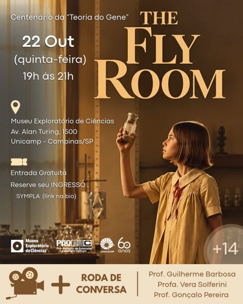
One hundred years ago, in 1926, Thomas Hunt Morgan published The Theory of the Gene, a landmark work that consolidated the physical basis of heredity and forever changed the landscape of modern biology. To celebrate this centenary, researchers and students gathered with the public on the evening of October 22, 2026, at the Exploratory Science Museum of the University of Campinas (UNICAMP), in Campinas, Brazil, for a special outreach event combining science, history, and cinema.
Designed as an open and free Cine-Debate for the general public, the initiative built a bridge across three core pillars: the historical centenary of the gene concept, the enduring legacy of Drosophila melanogaster as a model organism, and the role of science museums as democratic spaces for public engagement.
Bringing the “Fly Room” to the Science Museum
For many outside academia, genetics can feel abstract and distant from everyday life. Hosting the event at UNICAMP’s Exploratory Science Museum allowed us to step outside the traditional walls of our research laboratories and invite the local community into an interactive, welcoming environment and open conversation (UNICAMP news).
The evening began at 7:00 PM with a brief historical contextualization introducing Thomas Hunt Morgan, the early days of chromosomal research, and the tiny fruit fly that became one of biology’s most powerful allies. This set the stage for the screening of Alexis Gambis’s feature film The Fly Room (2014).
Told through the eyes of ten-year-old Betsey as she spends a day in the genetics laboratory of her father, Calvin Bridges (one of the key pioneers in Morgan’s group at Columbia University) the film offered the audience an intimate look into both the scientific breakthroughs and the complex human dynamics behind the birth of modern genetics. Throughout the screening, the sensory imagery of the flies and the delicate father-daughter narrative captivated attendees, bridging the gap between historical science and everyday emotion.

Exhibit poster of the movie night showing the The Fly Room at the Exploratory Science Museum, in celebrationg of 100 years of “The Theory of Gene” by Thomas H. Morgan.

“From Flies to Genes: 100 Years of a History That Transformed Biology”

Following the screening, the evening transitioned into a lively roundtable discussion. The panel was intentionally assembled to reflect how the foundations laid in Morgan’s fly room have branched out across generations, model organisms, and biological scales at UNICAMP:
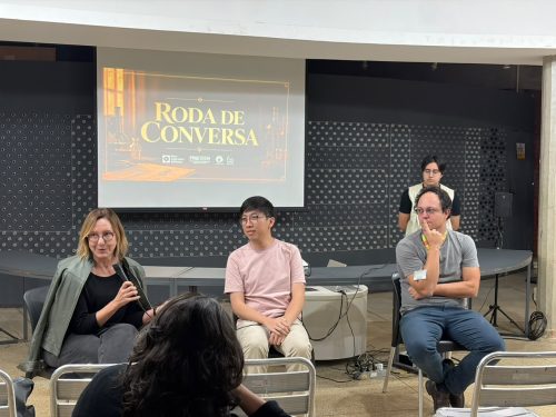
Prof. Dr. Guilherme Oliveira Barbosa, a researcher in the Fly Lab at UNICAMP’s Institute of Biology. Holding a PhD in Cell Biology, Guilherme investigates morphogenesis using Drosophila melanogaster and actively works in science education and public outreach. He anchored the discussion in the day-to-day reality of working with fruit flies today and how basic developmental questions continue to drive discovery.
Prof. Dr. Vera Nisaka Solferini, a leading reference in genetics and evolutionary biology in Brazil. Whose research explores the historical biogeography of Neotropical biomes and the adaptive processes driving biological diversification, Vera expanded the conversation from the classic laboratory bottle to natural populations and evolutionary history.
Gustavo Seguchi, a Master in Genetics and Molecular Biology from UNICAMP and current PhD candidate at the Laboratory of Genomics and bioEnergy (LGE), where he studies the potential of non-conventional yeasts for biofuel production from diverse biomasses. Gustavo brought the perspective of early-career researchers applying century-old genetic principles to modern genomic and biotechnological challenges.

Prof. Vera N. Solferini, Gustavo Seguchi and Prof. Guilherme O. Barbosa sharing their perseption of classic genetics to modern perspectives based on their current research. Dante Albino Idesti (in the back) was the host of the night.


Bridging Past and Present: The Human Side of Science


During the conversation, the speakers unpacked the scientific and historical nuances portrayed in The Fly Room, sharing behind-the-scenes curiosities of working with Drosophila and tracing how the concept of the gene evolved over the last century — from classical linkage maps to modern developmental biology, evolutionary ecology, and synthetic genomics. The dialogue with the audience flowed naturally, sparked by curiosity about how routine work with tiny model organisms translates into major biological concepts.
Towards the end of the discussion, Dr. Guilherme Barbosa offered a poignant reflection that captured the spirit of the entire evening. He highlighted how powerfully the film portrays the human side of scientists, reminding everyone that researchers are not detached figures isolated in lab coats, but ordinary people driven by the same wishes, passions, insecurities, and desires as anyone else, and that these personal realities inevitably intertwine with and shape their scientific work.
Looking back on the evening, celebrating the centenary of The Theory of the Gene through cinema proved to be an inspiring way to demystify academic research. By bringing Drosophila out of the lab and onto the big screen at the Exploratory Science Museum, the event reminded both seasoned researchers and first-time visitors that the history of genetics is not just a collection of textbook facts, but an ongoing, deeply human story fueled by curiosity.

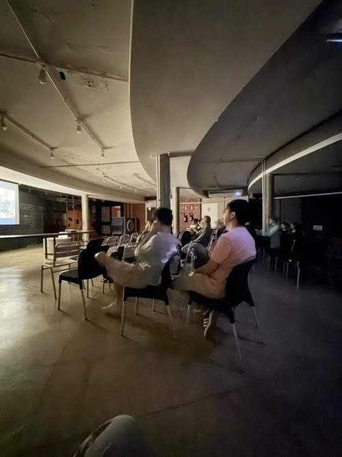
The Fly Room, movie night at the Exploratory Museum of Science (PROEEC-UNICAMP), Campinas-Brazil
(1 votes)

Tags: , , , , , , , ,
Categories: Outreach

What is the cost of knowledge?

Posted by , on 23 October 2026

To mark International Open Access Week 2026, The Company of Biologists has been reflecting on this year’s theme: ‘The Cost of Knowledge’.

In our latest blog post, ‘The true cost of knowledge: exploring value in publishing‘, we reflect on what researchers value in publishing and who bears the cost of creating that value. We also discuss how, through initiatives like Biology Open’s Fast & Fair peer review, we are continuing to innovate to improve the author experience.

To read the full story and other similar stories about our journal and charitable initiatives, you can visit The Company of Biologists website.

(No Ratings Yet)

Tags: , ,
Categories: Discussion, News

Meet Bill Hinchen, winner of the 2026 writing challenge

Posted by , on 22 October 2026

During August, we ran a writing challenge, giving contributors to the Node a chance to win £200 for publishing a blog post on our website. We really appreciate the effort that all of our contributors put into sharing posts on the site, and we hoped that the challenge would help motivate and encourage writers to share their ideas. The Node is a community for all, regardless of experience, background, skills or career stage. For this reason, we decided to award the prize by randomly selecting the winner rather than a competitive format. We hope this approach reassured authors to share on the Node without apprehension that their work would be judged. 

The winner of our writing challenge was Bill Hinchen with the post ‘The little beach hopper that could’. This entry explores the story of the unsung hero, the beach hopper. Check out our interview with Bill.

Bill Hinchen

Tell us about yourself.

I’m a freelance science writer and copywriter, working mostly for those in the biotech industry. They have cool science but struggle to get that across to clients or the public, and I can understand the science well enough, and write well enough, that I can bridge that gap. I’m a wordy bridge, essentially. I started writing back in 2011, and went freelance in 2019, but before all that I was in a lab at Cambridge poking crustacean embryos to see what on earth cells are doing in the stages leading up to gastrulation.

Can you describe your research journey? 

My first taste of proper research was at the University of Plymouth, where I was looking at copper-induced neurotoxicity in catfish. I’d always loved ecotoxicology, and with a marine biology background, it was a perfect step into research. I quickly realised that having the lab to myself, with experiments to plan, failures to recover from and just a whole world of independence afforded to me, research was the one.

From there I started a PhD at Cambridge with Cassandra Extavour in the lab of Michael Akam. Bit of a subject shift, for sure, but dev bio was another thing I was always in awe of (after looking at On Growth and Form, by D’arcy Wentworth Thompson – go read it!) and I’m not a fan of limiting myself to one topic forever.

Starting out felt like the very deepest of deep ends and I had lead weights made of solid imposter syndrome tied around my ankles. But, I eventually settled and got into the swing of things, from peeling Parhyale embryos with tungsten needles under the microscope, to running endless antibody stains on the hunt for beta-catenin and finally into making lots, and lots and lots of 4D time-lapse movies of these embryos. By the end, I felt fully confident about how research works, and, like anyone studying something long enough, just how little I knew in the grand scheme of things!

After that, I spent a short spell in a biophysics lab with Susan Cox at King’s College London. They needed someone who could work with embryos for a microscopy experiment they were running (controlled light exposure microscopy), so I jumped in. Absolutely incredible! I had to pick up C. elegans embryology super quick and work alongside some brilliant physicists. It’s rather impressive how much our academic backgrounds didn’t overlap! But interdisciplinary research is a true joy, and I encourage everyone to get out of their comfort zone and go get cosy with other scientists from other fields.

What does Parhyale hawaiensis mean to you on a personal level? What inspired you to write this story? 

I’ve been a science writer for a long time now, and a common theme in what a lot of folks are saying is just how much their work is going to “accelerate innovation”, cause a “paradigm shift” or some other hyperbolic marketing sentiment. 99% of the time it’s not true, but the emphasis on practical, tangible outputs that will have a huge impact is consistently (over)stated.

Parhyale for me is plain old humble. It’s a tiny little crustacean that looks a bit like a shrimp and at the time, was worked on by just a handful of people who all knew each other on a first-name basis. It wasn’t claiming to change the field or revolutionise medicine; it just had some cool embryos that were amenable to experimental manipulation. But because of its flexibility, it was slowly adopted by a new lab, then another, then another, and then it suddenly crossed disciplines. Such an innocuous little critter, making no grand claims, but being used to develop new tools and answer new questions by new scientists from new fields – like a tiny little baton of research. And all without the fanfare that usually accompanies this sort of thing. I like that.

How did you pivot from research to science writing?

Honestly, it was an accident. Academia was the only career I ever wanted, but that all went a little awry and I needed work. Someone suggested medical writing, which I’d never heard of, and the rest is history.

What are your favourite things about your career as a scientific writer?

As a freelance writer, it’s freedom. I don’t have to waste time in meetings or fill my time with pointless jobs just to keep busy during the day. I take on clients I like, I write in bursts, take my son to and from school and go surfing when the waves look good. Being able to choose my own schedule and my own clients has been my favourite aspect of this by far.

But in general, I’d say it’s dipping my toes in all the different fields of biology. Admittedly, sometimes it feels a little shallow because it’s always someone else’s research, and I don’t think I’ll ever not want to be at the bench, but it can be fascinating to read and write about something you’d never even considered up until you start researching it!

Do you have any advice for researchers who are interested in a career in science writing?

Figure out the type of writing you want to do. Do you want to translate science for the public, or do you want to help companies articulate the benefits of something? Do you want to do journalism or editing? Once you’ve got an idea, start doing it. Start on a blog or LinkedIn and start putting your writing goal into practice. Show people your voice – don’t waste your time trying to sound professional, just be honest. Also, read the terrible things people write across their webpages and write about why they’re wrong and what you’d do differently! I kid you not, in no time at all you’ll have a better understanding of what not to do when it comes to science writing.

Where can people find other pieces written by you?

You can find a whole range of subjects I write about for fun over on my blog, rant & reason: https://billhinchen.substack.com

Want to take Bill’s advice about writing? The Node, as well as our other community sites, preLights and FocalPlane, give you a place to share your writing and provide a network that supports you through the writing process. With this in mind, please reach out to any of our Community Managers if you would like to discuss any ideas or drafts for the sites, or if you would like to join the preLights community team. In addition to written posts, the Node welcomes other types of audio or visual communication, including images, illustrations and videos. With challenges such as this one, we also want to remind our readers and authors that anyone can write and publish a blog, and you don’t need an invitation to do so.  

(No Ratings Yet)

Categories: News

Register for a free scientific storytelling workshop

Posted by , on 16 October 2026

Storytelling is an art, and scientific storytelling can often seem daunting. Join us for tips on making your research presentations more impactful, communicating the most interesting aspects of your work and crafting compelling scientific stories through visuals. Join us on 10 November 2026 from 13:00-17:00 GMT for ‘Scientific Storytelling’, a free half-day workshop designed to help researchers communicate their science with greater creativity and impact.

Duncan Yellowlees will share advice on making science presentations more thoughtful, purposeful, and creative. Following this, there will be a two-minute research talk competition. Then, Dr Kathryn Knight, News & Views Editor at the Journal of Experimental Biology, will begin the storytelling sessions with practical advice on spotting the most interesting stories within scientific research and communicating them effectively. Drawing on her experience writing ‘Inside JEB‘ articles and press releases, she will share strategies that attendees can put into practice straight away. Finally, Dr Sonhita “Son” Chakraborty will lead the part of the event focused on visual storytelling in science. Through examples from her own work, including early drafts and iterations, she will share practical insights into the creative process behind crafting compelling scientific stories through visuals such as graphical abstracts and cover art. The session will conclude with time for discussion and questions, giving attendees the opportunity to reflect on how to apply these principles to their own research communication.

Overall, this event will provide a unique opportunity for participants to improve their storytelling skills, network with peers, and showcase their own research.

There is no fee for this workshop, thanks to support from The Company of Biologists.

To register, please complete the registration form. Registration for the two-minute research talk competition has been extended and closes on Sunday 1 November 2026, while general registration for the workshop remains open until Sunday 8 November 2026.

Agenda

  • Welcome and introductions
  • Making science talks thoughtful, purposeful and creative – Duncan Yellowlees
  • Two-minute research talk competition
  • Finding the story in scientific research – Dr Kathryn Knight
  • Visual storytelling in science – Dr Sonhita (“Son”) Chakraborty
  • Closing remarks

Research talk competition

To be considered for the two-minute talk competition, please submit one of the following (as indicated in the registration form):

  • A short, engaging headline about your work
  • A one-minute video or audio recording explaining your research
  • A graphic representing your research

We look forward to seeing you there!

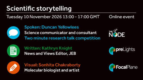
Scientific storytelling
Tuesday 10 November 2026 13:00 – 17:00 GMT Online event
 
Icon of a speech bubble
Spoken: Duncan Yellowlees
Science communicator and consultant
 
Two minute research competition
 
Icon of a paper
Written: Kathryn Knight
News and View Editor, JEB
 
Icon of a pencil
Visual: Sonhita Chakraborty
Molecular biologist and artist
 
Logos of the Node, preLights and FocalPlane
(2 votes)

Categories: News

The laboratory I returned to was not the laboratory I remembered

Posted by , on 15 October 2026

Between school biology, DNA sequencing, and a research career still waiting to happen

The first thing that surprised me when I returned to university was not a sophisticated laboratory instrument. It was the students.

Many of them were considerably younger than I was. I had already spent years teaching biology, preparing lessons, marking assessments, managing classrooms, and trying to help students with very different abilities. Suddenly, I was sitting among students who belonged to another generation, asking questions and trying to learn again.

For a moment, the distance between us felt enormous.

Then something unexpected happened. I began to enjoy being a student again.

I wanted to examine every instrument, understand how it worked, discover its limitations, and learn where mistakes could enter an experiment. I was curious in a way that reminded me of my earliest university years.

But I should explain why I returned in the first place.

The truth about why I went back

It would be easy to say that I pursued a Master’s degree because of my lifelong passion for science. That would be true, but incomplete.

I was also exhausted.

Teaching is a demanding profession, and much of its work is invisible to people outside the classroom. There are lessons to prepare, assessments to design, paperwork to complete, classrooms to manage, and students who need different kinds of support. You must challenge those who are doing well without leaving behind those who are struggling.

I had been teaching biology for years. I valued the work, but I could not ignore how tiring it had become.

Long before I became a schoolteacher, I had studied biology with a specialization in biochemistry. Like many students in my field, I had imagined a future in laboratories, research institutions, or scientific industries. Teaching was not the only career I had pictured for myself.

Returning to university was partly an attempt to recover that earlier ambition. It was also a response to a problem I had noticed in my classroom.

The biology we teach in schools has changed.

Students encounter increasingly sophisticated concepts, from cellular respiration and molecular biology to the principles behind DNA sequencing. A teacher may know how to explain the Krebs cycle or the Bohr and Haldane effects, but knowing the textbook explanation is not quite the same as understanding how scientists investigate the underlying processes.

I began to feel that my students deserved more than definitions and diagrams. I wanted to show them where scientific knowledge comes from.

And I knew I needed to learn more myself.

A laboratory that had changed its language

When I first studied at university, much of our laboratory work relied on manual techniques. Computers were not integrated into many of the instruments we used. Where they were available, their role was often limited to recording results or processing data in spreadsheets.

The laboratory I returned to was different.

Modern instruments could generate enormous quantities of digital information. Understanding their output required familiarity not only with laboratory procedures but also with software, databases, computational analysis, and statistics.

Learning to operate an instrument was no longer enough.

I needed to understand what happened to the data afterward. How were sequences compared? What assumptions did the software make? How reliable was an apparent match? What did the analysis reveal, and what could it not tell us?

From sequence alignment and biological databases to statistical tools and Oxford Nanopore sequencing, each answer seemed to produce another question.

It felt as though I was learning two sciences at once: biology, which I had studied for years, and the computational language through which so much modern biology is now understood.

There was a challenge behind every challenge.

Strangely, I enjoyed it.

When a sequence is not yet an answer

My Master’s research brought me into contact with questions of particular clinical importance: the potential use of DNA sequencing to investigate bloodstream infections in premature newborn infants.

The prospect was compelling. Sequencing technologies can detect microbial genetic material, including signals that conventional approaches may not readily reveal. But the excitement of seeing a microbial name appear in an analysis must be balanced by a more difficult question.

What does that result actually mean?

In low-biomass samples, where very little microbial DNA may be present, contamination and other technical factors can complicate interpretation. Detecting a sequence does not automatically establish that an organism was present in the patient’s bloodstream, much less that it caused an infection.

I came to appreciate that a sophisticated instrument does not eliminate uncertainty. Sometimes it gives us more information than we are immediately able to interpret.

That lesson matters far beyond any one sequencing platform. Technology can make observations possible, but scientific judgment is still needed to decide what those observations support.

I remain proud of the research and grateful for the people who helped me complete it.

Dr. Hadi Yassine and Dr. Fatiha Benslimane were among the academics whose support and encouragement I deeply appreciated during my scientific journey. I am grateful, too, to the colleagues, lecturers, and friends who helped make returning to academic study possible.

Research may depend on instruments and data, but it also depends on people willing to share their knowledge.

The result I did not expect to find in a classroom

Something changed when I returned to teaching.

I had been explaining some biological concepts since 2013. The subjects were familiar, and I knew how to present them. But after my laboratory experience, I noticed a difference in the way I spoke about science.

When I introduced a technique, I could describe the instrument behind it. I could explain what happened to the sample, why a researcher performed a particular step, and where difficulties might arise.

I was no longer relying solely on descriptions I had learned from books. I could bring my own experience into the explanation.

And the students seemed to notice.

When I described an experiment step by step, I could see them becoming more attentive. They followed the procedure with curiosity. They seemed to imagine the instruments, the samples, and the researcher making decisions along the way.

For a few minutes, I felt I could transport them from their classroom desks into a university laboratory without anyone leaving the room.

That was one of the most rewarding surprises of my Master’s degree.

The laboratory had made me a different kind of teacher.

But I do not want to turn that observation into a sentimental ending.

The dream I have not abandoned

Almost two years have passed since I completed my Master’s degree.

Sometimes I still look at the cover of my printed thesis.

I feel an enormous sense of pride.

Not because I believe the research answered every question, or because earning the degree made me an expert in everything I wanted to learn. I feel proud because I returned to university after many years, faced unfamiliar technologies, and completed a demanding scientific project.

And yet, there is another truth I do not want to hide.

If I were offered a stable position in a research laboratory, with the opportunity to grow professionally and contribute to meaningful scientific work, I would seriously consider leaving teaching.

That does not diminish my respect for my students or for education. It simply acknowledges something I have felt for a long time: teaching is important, but it is not the only future I want for myself.

I still hope to pursue a PhD in bioinformatics. I continue working to strengthen the skills that might make that possible.

I am older than I was when I first imagined a career in research. I know that returning to the laboratory permanently may not be easy. There are practical questions about opportunity, stability, and finding a place where I can contribute.

But I have not stopped looking.

Perhaps the most unexpected part of this journey is that research helped me become a more engaging teacher, even as it reminded me how much I still want to be a researcher.

I am proud of what teaching has allowed me to give.

But I am not ready to give up on what I still hope to become.

You can find me on LinkedIn.

(3 votes)

Tags:
Categories: Discussion

Gene expression in the formation of the avian hypoblast

Posted by , on 12 October 2026

This summer, I was able to gain insight into where evolution and development meet during my time working in the laboratory of James DiFrisco. My project focused on understanding the evolution and development of the avian hypoblast and investigating the role of gene expression in its organisation and development.

The hypoblast is an embryonic cell layer that gives rise to extraembryonic tissues. Despite not directly forming the embryo, the hypoblast formation is an essential step before gastrulation – the process which forms the 3 germ layers (endoderm, mesoderm and ectoderm). These germ layers give rise to major tissues and organs in our body. Therefore, understanding the role the hypoblast plays in early development can provide insights into potential causes of developmental failures in the chick and early human development.

A question in evolutionary developmental biology is whether the hypoblast is conserved. This means asking whether there are key characteristics that are maintained during evolution. These characteristics could include signalling pathways or gene expression patterns. We are trying to understand, in this case, whether the hypoblast shares similar molecular signatures across vertebrates.

The early stages of endoderm development are closely assoicated with the hypoblast and can be split into two waves. The first wave begins with cells becoming specified as primitive endoderm prior to gastrulation. This has a role in axis and yolk sac formation. The second wave occurs as a result of gastrulation, allowing cells to form the definitive endoderm.

With R, we used UMAPs to see how cells are organised into clusters based on gene expression similarities. This visualisation was done by condensing single-cell RNA data from chick embryos into a 2D format for easier visualisation. After obtaining UMAPs for several markers, we chose to focus on two well-known endoderm markers Gata6 and Sox17. These markers play key roles in early embryonic development, and their activity as transcription factors allows endodermal identity to be maintained.

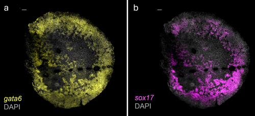
Using existing data sets, I first used spatial data to get an idea of how we would expect the expression of our chosen markers to look. My first step was the collection of chicken embryos after incubation at 38°C. HCR-FISH was then carried out, where probes bind to the RNA of a gene of interest during detection. These embryos were then mounted, imaged using spinning disk confocal microscopy to see exactly where the gene expression was and analysed using Fiji to give our final images (Fig. 1).

Fig. 1: HCR data images to visualise gene expression at stage EGK XI. Images obtained after completing HCR-FISH on chicken embryos and imaging using spinning disk confocal microscopy (a) Gata6 (b) Sox17 

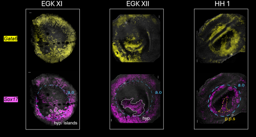
Fig. 2: Expression of Gata6 and Sox17 shifts from the area opaca to embryonic regions in later developmental stages. a.o: area opaca, hyp.: hypoblast, p.p.s: pre-primitive streak. (a) Stage EGK X: area pellucida consists of an upper layer and clumps of cells (hypoblast islands) which indicate a developing lower layer. (b) At stage EGK XI, there are high levels in the area opaca and the hypoblast islands which are moving anteriorly to later merge to form the hypoblast (c) Stage HH 1: Sox17 and Gata6 expression is reduced in the area opaca, shifting to the pre-primitive streak. Timepoints are from post-egg laying, growing in 38 °C incubator.

In the end, I was able to successfully visualise gene expression of markers Gata6 and Sox17 in the chick embryos and to trace the progression of these endodermal signatures to see how it shifts between different regions of the embryo (Fig. 2). In future, this project would benefit greatly by including more replicates and timepoints for validation. Extending this research to other vertebrates such as fish and frog would allow comparative work to begin allowing us to test the hypothesis that the hypoblast is conserved across vertebrates.

I would like to give a huge thank you to the DiFrisco Lab as well as the Briscoe Lab, where I was warmly welcomed and given a space to further enrich my knowledge in developmental research. I am especially grateful to my supervisor, Arsham Nejad Kourki, and to Sumit Garai for their invaluable guidance over the 9 weeks. Furthermore, I would like to thank the Francis Crick Institute for providing this insightful experience, and to the Rosa Beddington Fund for funding my project.

(8 votes)

Categories: Lab Life, Research

2026 writing challenge: your words matter 

Posted by , on 7 October 2026

Last month, we held our 2026 writing challenge. We had 14 entries in total, and it was great to see so many people from our community share their diverse perspectives on topics that matter to them. We really appreciate the effort all our contributors put into sharing posts on the site, and we hoped the challenge would help motivate and encourage writers to share their ideas. The Node is a community for all, regardless of experience, background, skills or career stage. For this reason, we decided to award the prize by randomly selecting the winner rather than using a competitive format. We hope this approach reassured authors to share on the Node without apprehension that their work would be judged. The winner of our writing challenge was Billy Hinchen, who wrote the post ‘The little beach hopper that could’. Billy celebrated the beach hopper, Parhyale hawaiensis, and taught us how it went from a mundane beach critter to a lab celebrity. We’ll feature an interview with Billy in a post soon. 

Several of our community members used this challenge to report on meetings and events that brought our developmental biology and stem cell communities together. The FlyPower team recounted their experiences at the Brazilian Society for Cell Biology (SBBC) meeting, showing how fruit flies continue to push the boundaries of biomedical science. Zainab Afzal reflected on attending the Society for Developmental Biology (SDB) meeting in Las Vegas, comparing her first meeting from 10 years ago to her experience today through two lenses: her scientific lens and her camera lens. Liliana Piñeros and Diego Sainz de la Maza shared insights from The Company of Biologists’ Workshop ‘Revisiting metabolic fundamentals’, while the Root Anatomy and Architecture Working Group of the International Society of Root Research (ISRR) reported on the “Rooting” conference on the Adriatic coast of Southern Italy, where many members of the group met in person for the first time.   

Our community members also shared research stories throughout the challenge, which took us on a journey across different research organisms, from cnidarians and fruit flies to plants and beach hoppers, tackling an equally diverse mix of research questions. Katarina Kurtović explored her fascination with the magic and beauty of plant developmental biology, while Vangeli Geshkovski discussed how ULTRAPETALA1 shapes plant development. Drawing on examples of cnidarians, Ian Velloso Randolph explored how embryogenesis may have emerged and evolved. Our challenge winner, Billy Hinchen, taught us about the beach hoppers’ meteoric rise to fame across different areas of science, while Saurabh Chand Sagar explored the story of Drosophila Malpighian tubules and the role caspase-3/Drice plays in their development and function beyond apoptosis. 

Our community members used this challenge to expand beyond the traditional research stories. Julia Ibe taught us how to start exploring single-cell data, making it accessible through publicly available datasets. Likewise, Friedrich Bliem reflected on the role of patterns as bridges between scientific, cognitive and artistic processes of understanding the world.  

Finally, authors turned their attention beyond the research itself and had honest conversations about the human side of science and academic life. Stefanie Williams recounted the burdens that first-generation students face and the importance of reminding them that they belong in academia. Alyssa Paparella highlighted the work of DisabledInSTEM and examined the gatekeeping of science, much of it invisible to everyone except those who are being kept out. Cherene de Bruyn reflected on one of the hardest stages of the PhD, the last 6 to 8 months, and the perseverance needed to cross the finish line.  

My favourite part of reading the entries was seeing each person write about their research, thoughts and experiences through their own unique lens. Each post had its own voice and flair. One phrase from Katarina has echoed in my mind as I was reflecting on this challenge: ‘Every seed matters’. I like to think the same is true for our community. Every member matters. Your science matters. Your experiences matter. Your words matter.  

The Node is always here if you have any research, thoughts or experiences that you would like to share. We also welcome audio or visual communication, including images, illustrations and videos. Challenges like this one remind us that anyone can write for the Node. You don’t need an invitation, just a story that you would like to share. 

You can find all the blog posts that were entered in the writing challenge here: https://thenode.biologists.com/tag/2026-writing-challenge/.  

(7 votes)

Tags: , ,
Categories: News

Loke CTR PhD studentship – applications open! Closing 3 November

Posted by , on 6 October 2026

The Loke Centre for Trophoblast Research (Loke CTR) offers fully-funded 4-year PhD studentship for home or overseas students

The Loke CTR provides a dynamic environment for PhD training in placental biology and reproductive science. Founded in 2007, it brings together 30 Principal Investigators with a shared interest in the scientific study of the placenta, early development and maternal-fetal interactions during pregnancy.

See our listing for more details and how to apply

PhD Studentships | Loke Centre for Trophoblast Research

(No Ratings Yet)

Categories: Careers, Education, Funding