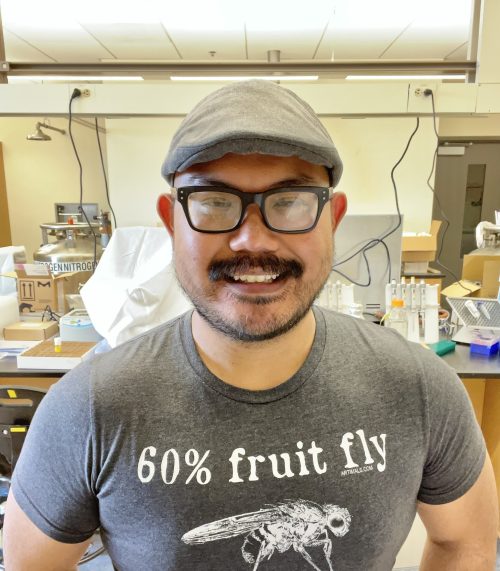
SciArt profile: Frank Macabenta
Posted by the Node, on 19 June 2023
In our latest SciArt profile, we spoke to Frank Macabenta, a developmental biologist who freelances as a scientific illustrator. Franks share with us his lab’s work on quality control mechanisms regulating organogenesis in the fly embryo, and how art influenced his decision to pursue studies in developmental biology.
Can you tell us about your background and what you work on now?
I am an immigrant and first-generation scientist. My family emigrated from the Philippines to the island of Guam when I was very young, which is where I lived for most of my early life. I studied biology at the University of Guam, initially with aspirations of going to medical school; however, after trying out research in Dr. Mari Marutani’s vegetable horticulture lab at the UOG College of Agriculture and Life Sciences, I decided to change my trajectory and focus on pursuing a career in research. I wasn’t sure what I ultimately wanted to study, but I was lucky enough to be selected for a summer research opportunity at Rutgers University just before starting my senior year, where I worked in the lab of Dr. Sunita Kramer. I fell in love with the Drosophila model system and developmental biology in Sunita’s lab, where we used confocal microscopy to study heart tube formation in the embryo. I ended up applying for grad school at Rutgers, where I conducted my doctoral thesis work in Sunita’s lab. After obtaining my PhD, I decided to stick with Drosophila as a model system (while also seeking warmer climes!) and worked as a postdoc in Dr. Angelike Stathopoulos’ lab at the California Institute of Technology, where I studied the genetic programs that guide the collective migration of a small population of muscle precursors in the Drosophila embryo called the caudal visceral mesoderm (CVM). CVM cells undergo collective migration along the length of the embryo to form the longitudinal fibers of the larval midgut muscles. We not only identified a number of complex multi-tissue interactions that drive CVM collective behavior, but also learned a lot about the intersecting signaling pathways that control growth and cell death to ensure stereotyped assembly of these muscles. I am currently an Assistant Professor at the California State University, Monterey Bay, where I teach both introductory and advanced courses in general and developmental biology, as well as run a research lab where we continue to study quality control mechanisms regulating organogenesis in the fly embryo.
Were you always going to be a scientist?
I definitely believe I was always going to be pursuing science in some capacity — since I was a little kid, I enjoyed going out in the yard to observe and collect various insects, or making a mess in the kitchen after seeing some experiment demonstrated in a TV show (often to my mother’s chagrin!). My parents encouraged and nurtured my love for science by buying every book, even gifting me with a simple compound microscope and slide-making kit — however, in my culture, children who have an affinity for science are typically expected to pursue a degree in medicine. I rationalized the choice of studying to become an MD as a career path that would still allow me to do science, and held onto this aspiration all the way up to college. However, my first research experience at the University of Guam under the auspices of an NIH Research Initiative for Scientific Enhancement (RISE) program, combined with some conversations with my professors convinced me to fully embrace scientific research as a career goal.
And what about art – have you always enjoyed it?
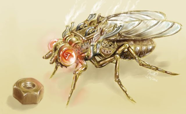
Art has always been a passion of mine. Growing up as an only child with few children my age in the neighborhood, drawing was an outlet that allowed me to express my imagination. Like my love for science, my parents also indulged my love for art — I’ve dabbled in pretty much every medium while growing up. Art also influenced my decision to pursue studies in developmental biology; I was awed by the beautiful high-resolution images that one can obtain of immunostained tissues. In a way, developmental biology research combines two of my greatest passions!
What or who are your most important artistic influences?
I would say I’m heavily influenced by detailed sci-fi/fantasy art. Growing up, I admired the works of artists like James Gurney, who combined meticulous depictions of dinosaurs with fantastical scenes for ‘Dinotopia,’ evoking a world where humans and dinosaurs coexist. As a cell biologist, I also greatly admire the work of Dr. David Goodsell — his ability to bring biomolecules and the spectacular, microscopic worlds they inhabit represent a truly elegant union of science and art!
How do you make your art?
I typically use black and white media like pencil and pen on paper for sketches and such. Some time back I bought myself a drawing tablet and started dabbling in digital art. I didn’t go through any formal training, so it’s been a lot of trial and error for me. I used to use the GNU Image Manipulation Program to make pieces, but I’ve since moved on to using Adobe Illustrator or Adobe Photoshop.
Does your art influence your science at all, or are they separate worlds?
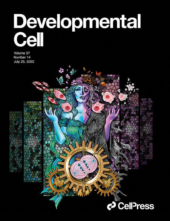
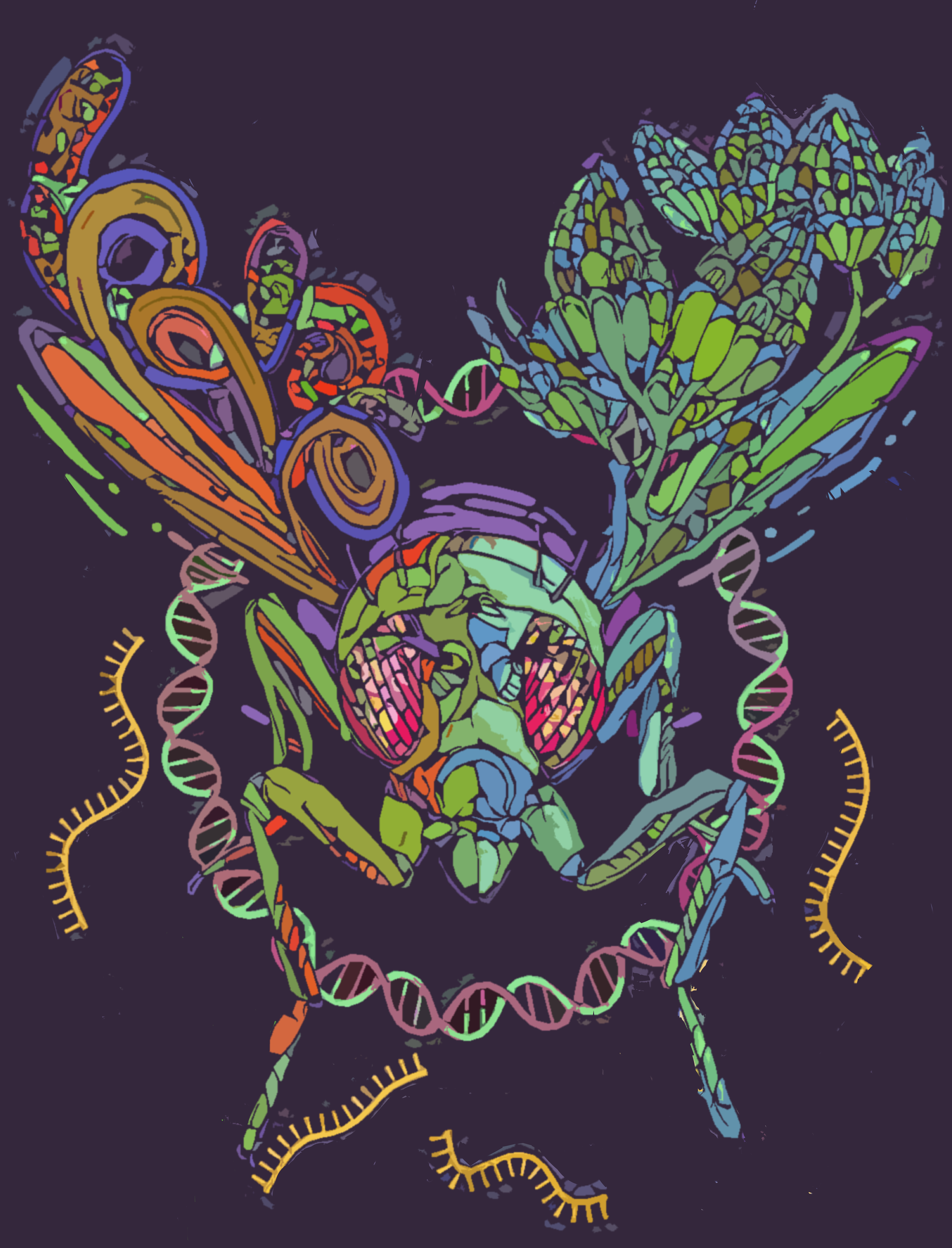
Art definitely has a profound influence on my field of research! Throughout the grad school and postdoc phases of my career, I often illustrated figures (and occasionally cover art) for manuscripts in addition to benchwork. My lab studies how embryonic muscles undergo precise and highly-stereotyped patterning, so my group spends a lot of time collecting images of immunostained cells. Scoring defects in muscle assembly takes quite a bit of observation and pattern recognition, which are skills that both artists and developmental biologists use regularly. Trying to understand the various phenotypes we observe also takes quite a bit of creative thinking — I think a lot of people underestimate how big a role imagination plays in planning and executing studies!
What are you thinking of working on next?
I’m thinking of creating more fruit fly-related work. I love the Drosophila research community, and want to inspire my students to see the beauty in studying developmental biology through model organisms like the fruit fly!
Find out more about Frank:
Personal website: macabentalab.com
Behance profile: https://www.behance.net/FDMacabenta
 (No Ratings Yet)
Get involved
Create an account or log in to post your story on the Node.
Sign up for emails
Subscribe to our mailing lists.