Stunning cysts
Posted by Erin M Campbell, on 6 October 2010
Hello to all of you Node readers! My name is Erin Campbell and I’m the blogger behind HighMag Blog, a blog that features cell biology images a few times a week. The great Eva Amsen contacted me about featuring some images on The Node, so I’m excited to be part of this growing community forum. The first image I’m blogging about is from a paper in the October 1 issue of Development, and features the biologically complex and visually stunning Drosophila ovary.
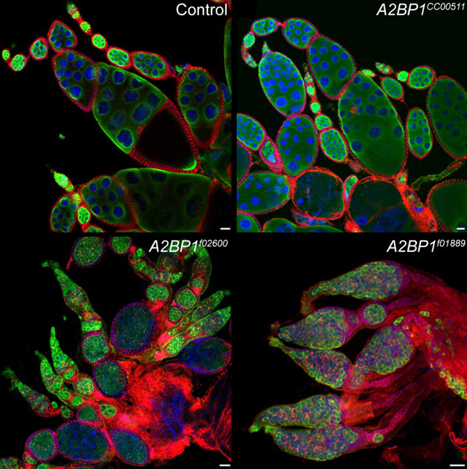
Ovarian cyst development begins in the germarium, with a stem division that produces the cystoblast, which then divides 4 more times. One cell in this 16-cell mass eventually becomes the oocyte, and the remaining cells serve as nurse cells to support the growth of the oocyte. This 16-cell cyst then becomes a separate egg chamber after being surrounded by follicle cells and budding off of the germarium. The hierarchy of signals and events that allow the differentiation of the cyst has been well studied, and a recent paper fills in the gaps in our understanding of this process.
Tastan and colleagues report that the Drosophila homolog of the human ataxin 2-binding protein 1 (A2BP1) gene functions in the intermediate stages of cyst differentiation, bridging the expression of early and terminal differentiation markers. Mutations in A2BP1 cause defects in cyst differentiation, as shown in the images above. Germline cells are labeled in green using anti-VASA antibodies, membranes are labeled in red using an anti-1B1 antibody, and DNA is labeled in blue. Compared with control ovaries, mutants exhibit a range of phenotypes: A2BP1CC00511 cysts have extra nurse cells, A2BP1f02600 cysts exhibit a mildly tumorous phenotype, and A2BP1f01889 cysts have an extreme tumorous phenotype.
Reference: Ömür Y. Tastan, Jean Z. Maines, Yun Li, Dennis M. Mckearin and Michael Buszczak (2010). Drosophila Ataxin 2-binding protein 1 marks an intermediate step in the molecular differentiation of female germline cysts. Development 137, 3167-3176. Paper can be found here.
 (10 votes)
3 thoughts on “Stunning cysts”
Leave a Reply
Get involved
Create an account or log in to post your story on the Node.
Sign up for emails
Subscribe to our mailing lists.
Fantastic images! Looking forward to more.
very nice pic`s in your site Erin !…I hope someday upload some of my pics there,…recommended in FB, :)
Shiny bead necklaces! they never cease to amaze me…great choice.