Visualizing stem cells at home
Posted by Erin M Campbell, on 13 June 2011
 The Drosophila ovary is stunningly beautiful, and a playground of wonderful biological questions. Within the germarium alone, developmental biologists can look at asymmetric division, stem cells and their niches, cell migration, and cell specification. A recent paper in Development describes a technique allowing the in vitro imaging of a fruit fly ovary, and opens the door for further studies of development and stem cells.
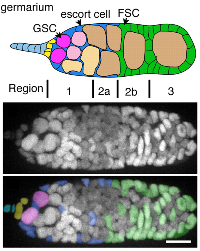
The Drosophila ovary is composed of about 15 ovarioles, which each houses an organ called the germarium that serves as the assembly line for egg production. In the germarium, two types of stem cells play important roles – germline stem cells and follicle stem cells. Germline stem cells divide to kick off the egg production process, while follicle stem cells divide to provide several different types of ovarian follicle cells. A recent paper by Morris and Spradling describes a technique allowing the live imaging of a developing follicle. After dissecting and imaging an ovariole in culture, Morris and Spradling were able to monitor cell division, orientation, and movement during follicle generation for a prolonged period. This technique allows biologists to address many unanswered questions about follicle generation and stem cell biology, as both populations of stem cells can be imaged simultaneously and in their own niches.
Images show a cartoon and high-resolution images of a fruit fly germarium, with germline stem cells (GSC) marked in pink and follicle cells in green (FSC are follicle stem cells). BONUS!! For a cool movie of a germarium showing cell divisions and dynamic movement, click here.
For a more general description of this image, see my imaging blog within EuroStemCell, the European stem cell portal.
Morris, L., & Spradling, A. (2011). Long-term live imaging provides new insight into stem cell regulation and germline-soma coordination in the Drosophila ovary Development, 138 (11), 2207-2215 DOI: 10.1242/dev.065508


 (1 votes)
Lucy Morris wrote a post about this work a few weeks ago, describing the procedure of setting up the experimental system, which was tricky at times.
A great technique, I’m glad it’s been featured twice!
Great image and nice descriptions, both the one here and the less specialist one on EuroStemCell. Thanks Erin!