Woods Hole images 2014 movie round- vote for a Development cover
Posted by the Node, on 2 March 2016
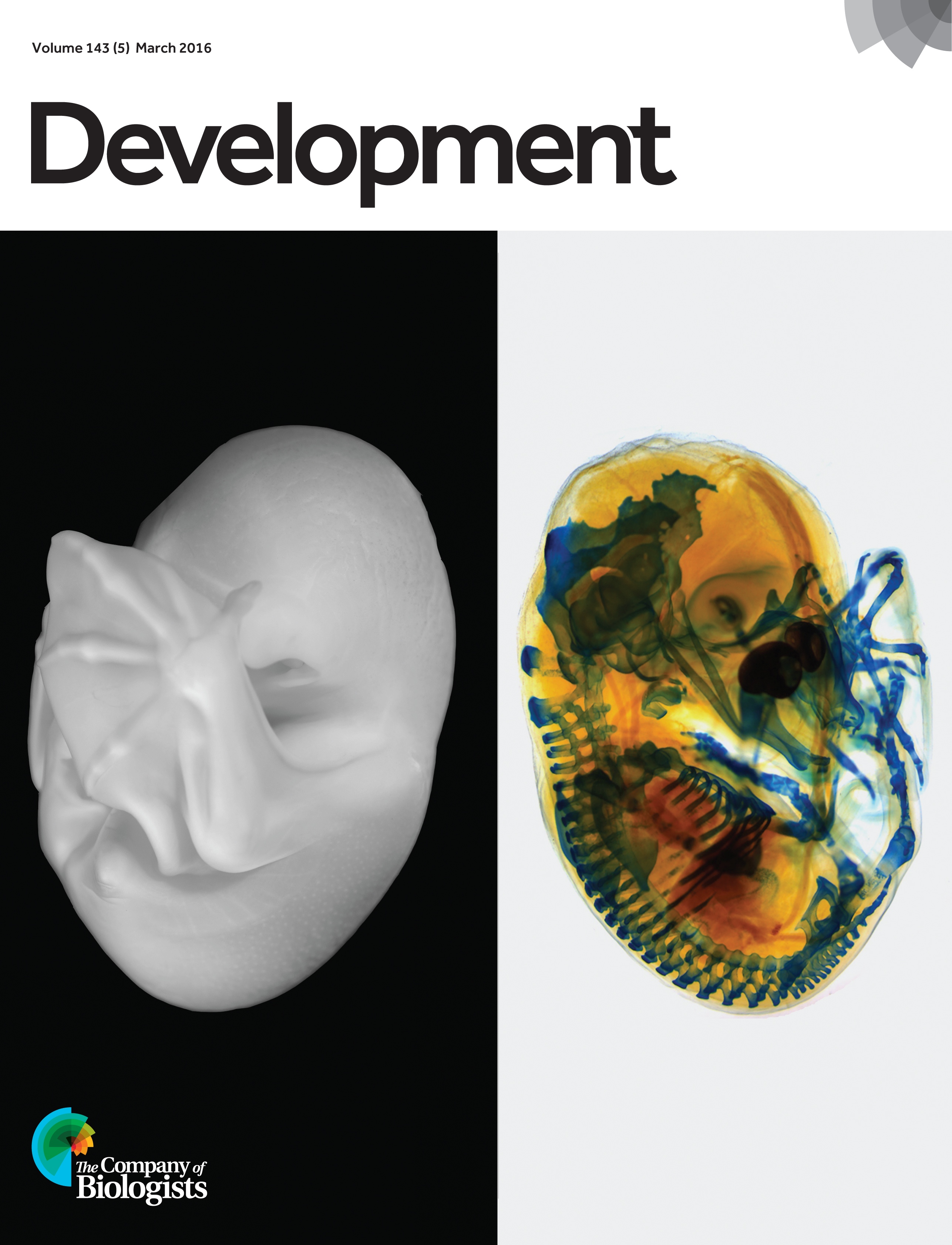
 Earlier this year we asked you to vote for your favourite image from a selection of 4 beautiful pictures taken by the students of the 2014 Woods Hole Embryology course. You chose a stunning image of a bat embryo, which features in the cover of the latest issue of Development.
It is now time to vote again, but this time we would like you to choose your favourite movie from the 4 below. The winning movie will feature on the homepage of Development, and a selected still will feature in the cover of the journal (click the links below each image to see what the cover would look like). The poll is set up to allow only one vote per person, so please stick to this rule to give all the images a fair chance!
Voting will close noon GMT on March the 30th.
Movie 1. Fly Embryo – Dorso-ventral Split
Drosophila melanogaster embryo stained for DAPI (blue, all nuclei), Elav (green, neural nuclei), anti-HRP (red, neural membranes and axons, and ring gland) and Fasciclin II (yellow, subset of CNS neuron cell bodies and motoneuron axons). Dorsal side at stage 16, ventral side at stage 14. Cover image. This movie was taken by Shane Jinson (MBL, USA) and Amber Famiglietti (Swarthmore, USA).
Movie 2. Fly Embryo – Sections
Drosophila melanogaster embryo, stage 16/17, stained for Tropomyosin (cyan, muscles), anti-HRP (magenta, neuron cell membranes and axons), Pax 3/7 (yellow, segmental patterns in ectoderm and CNS, MAb DP312), Engrailed (red, segmental patterns in ectoderm and CNS), and Twist (green, subset of neural and muscle nuclei). Imaged with a Zeiss LSM 780 Confocal. Cover image. This movie was taken by Carolyn Kaufman (Stowers Institute, USA).
Movie 3. Fly Eye Disk
Drosophila melanogaster third instar eye disk. DAPI (dark blue, nuclei), anti-HRP (magenta, photoreceptor cell bodies and axons), Elav (cyan, photoreceptor nuclei), Repo (yellow, glia cell nuclei), Expanded (white). Disk also contains Tie-Dye clones – EGFP (green), RFP (red), and LacZ (medium blue). The EGFP clone is in patches of photoreceptors, while the RFP and LacZ clones are largely overlapping in a patch of peripodial membrane (RFP being mostly a subset of the LacZ clone). Imaged with a Zeiss LSM 780 Confocal. Cover image. This movie was taken by Jiajie Xu (University of Chicago, USA).
Movie 4. Fly Embryo – Seven Channels
Drosophila melanogaster embryo, stage 17, ventral view. DAPI (blue, nuclei), Elav (green, neuronal nuclei), Spalt (yellow, subset of neuron and muscle nuclei), BP102 (red, CNS axons), Even-skipped (magenta, subset of CNS nuclei, and ring of nuclei around anal pad), and anti-HRP (gray, neuronal cell bodies and axons). DIC image also collected during the scan. Music begins about 40 seconds into the video. Imaged with a Zeiss LSM 780 Confocal. Cover image. This movie was taken by Connie Rich (University of Cambridge, UK).


 (4 votes)
Number 3 wasn’t working/ playing for me. Also, any info on what software they used to make the movies?
Hi Sarah,
The movies were put together by Nipam Patel, one of the lecturers at the course. I have emailed him to ask him what software he used, and I will let you know as soon as I hear back from him!
And the software is….?
Hi Stephen,
The movies were put together by Nipam Patel, who is one of the lecturers at the course. I have emailed him to ask him what software he used, and I will let you know as soon as I hear back from him!
The software used to generate the movies is Volocity (Perkin Elmer). A big thanks to them for making their software available for use in the MBL Embryology Course, and of course the microscope companies that make the scopes available.