The first Xenopus imaging course
Posted by Gary McDowell, on 17 February 2014
The first Xenopus imaging workshop was held at the Xenopus Resource Centre at the Marine Biological Laboratory in Wood’s Hole, MA from November 17th – 22nd.

Kymograph of a beating heart in a Xenopus tadpole expressing GFP under a cardiac actin promoter. By Kyle Jewhurst, Tufts University.
20 international frog researchers, from Japan to the UK to the US, all looking to use microscopy for many different applications, attended the workshop. Stages of frog egg and embryo development, from late-stage oocytes to late tadpole stages, were of interest to the students attending; there were those wishing to dissect tissues for high resolution imaging compared to those wishing to image whole embryos; those wanting to do live imaging and those wanting to figure out how to get pictures of fixed samples used for in situ hybridisation. Although there were a wide variety of research goals, everyone was looking to use a similar set of techniques geared towards their own individual needs. I have included some examples of images taken at the course throughout this blog, so you can see what we got up to.
The course was very free in structure, which meant people with their own projects had time to prepare samples, whilst those with no agenda could try to learn how to use the microscopes and just play around with things. mRNAs for microinjection, and embryos from both wild type and various transgenic frog lines were provided, to give people an opportunity to try out experiments at the course. Microscopes from Zeiss, Nikon and Leica were made available by those companies, with reps from Nikon and Zeiss in attendance to help out and provide guidance on microscope use.
We were lucky to have a number of experts at the course: from the University of Southern California, Scott Fraser; from the University of Texas at Austin, John Wallingford, his graduate student Eric Brooks and postdoc Asako Shindo; and from the University of Pittsburgh, Lance Davidson and his graduate student Jo Shawky. With such a high teacher:student ratio, there was a lot of opportunity to get time with each of the instructors. Each instructor also gave talks about theory (for example, Scott Fraser gave an excellent overview of the theory of microscopy; whilst John Wallingford went through the do’s and don’ts of Photoshopping) and their own work, highlighting examples of the use of imaging in their own science. Some excellent stories were told – we got a sneak preview to the work of Asako Shindo, published just recently in Science (Science 2014, 343, 649-652).
In addition to the showcasing of developed scientific stories, we also had the opportunity to showcase our own work each day, with an evening show-and-tell of everyone’s best images. Not only did we get the opportunity to see what everyone else was working on – we also were able to critique the work of others and ourselves, to figure out how to improve the imaging technique for future use.
There was also the opportunity to make tools for frog manipulations – I now have my very own hair loops and eyebrow dissecting knife – staples for any frog researcher!
At the end of the day, after downing tools and critiquing images, we would get together for a quiet drink and a chat, an opportunity to talk to other researchers we might not necessarily meet at the conference. In addition to what you would normally talk about or see at conferences, because we were spending the day doing practical things, we were often able to look directly at someone else’s research as it happened and have discussions about what they were doing, looking at actual samples.
Video of Stage 8 Xenopus laevis embryo with EB3-GFP labeling microtubule + ends and H2B-RFP labelling histones magenta. By Romain Gibeaux, University of California, Berkeley.
Confocal z-stack image of sectioned Stage 45 tadpole, injected with tomato-Sert, showing nerve bundle connecting to otic placode. By Gary McDowell, Tufts University.
The course was the first of its kind that the frog community has held but it was very well received and all of us students had a great time. In particular there was a lot of help provided by the National Xenopus Resource staff, Esther Pearl and Christy Lewis, who provided frog and microinjection support for the students, as well as ensuring mRNAs were ready and available for people to try. Hopefully the course will be run again as we all appreciated what a great resource this could be for our fellow researchers in the frog community. Also, where else are you going to be able to generate the images for your own t-shirt!
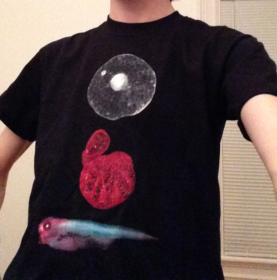
T-shirt designed by Esther Pearl of National Xenopus Resource; images by Gary McDowell, Kyle Jewhurst and Emily Pitcairn, all of Tufts University.
Gary McDowell, Center for Regenerative and Developmental Biology, Tufts University
You can follow the National Xenopus Resource on twitter at:
You can also follow more frog posts at:
bewareofthefrog.tumblr.com
 (5 votes)
One thought on “The first Xenopus imaging course”
Leave a Reply
Get involved
Create an account or log in to post your story on the Node.
Sign up for emails
Subscribe to our mailing lists.
Read the latest Development issue
Most-read posts in May
- From comparison to mechanism: decoding heart regeneration
- A day in the life of a Reviews Editor (at Development)
- What does a Reviews Editor do?
- New evo-devo textbook ‘Eco-Evo-Devo: The Environmental Regulation of Development, Evolution, and Health’
- preLighters’ choice – A curated selection of recent preprints
Hey Gary, how nice to see your post here! I agree that the imaging workshop was great, and we all had a wonderful time there. Cool design of the T-shirt, isn’t it? :)