

From killing cells to shaping them: enigma of Caspase-3 beyond apoptosis
Posted by Saurabh chand Sagar, on 1 September 2026
Small historic background:
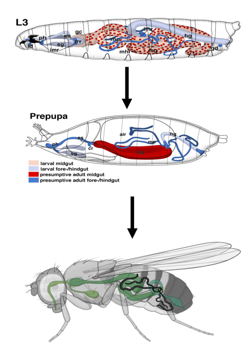
In an era when caspases are known as killer proteins, Drosophila Malpighian tubules state otherwise. Drosophila is a holometabolous insect, meaning that it goes through three life stages: larval, pupal and adult. The pupal stage of insect development is when the major transformation occurs. Most of the larval tissues get histolysed, while adult tissues are reformed from imaginal discs that remain quiescent and isolated during the larval stage. Surprisingly, during metamorphosis, some larval tissues, such as Malpighian tubules (MTs), tracheal tubes, ventral nerve cord and some larval muscles, skip the histolysis process. However, why these tissues escape histolysis during metamorphosis has remained a question for more than a century. On the other hand, Caspases have been well established as killer proteins since their discovery; in Drosophila Caspases often compliment programmed cell death (PCD) during the histolysis. They were considered primarily as executioners of cell death until the early 2000s, when evidence began to accumulate that caspase functions extend well beyond cell death, including roles in development and tissue morphogenesis. What if caspases do much more than just kill cells?
The question asked?
While working on Drosophila melanogaster, my supervisor, Prof. Madhu G. Tapadia, wondered why insect kidneys (Malpighian tubules) escape histolysis during metamorphosis. What is so special about the Malpighian tubules in insects?
And the story began…
The story began with this very simple question, which was then taken up by two of her PhD students in 2011. Both of them started working on the expression and localization of caspases in the Malpighian tubules that is essentially reuired for the PCD. They established a landmark finding that eventually led me to my current work. Both of them reported that caspases are expressed in the cells of Malpighian tubules. However, their exact role and why MTs do not undergo histolysis during metamorphosis remained unanswered. One of them suggested that apoptotic proteins are translated in the Malpighian tubules; however, they are sequestered in their pro-apoptotic form within the nucleus and therefore coldn’t execute the cell death. Other one went one step further and reported a possible role of apoptotic proteins (reaper, hid and grim; often referred to as RHG proteins) in tissue morphogenesis and polarity maintenance.
The candidate protein Rho1GTPase!
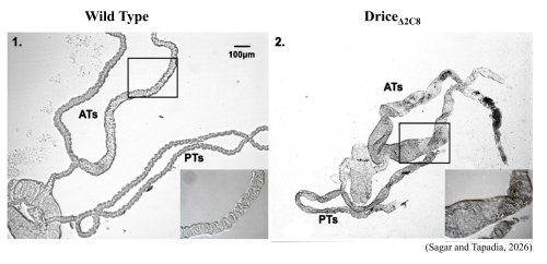
Next came another of my senior, who carried this question through her PhD journey at Prof. Tapadia’s lab, Department of Zoology, Banaras Hindu University. She took the lead from previous studies and started examining the morphology and physiology of the Drosophila renal tubules. She discovered that executioner caspase-3/Drice (in Drosophila) is activated in both larval and pupal MTs, yet they still escape histolysis. She reported that Caspase-3 deletion mutants (Drice mutants) show cystic MTs containing multiple cyst-like structures. Additionally, cytoskeletal and polarity proteins are highly disorganized, tubules are shorter, and cell number and cell shape are affected, along with a significant reduction in tubular secretion by the Drosophila kidneys in the Drice mutants. She also identified Rho1GTPase as a key candidate protein and a master regulator of actin dynamics and polarity establishment. Rho1GTPase was significantly upregulated at the protein level in the MTs of Drice mutants, suggesting a negative correlation between Drice and Rho1.
That’s where I came in the picture…..
Finally, I joined the lab back in 2021. Since, Caspases are essentially required during metamorphosis, however, in case of Drosophila MTs, caspase-3 activity was present yet they escapes histolysis completely. Therefore, the core question had evolved significantly from how MTs evade histolysis to what role caspase-3/Drice performs in the MTs, if not cell death. I began my work with this very question. By then, it was already established that Drice is essentially required for normal tubular architecture and physiology; I also had a candidate protein to work with, viz., Rho1GTPase.
Instead of jumping directly to Rho1, I decided to look at the RhoGTPase family. In order to do so I planned to check protein expression as well as transcript levels of many targets. I examined the transcript levels of more than 20 genes for this study.
Trouble with RT-PCR….
During my RT-PCR era, my lab once received a faulty batch of SYBR Green. I was so unlucky that I got to work with that faulty batch totally unaware of what was coming. Initially, I thought the problem was with me because there was too much variation in the results. I tried again and again. At one point, my colleagues started doubting my experimental capabilities, but I couldn’t accept that and kept doing it again and again refusing to give up. I even recalibrated the machine, but the problem remained, and finally, after countless PCRs, I almost gave up. Then came the idea of trying an alternative SYBR Green, and that’s when the problem was discovered that SYBR was the actual problem. Later, even the manufacturer accepted that the batch was faulty. Anyway, it cost me around 3–4 months and a great deal of frustration. But I learned one thing from the experience: if you are doing it correctly, you will eventually get it done.
CDC42 is also affected by Caspase-3 absence along with Rho1 in the MTs….
Since Rho1 was already known to be involved, I next examined the other RhoGTPases, Rac and CDC42. I found that CDC42 was also dysregulated in the MTs of Drice mutants, whereas Rac remained unaffected. This made me wonder: how were Rho1 and CDC42 affecting the actin cytoskeleton, the internal framework of the cells?
I first followed the Rho1 pathway and found something unexpected: Rok, a downstream effector of Rho1, was significantly reduced despite high levels of Rho1 in Drice mutants. When I knocked down Rok, the MTs developed defects similar to those of the Drice mutants, including disorganized actin and polarity proteins. This suggested that reduced Rok could contribute to the tubule defects.
I then turned to CDC42. CDC42 and its downstream effectors were increased, and CDC42 is known to promote actin thickening through the Arp2/3 complex. Interestingly, I observed similar actin thickening in Drice mutant MTs. To test this idea, I reduced Arp2 and Arp3 in Drice mutants, which restored the excessive actin thickening. Together, these findings pointed to Rok and Arp2/3 as two important downstream components through which Drice influences actin organization.
Actin polymerization vs depolymerization!
Now the next problem was densely packed actin: what was actually happening? Was actin being hyper-polymerized or depolymerized in the MTs? The best way to answer this was to look at the levels of F-actin and G-actin separately. Very high G-actin and low F-actin would suggest depolymerization, while the reverse would indicate actin polymerization. Following this, I found that in Drice mutants, F-actin was significantly higher and G-actin was very low compared with the wild-type MTs. We therefore hypothesized that actin was undergoing hyper-polymerization in the MTs of Drice mutants, and we moved ahead with this hypothesis.
But until now one major question remained: how was Caspase-3/Drice controlling these in the first place? That was the next question I had to answer.
What exactly Caspase-3 is doing in the MTs?
Until now, we had made significant progress. We knew that the morphological defects in the MTs were most likely due to Rok dysfunction and Arp2/3 overexpression. However, one crucial question remained: How was Caspase-3/Drice regulating actin dynamics in the MTs? Was Caspase-3 interacting with Rho1? If so, was this interaction direct, or was it mediated indirectly through some modulator?
Three months of frustration….
To address this crucial question, I initially targeted a few candidate proteins and started checking whether they had any effect on actin expression and organization in the MTs. Growing flies, dissecting MTs and performing immunostaining for multiple targets was not an easy task. Also, once the immunostaining was done, there was still slide scanning, figure panel preparation and image analysis—oh gosh! I wish it were as easy as I have written it here. Target after target! I could not make significant progress and kept changing the target proteins. There was a time during this period when I seriously hated the lab and my work. The solution to this problem was actually very simple: a new technique that I learned quickly.
My saviour: immunoprecipitation – the answer was finally found.
After realizing that one-by-one targeting was never going to solve my problem, I performed immunoprecipitation (IP) to check the protein–protein interaction partners of Rho1. Guess what? Out of 75 proteins, there was only one that linked Rho1, Actin and Caspase-3 together—and that was Gelsolin, the protein I had been looking for all along. IP suggested an interaction between Rho1 and Gelsolin in wild-type MTs, which was absent in the Drice mutants. Gelsolin also showed Caspase-3-mediated regulation of actin dynamics. I further confirmed this finding to be fully assured that it was reproducible, and I obtained similar results repeatedly. That was when I became convinced that we had finally found the missing link.
Gelsolin contributes to actin filament turnover and severing and thereby helps regulate the F-actin pool. Therefore, the absence of Gelsolin provides a possible explanation for the elevated F-actin levels and altered actin dynamics in the MTs. I finally had my answer, and we could finally move on to the publication part.
Paper communication: make or break point of the story!
Scientific publication always feels heavier and harder than the research itself. Also, by this stage, my supervisor was more convinced of my work, and I was also satisfied with what we had achieved. She encouraged me to send the work to prestigious journals, and I did so. After being rejected by two journals, the manuscript finally landed in Cell Death & Discovery, where they agreed to send it for revision.
Reviewer’s comments: Is it going to be accepted?
This work was reviewed by three reviewers in total. The first two agreed to review the manuscript within a week, while the third one took his time. After waiting for almost three weeks, the first wave of reviews finally arrived in my mailbox. I was anxious about what the reviewers would say. After reading the first reviewer’s comments, I was completely frozen. The first reviewer had rejected my work outright, mostly because I had not cited a particular paper and he did not seem convinced by the work. My anxiety levels were beyond words. Then came the second reviewer. What was it going to be? The second reviewer was very optimistic and seemed to like the concept I had presented in the manuscript. He asked several questions, which I was happy to answer. I had already anticipated three or four of those questions and had performed the experiments in the background, and when the second reviewer asked two of them, my morale was significantly boosted.
Finally, after another two weeks of waiting, the comments from the third reviewer arrived. He really liked the work and suggested only minor revisions. The third reviewer summarized my manuscript so well that I even added a few of his lines to the Discussion section of the paper. And finally, with the publication of the paper, the story came to a happy ending.
What I learned during this journey:
Looking back, this journey taught me that science is rarely a straight path. There were times when I questioned my experiments, my approach and even myself. But every failed experiment, every unexpected result and every setback pushed me a little closer to the answer. The faulty SYBR Green taught me not to blame myself too quickly; the failed candidate-protein approach taught me to change my strategy; and Gelsolin taught me that sometimes the answer is hiding among the possibilities you have not yet considered.
Most importantly, I learned that if you believe in the question, stay honest with your data and keep looking for the answer, you will eventually get there. So, the take-home message is:
Ask good questions.
Believe in yourself when things go wrong.
Change your strategy when the evidence tells you to.
And never mistake a setback for the end of the story.
Get involved
Create an account or log in to post your story on the Node.
Sign up for emails
Subscribe to our mailing lists.