From our sister journals- May 2015
Posted by the Node, on 19 May 2015
Here is some developmental biology related content from other journals published by The Company of Biologists.

Xenopus as a developmental model of neuroblastoma
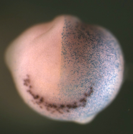 Neuroblastoma (NB) is a paediatric form of cancer derived from the sympathetic nervous system. Recent genome-wide sequencing data suggest that often NB does not have a clear genetic cause, leading the authors to hypothesize that NB results from aberrations of normal development. To test this hypothesis, Anna Philpott’s group used a population of anteroventral noradrenergic (AVNA) cells from Xenopus embryos. These cells share several features with mammalian sympathetic neurons, including the expression of noradrenergic-associated genetic markers such as the achaete-scute complex-like 1 (Ascl1) gene, which encodes a transcriptional driver of neurogenesis. By comparing AVNA and NB cells, the authors found that, whereas Ascl1 is only transiently expressed in AVNA cells, it is aberrantly maintained in NB, where it is phosphorylated on multiple serine-proline sites. The authors then show that differentiation of AVNA cells is enhanced by dephosphorylated Ascl1. Moreover, this process is inhibited by experimental manipulations of NB-associated genes, but, interestingly, dephosphorylation of Ascl1 is able to overcome this inhibition. This work demonstrates that Xenopus AVNA cells represent a unique system to study sympathetic nervous system development and its relationship to NB. Moreover, it suggests that Asc11 phosphorylation might promote stalled differentiation leading to NB, thus identifying a potential target for therapeutic purposes. Read the paper here (Open Access) and the authors’ Node post here.
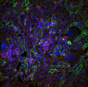
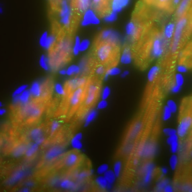
Modelling DMD-associated cardiomyopathy in patient-derived iPSCs
 Duchenne muscular dystrophy (DMD) is a genetic muscular disorder characterised by progressive muscular weakness and wasting, with cardiac complications (such as dilated cardiomyopathy) and respiratory muscle failure arising in the late stage. To investigate mechanisms of dilated cardiomyopathy in DMD, Lei Yang and collaborators derived cardiomyocytes (CMs) from DMD-patient-specific induced pluripotent stem cells (iPSCs). Compared to control cells, DMD iPSC-CMs exhibited elevated cytosolic calcium, mitochondrial damage and increased cell apoptosis. To further dissect the mechanisms underlying these alterations, the authors performed transcriptional and translational analyses and identified a mitochondrially initiated molecular cascade – which involves CASPASE3 (CASP3) activation – as being responsible for the increased apoptosis in DMD iPSC-CMs. Notably, the application of the membrane sealant Poloxamer 188 could prevent calcium overload and CASP3 activation, significantly reducing apoptosis in these cells. Thus, the authors established a useful in vitro system to disclose mechanisms of cardiomyopathy in DMD and to identify molecular targets that could be pharmacologically manipulated. Read the paper here (Open Access).

Endocytosis and micropinocytosis internalise cadherin-6B in EMT
 Epithelial-to-mesenchymal transition (EMT) is an integral developmental and physiological process, but can also be utilised by cancer cells at the initiation of metastasis. A requirement for EMT is the post-translational removal of adhesion proteins from the plasma membrane. Here (p. [164426]), Lisa Taneyhill and Rangarajan Padmanabhan study cadherin-6B (Cad6B) internalisation to elucidate the mechanisms of EMT in chick cranial neural crest cells. The authors found that in neural crest cells that are initiating EMT, Cad6B was detected in cytosolic puncta that were endocytic, rather than exocytic, in nature. They then identified two intracellular motifs that were potentially important for regulating Cad6B internalisation. Mutating the p120-catenin-binding (EED) motif, but not the dileucine (LI) motif, significantly increased Cad6B internalisation, supporting the idea that Cad6B is removed from the plasma membrane through endocytosis. However, although Cad6B colocalised with clathrin, the colocalisation was not exhaustive, suggesting that an additional mechanism is involved in Cad6B internalisation. Therefore, the authors used an array of pharmacological treatments to show that Cad6B was removed from the plasma membrane through both endocytosis and macropinocytosis, and that both of these processes depended on dynamin. This study demonstrates that EMT and neural crest migration require Cad6B internalisation through endocytosis and macropinocytosis. Read the paper here.

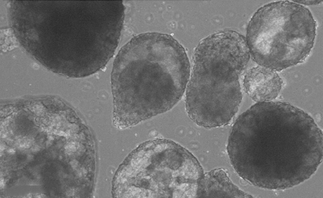
Generating an in vitro 3D cell culture model from zebrafish larvae for heart research
 Spontaneously beating 3D ‘heart’ structures can be developedin vitro from larval zebrafish using a novel, fast and inexpensive method that could be employed in ecotoxicology and biomedical safety testing. Read the paper here.
 (2 votes)
Get involved
Create an account or log in to post your story on the Node.
Sign up for emails
Subscribe to our mailing lists.