This year’s venue will be the CRUK Scotland Institute in Glasgow from Tuesday 23rd to Friday 26th of September and will be the 7th since the meeting was first held at the Doherty Institute in Melbourne in 2018, the name having been coined by Prof Elizabeth Vincan at the time. The Glasgow meeting this September will be the first to be held in Europe!
Plenary, Keynote and Symposium sessions, including rapid-fire short talks and poster sessions, make up the programme, which includes many well-known international speakers.
Early bird registration closes on the 15th of June and includes discounted rates for students.
Organisers are very excited to be hosting this meeting, where cutting-edge technologies meet unanswered biological and clinical questions.
Prof Elizabeth Vincan, Clinical Scientist and Medical Researcher in the Department of Infectious Diseases, Melbourne Medical School, University of Melbourne says: “Organoids Are Us brings together delegates from diverse fields and disciplines to show case and learn about the latest advances in organoid technology, and its application to fundamental and clinical research to fast track discovery and translation. This is the seventh conference in this highly successful series, and the first in the UK.”
Prof Ramanuj DasGupta, Professor of Cancer Systems Biology at the School of Cancer Science, University of Glasgow and CRUK Scotland Institute says: “The Organoids Are Us conference aims to bring together the key opinion leaders in the fields of cancer, infectious disease and regenerative biology, who utilise organoid models to address key questions in human disease biology and development of novel therapies.”
This week we will meet Dr Cassandra Koh, who is a new faculty at Institut Pasteur. Cassandra is driven by curiosity and is passionate about decoding the molecular choreography between viruses, their insect hosts, and the lipids that entwine them. Her research traces the intricate ways arboviruses hijack mosquito metabolism to fuel their replication and has delved deep into how symbionts like Wolbachia rewire these metabolic pathways. Her brand-new lab will focus on how virus–virus–host interactions offer a nuanced view of disease transmission and microbial co-evolution. But science for Cassandra is more than experiments — it’s a way of asking questions about the world. She credits strong mentors and surprising inspiration from artists and storytellers for shaping her journey. Whether she’s tracing lipid dynamics in mosquito cells or hosting a long lunch for friends, Cassandra believes in curiosity as a compass. She’s currently welcoming collaborators and postdocs interested in exploring the metabolic intersections of immunity, infection, and symbiosis. Check out her work here ! Give her a follow over Bluesky and Twitter.
What was your first introduction to the fields of metabolism and viral infections?
It started with cholesterol. The fruit fly Drosophilamelanogaster carries a bacterial endosymbiont called Wolbachia that suppresses pathogenic viral infections in its host. This was a very exciting finding that resulted in a biological intervention strategy against mosquito-transmitted viral diseases based on the stable introduction of the Wolbachia wMelstraininto natural populations of a major mosquito vector species, Aedesaegypti. Eric Caragata had showed that Wolbachia-induced viral suppression became weaker when Drosophila flies had more cholesterol in their diets, leading to the conclusion that Wolbachia and Drosophila viruses were competing for host cholesterol (PMID: 24337107).
Naturally, this led to the question of whether Wolbachia and mosquito-transmitted viruses (also called arboviruses) also compete for host lipid resources. During this study, I learned a lot about the role of lipids in viral infections in mosquitoes from a key paper by Rushika Perera (PMID: 22457619). Her work had shown that dengue virus replication in mosquito cells relies on the activity of fatty acid synthase. In addition, as a flavivirus, dengue virus re-organizes the endoplasmic reticulum membranes to form replication complexes, and this is reflected in an enrichment of lipids that promote membrane curvature and permeability.
You use the term “virus-virus-host” interaction for describing the theme of your lab. Could you share your journey into studying this and using mosquitoes as one of your host model systems? Tell us how you got inspired to further branch out into metabolic aspects of viral infections?
The interaction between arboviruses with their mosquito host is a fascinating subject in itself. Mosquitoes have evolved interesting ways to fight off and tolerate viral infections and the virus seeks to complete its transmission cycle while minimizing virulence to its vector host. With the recent appreciation that mosquitoes harbor a multitude of other viruses that constitutes its resident microbiota, virus-mosquito interaction is no longer a two-player game. These resident viruses are called “mosquito-specific viruses” to distinguish them from the ones that infect and cause disease in humans and animals. They have entered the research spotlight as many studies have reported their ability to reduce or enhance arbovirus infection in mosquitoes, which implies that they have a role to play in disease transmission. Many questions have since sprung up about how they interact with arboviruses and the mosquito host. I am curious to see whether these interactions can be observed in the metabolic dimension.
Your work intersects virology, metabolism, immunology and RNA interference – tell us what studying immune-metabolism means for you. How do you integrate these disciplines in your research, and what unique insights have emerged from this approach?
Immune responses cost energy. Viral infections cost energy. A virus infection is therefore a disruption to immune and lipid homeostasis. There is already some evidence of crosstalk between immune signaling and lipid metabolism modulations in bacteria-infected Drosophila (PMID: 30902902, PMID: 33227003). On top of that, viruses hijack host lipid membranes and reconfigure phospholipids to form viral replication complexes (PMID: 33087565), adding to the toll on the metabolic burden of virus-infected cell. Immune and metabolic regulation therefore go hand in hand and would provide a more holistic view of cellular responses to viral infection.
How has Wolbachia evolved in Drosophila and mosquito species – are a lot of mechanisms of symbiosis evolutionarily conserved? How can studying Wolbachia-insect interactions help us understand symbiont-driven metabolic changes? Could you shed more light on these emerging areas of research.
Wolbachia is a common endosymbiont among arthropods including insects like Drosophilamelanogaster and some mosquito species like Aedes albopictus. When it was observed that some Wolbachia strains protect their Drosophila host from viral infection, the notion that the endosymbiont could be introduced into Aedesaegypti mosquitoes, a non-native host, led to the development of Wolbachia-based intervention strategies to limit the spread of viruses transmitted by this major vector species. That Wolbachia is vertically transmitted through the maternal line was a very useful property for a sustainable and self-driving intervention.
Studying how Wolbachia interacts with its hosts would reveal its mechanisms of symbiosis, which would say something about the directions of evolutionary pressures in natural or introduced hosts. These mechanisms might take the shape of metabolic mutualism, immune priming, or something else entirely.
What are the key differences in how Wolbachia and DENV-3 (Dengue virus serotype 3) alter host lipid metabolism?
In our work comparing how Wolbachia and DENV-3 modulate Aedesaegypti lipids, we found that the endosymbiont and the virus individually produce very different lipid alterations. While DENV-3 produced strong elevations, Wolbachia-infected mosquitoes exhibited much milder perturbations of different lipid species, which does not support the Wolbachia-virus competition hypothesis.
Tell us about your findings on association of DENV3 infection with lipid droplet growth and hyperglycemia in mosquitoes? Tell us about your exciting finding of how Cardiolipins are involved in DENV infection.
DENV-3 infection alone strongly elevated levels of triacylglycerols, glycerophospholipids high in polyunsaturated fatty acids, and Amadori-glycated phosphatidylethanolamines in Aedesaegypti mosquitoes. These lipid classes indicate lipid droplet accumulation, cellular membrane remodeling, and viral infection-induced hyperglycemia. The latter is especially interesting as it suggests that this cellular phenomenon, which has previously been observed in human cells infected by dengue viruses, could also occur in mosquito cells.
Cardiolipins were an interesting class of lipids to notice in our dataset because they have not been previously associated with infection. Given its role to maintain mitochondria function, and our finding that cardiolipin depletion disfavors virus replication, we surmised that cardiolipins help to buffer the effects of cellular stresses from viral infection that would otherwise lead to the triggering of the apoptosis regulation pathways.
Building upon your findings in lipid metabolism, what are your upcoming plans? Are there particular metabolic pathways or hormonal regulators you aim to investigate further?
It seems that viruses are strong remodelers of the lipid landscape in mosquitoes. I am keen to see how these modulations take place in different virome backgrounds. The crosstalk between immune and metabolic pathways is something I wish to dive into deeper. It would tell us something about the mechanisms through which host microbiota influence arbovirus infection and transmission.
What role does curiosity play in your life, both within and outside of science? How does studying viral manipulation of host lipid dynamics have implications on evolution of metabolism and its connection to therapies?
Interesting question, because I think I view and approach life in general with curiosity. The fact that I can make a living from finding out stuff is something I am very thankful for.
The insights from host lipids modulation could inform on long-term co-evolutionary dynamics between mosquitoes and their viruses. On the clinical side, understanding what host lipids and metabolites are co-opted by arboviruses, for both the arthropod and vertebrate side of the story, may lead to identification of disease severity risk factors or new therapeutic targets.
Tell us about how you see the future of immune-metabolism evolve with the new upcoming tools – what techniques have you used and which tools are you most excited about?
Single-cell transcriptomics and metabolomics are currently the ‘shiny new things’ in the omics space. As viruses don’t infect every single cell within a tissue, this unprecedented level of resolution would allow us to pinpoint cellular virus-modulated metabolism so much more accurately. I look forward to some paradigm shifting revelations that these techniques will bring about.
What advice would you offer to students and early-career scientists interested in exploring the intersections of metabolism and host-virus interaction? What’s an unexpected place you’ve found inspiration for your work?
Finding the right mentors who showed me what it means to be a scientist has been instrumental in my career path. I have also surprisingly found inspiration from actors and singers, like Hugh Jackman, who (according to a podcast interview I heard) gave himself five years after graduating from drama school to see where his career would take him, or Sabrina Carpenter, who debuted in 2014 and kept going until she produced chart-toppers ten years later.
Science-wise, I think more people should know about this thorough review about virus-vector metabolic interactions (PMID: 37360524).
How do you maintain a balance between your rigorous research activities and personal life? Are there hobbies or practices you find particularly rejuvenating?
Maintaining balance will be always a challenge for me but it helps greatly to know where I draw fulfillment and contentment from. I love my work as a researcher, but personal relationships are what brings me the most joy. I am the grandma friend, so I enjoy hosting long lunches and dinner parties.
If you hadn’t embarked on a career in biological research, what other profession might you have pursued, and why?
I think I would have been a food historian. I like learning about how cultures and geography influence the flavor profiles and methods of diverse cuisines. It is the intersection of anthropology and culinary science. I have questions like: Why do cuisines from warmer climates tend to be rich in spices? And what inspired someone to turn spent brewery yeast into a sandwich spread?
Anything else you’d want to highlight?
If immune-metabolism in vector mosquitoes sounds like your kind of vibe, please do get in touch. I would love to hear from postdoc candidates and/or grant co-writers.
Last week we learnt about treating rare metabolic disorders using Drosophila genetics and basic science research via precision medicine, check out the article – Finding Fruit in Flies (Holly Thorpe)
Prof. Wendy Bickmore elected as an International Member of the US National Academy of Sciences (NAS).
Image of Prof. Wendy Bickmore
Prof. Dr. Andrea Rentmeister receives 2024 Franco-German “Georg Wittig – Victor Grignard” Prize, awarded by the Société Chimique de France.
Image of Prof. Dr. Andrea Rentmeister
Prof. Edan Foley relocating her group from the University of Alberta to the Department of Life Sciences at University of Bath.
Prof. Dr. Ruben Portugues moving to the Department of Neurobiology and Behavior at Cornell University.
Dr. Sumeet Pal Singh joins the School of Natural Sciences at Shiv Nadar University as an Associate Professor.
Dr. Shawn Burgess named Associate Editor for GENETICS Journal.
Dr. Laurie Nemoz Billet awarded Accessit 2025 from SFBD for her thesis on the role of the ECM in motor nerve development and regeneration in zebrafish.
Zebrafish Rock! Slack tankspace surpassed 500 active monthly users. Register your interest, if you are keen to join at https://linktr.ee/zebrafishrock
Dr. Luís Hernández-Huertas of Pablo de Olavide Lab
Drs. Briana Davis & Maggie Morash of John Rawls Lab
Now you can submit news, jobs and research to the #DanioDigest without a social media account! Get direct access to the #zebrafish community and beyond by filling out our Google Form with the details: https://forms.gle/H4nFUYqY5feMhBgQ8
Special thanks to Maddie Ryan, Charli Corcoran & Michaela Noskova Fairley for putting this digest together! If you would like to thank the Zebrafish Rock! team for their time & effort, you can buy us a strong cuppa at the link below. Every little bit keeps us caffeinated and motivated! We appreciate your support 🙂
No such thing as a standard career path – an interview with Eve Seuntjens
Eve Seuntjens is currently the Principal Investigator of the Developmental Neurobiology Lab at the University of Leuven (KU Leuven), Belgium. Originally trained as a pharmacist, Eve decided to embark on an academic career but spent almost 16 years as a postdoc before landing an independent position. We chatted to Eve to learn more about her academic journey, how she grabbed hold of opportunities to advance her career and her advice to people currently in the endless postdoc period.
You were originally trained as a pharmacist. What made you decide to pursue an academic career?
We had a pharmacy at home, and out of the 8 siblings, I was the only one who studied pharmacy, so my mother was expecting me to take over her pharmacy. But during my studies, I got more interested in doing science, so I wanted to do a PhD first to know whether academia was for me. I always had this backup plan that I could go back and take over the pharmacy business. In that sense, I think I was a bit naive, because I wasn’t really very purposeful when I entered academia. My training had been very focused on running a pharmacy and not really doing science. I didn’t know the academic world when I started. It was a big jump into the unknown.
How did you end up doing a PhD in developmental biology?
For my PhD, I just picked a lab that somebody told me might have had an open position. It was a pharmacology lab, but in reality, they weren’t doing much pharmacology anymore. They were more into understanding paracrine signalling in pituitary function. My project was different, as I got to study how paracrine factors affected embryonic development of the pituitary. When I started by PhD, I was the first batch of students in our university’s formal doctoral training programme. They organised a few specific courses – one of them was on developmental biology. The professor of that course, Danny Huylebroeck, was super enthusiastic, and he really lit the fire of developmental biology in me. Even though he wasn’t my PhD supervisor, he later supported me to go for a postdoc.
How was your career path after your PhD?
By the end of my PhD, I’ve let go of the pharmacy idea, and I really wanted to go for an academic career. For my postdoc, I was more purposeful, and I went for EMBL in Heidelberg. By that time, I already had my first kid. I thought that raising a family and doing an academic career had to be combinable. It shouldn’t be because you’re raising a family that you’re a worse scientist. In Belgium, there already was a lot of support for women that wanted to pursue careers – universities had daycare and there were schemes for both parents to stay at home for your kids part time. But when I went to EMBL in 2001, I realised that people looked at having a family differently. EMBL was really great as a community with exciting science, but my PI didn’t really understand my viewpoint of having kids and an academic career. When I became pregnant again, he openly questioned why I bothered to have kids if I was putting them in daycare instead of taking care of them myself. I was shocked, because I came from a very different environment during my PhD. After two years, we decided to move back to Belgium; a bit earlier than anticipated, because of a job offer my husband had. I extended my postdoc in Belgium with Danny Huylebroeck and I continued working on developmental biology. He was very supportive of me while I was establishing my career. He also had the financial means to support me for a longer period of time. His lab was like a bio-incubator for postdocs who wanted to start their independent line of research.
You had three children during your postdoctoral period. How was your experience managing the various career breaks?
I didn’t really feel like, scientifically, there was that much impact. Of course, I lost time, because I was a postdoc running my own projects and had to pause them. I also stayed at home for one day a week for a long period. This impacted the speed of my publications, that’s for sure, but my publications were of high impact, and that was why I eventually landed my independent position. It just took me more time. More importantly, at key moments in my career, there were people that stood up for me and pushed me forward. They were not necessarily always the PI that I was working for, but influential people that would write reference letters for me and prepare me for an academic interview.
In the current academic system, where there’s often a time limit to how long a person can be a postdoc, what advice would you give to postdocs who are uncertain and anxious about their next career step?
I think how academia works is that there are windows of opportunity, and these windows open and close. For example, you just published a key paper, then a window opens for you to go to the next step of your career. But if you don’t manage to land a job in that window, maybe you have to go and get another experience somewhere else, make another contribution, and then a new window opens. Institutes are hiring at different levels and looking for different people at different times. I was super slow in figuring out what I wanted to do for my research. I didn’t have a plan to start with, so having more time to develop my plan was useful. Because I had so much incubation time, and worked in different environments and circumstances, I’m a bit more resilient to the stress that comes with an academic career.
During my longest postdoc period in Danny Huylebroeck’s lab, I was really given the freedom to build my own research line and network. I went to conferences and started collaborations on my own as a postdoc. And it was with this network where I shared my anxiety of this endless postdoc period. These people from that network would stand up and support me.
I was super slow in figuring out what I wanted to do for my research. I didn’t have a plan to start with, so having more time to develop my plan was useful. Because I had so much incubation time, and worked in different environments and circumstances, I’m a bit more resilient to the stress that comes with an academic career.
How was your experience applying for an independent position?
I applied to many open positions within and beyond our university in the broader area, because it was difficult to move away from Belgium with my family. I didn’t have many options, and everything failed. I was quite independent already, because of the leadership style of my PI. People in the hiring committees couldn’t really see that, as formal options to show independence, like obtaining grant funding, were not accessible to postdocs. When my contract ended, I didn’t know what to do, so I consulted my network, by sending an email to every PI that I knew, asking if anybody had any bridging money. Luckily, Laurent Nguyen from GIGA/University of Liège said he could pay me for a year. He was one of the key people who were there for me at the right time, at the right place. He helped me prepare for my final academic interview in which I landed the position that I have now.
After almost 16 years as a postdoc, you finally got a PI position at the University of Leuven (KU Leuven). How did you find the first few years as a PI?
The Department of Biology at KU Leuven was the perfect place for me to start my own group, because it was close to home, but in a new environment within a different group of people who saw me as somebody that brought something new. They also welcomed somebody who is Dutch speaking, because of the teaching language of our university in the Bachelor’s programme. I arrived in an environment that was very friendly, welcoming and sharing. It was an eye opener to me, because I was in a more competitive environment before. Having a close group of people that support you is so important at the beginning. Throughout the years I have been in different labs, institutes and universities, and these experiences have given me the impression that in times and places where there is not a lot of money, everyone collaborates more, shares more, and makes things work with the limited funding. That sense of solidarity I also experienced in my current environment, and it gave me peace of mind to endure times when grants wouldn’t come through easily.
Can you briefly talk about what your research is about? How did you find your niche?
I was always very interested in how nervous systems are built. For a long time, I collaborated with centres for human genetics. We would get genotype phenotype correlations and then build mouse models for human disorders. But when I started my lab, I had this crazy idea of going more in the evolutionary direction. Instead of using mice to study the mammalian brain, I got interested in cephalopods, which have a very unique and independent way of building a large nervous system. I didn’t have funding for it, but I thought it was fun and found somebody who got very excited very quickly. That was Graziano Fiorito (Stazione Zoologica Anton Dohrn in Napoli, Italy) but he was working on adult cephalopods, whereas I wanted to look at how the brain is built. Through a COST action network I met Eduardo Almansa (IEO, CSIC), who had access to octopus embryos. It took me a while to get funded for the octopus work, but I convinced collaborators and took bits and pieces from my other funding to start it up. Now, I almost only have money to do octopus research. I completely stopped working on mice because it was too much to keep the colonies running at the same time in my relatively small group.
How did you convince your institute to provide the space for raising octopus??
In my department, I can just ask, and we will get together and try to find a solution. My colleagues did ask if I was really sure to make that investment, but otherwise everyone was just helpful and excited for me. They were fine as long as I would pay my bills, and I wasn’t really asking for tons of space. A professor who worked on chicken just retired, so the chicken room became free. I asked whether I could put my system there. It was very small. I’ve always taken baby steps – nothing too radical or drastic. Gradually we started getting grants and papers on our cephalopod work.
You seem to be very good at reaching out to people and finding a supportive network. How do you manage that?
What I think saved me a lot of times, is my naivety and optimism. I always try to see the good in people, and in return, I feel like people are also more supportive of me. For example, during my inaugural lecture, I laid out my plan to study protocadherins in brain development in mice. Then as a sidenote, I said, but weirdly, there is also this protocadherin family expanded in cephalopod, so maybe it’d be nice to look at this in cephalopods as well. During the drinks after my lecture, a colleague from ecology (ecology evolution wasn’t really on my radar at the time) asked whether I was serious about working on cephalopods. He said he could introduce me to Graziano Fiorito. So, I got in touch with him, who invited me to join the COST network CephsInAction, a network of people interested in the research on cephalopod biology.
Have you ever revisited topics from your pharmacist background? Did you take anything away from your pharmacist training?
I moved away from pharmacy completely. The only thing I got from it is the way I work in the lab. As a pharmacist, you have to be super attentive. You cannot make any mistakes, because you could kill someone, so I’ve learned to be very precise. Coming from a pharmacist background into the developmental biology field, I was not too bothered by the dogmas of the field and that made me see different perspectives that other people don’t see, which I see as an advantage.
What’s the most memorable piece of career advice that you’ve heard/received?
I have spent a long time in this postdoc anxiety phase, where you have no idea where you’re going to land, and people keep asking whether you have a plan B, C, D and E. This anxiety is so draining. At some point someone asked me: “Where do you get your energy?” It’s a simple question, but to me it was an eye-opener, so my advice would be: try to identify what gives you energy and where your intrinsic motivation can be found. I tried plan B and C and D as well, but I kept failing in my applications for non-academic jobs, because during the job interviews, it would turn out that my heart was in academia. I think identifying the source of your energy is very important throughout all the steps of your career.
I kept failing in my applications for non-academic jobs, because during the job interviews, it would turn out that my heart was in academia.
What were your plan B and C? What other jobs did you explore?
They were all science related. I applied for a role as a grant advisor in a funding institution. There was another opportunity in the university for a role that scouts for international funding opportunities. I also interviewed for a company that was doing probiotics, because of my pharmacy background. After all those interviews, I figured out I was very poor in those things. I also found that going through the job application process is super draining for me, because you have to envision yourself in another job that might be very different from what you’re doing now. I massively underestimated the energy and time it took to apply for those jobs. In the end, the academic interviews were much easier for me.
Finally, what do you like to do in your spare time?
I like to swim and snorkel. I also like gardening and being outside. But in general, I don’t have any regular hobbies really! There is so much planned in my agenda during the day that I really like to have the freedom of doing just nothing.
In this SciArt profile, we meet Queralt Tolosa, who has a background in biochemistry and developmental biology. After her PhD, she transitioned into being a freelance scientific illustrator and animator. Check out this interview where we find out more about her scientific and artistic influences.
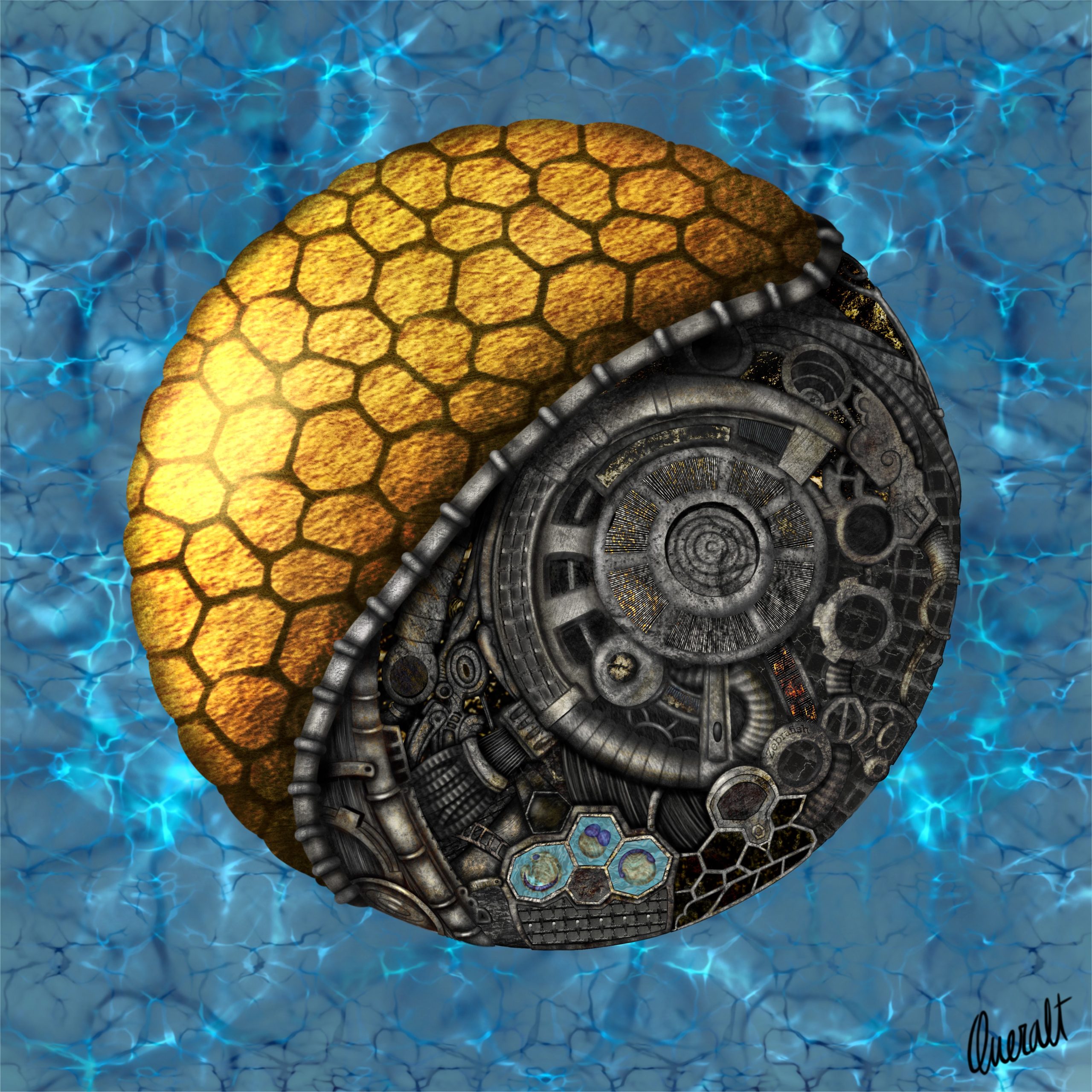
Steampunk Zebrafish Embryo – Digital artwork representing the dynamic world of Cell and Developmental Mechanobiology, blending biological accuracy with a steampunk aesthetic.
Can you tell us about your background and what you work on now?
I have a background in Biochemistry and earned both a Master’s and a PhD in Biomedicine. My doctoral research focused on how mechanical forces influence the formation and function of epithelial tissues during early development, using zebrafish embryos as a model.
Throughout my academic journey, I developed a growing interest in visual communication. As a visual learner, I found that graphical representations helped me focus and retain information more effectively from a young age. This realization led me to value the visualization of complex concepts, not only for my own understanding but also as a tool to communicate information and ideas to others. I quickly recognized how impactful clear, engaging visuals could be in simplifying difficult concepts. People appreciated them, and during my PhD was when some colleagues started to reach out to me for help or feedback on creating their own.
Encouraged by my colleagues, I began to explore scientific illustration more seriously as a potential career, rather than just as a tool I used in my research. After completing my PhD, I transitioned into freelance work as a scientific illustrator and animator, largely through self-teaching. Today, I create both 2D and 3D illustrations and animations, combining my scientific expertise with visual storytelling to make science more accessible and engaging.
If you need help transforming complex science into compelling visuals, feel free to reach out. I’m just an email away! :)
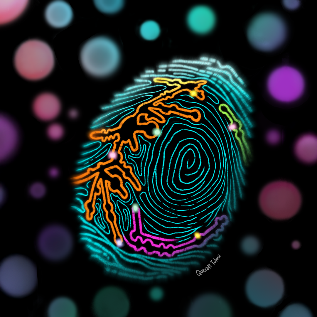
Ribosomal RNA Modifications Fingerprint – Cover illustration for Molecular Cell, Volume 85, Issue 1 (2025), depicting findings from the study “Epitranscriptomic rRNA Fingerprinting Reveals Tissue-of-Origin and Tumor-Specific Signatures” by Milenkovic, I. et al.
Were you always going to be a scientist?
In many ways, yes. As a kid and teenager, I was completely captivated by medicine, especially surgery. The idea of understanding what was happening inside the human body and being able to repair something so complex really intrigued me. TV shows like House, Grey’s Anatomy, and Bones definitely fueled that fascination, although I’ll admit I may have started watching them a little earlier than I probably should have.
By the time I reached the later years of high school, my interests shifted towards genetics and epigenetics, which ultimately led me toward a research career. It felt like the natural progression, combining my love for science with a deep curiosity about how the body works at a molecular level.
Of course, I had other strong interests, like cinema, literature, and drawing. However, when it came time to choose a career, none of them felt like something I would want to pursue professionally. There’s a clear difference between enjoying something as a hobby and envisioning it as a career.
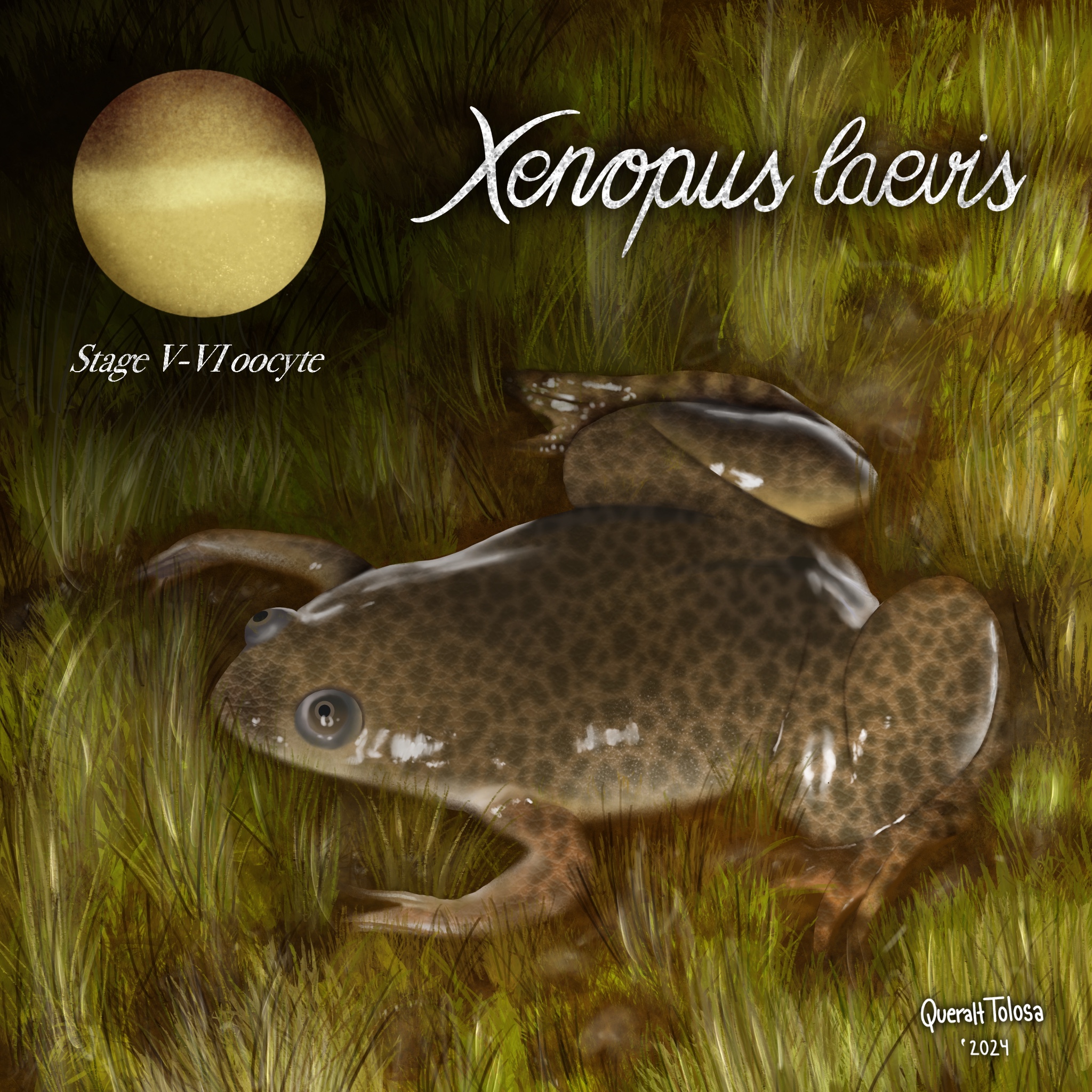
Xenopus laevis – Digital depiction of the vertebrate model organism Xenopus laevis (African clawed frog), featuring a stage V–VI oocyte, fully grown and primed for ovulation.
And what about art—have you always enjoyed it?
Absolutely. Art has always been a part of my life. I’ve been passionate about it for as long as I can remember, exploring every form I could, from pottery to drawing to acrylic painting. I took classes throughout school and high school, constantly doodling or sketching whenever I had the chance. It became my go-to way to relax, reflect, and express myself.
At the time, though, I never considered art as a career. The idea of having to create for a living felt overwhelming, almost like it would take the joy out of it. I wanted to keep art as a personal, stress-free outlet and do it when inspiration struck, not because it was expected of me.
But over time, things changed. Now, I’ve found a way to blend my love for science with my creative side. I’ve also learned how to maintain a healthy balance between professional work and personal art. Keeping a clear distinction between the two has allowed me to preserve the joy and freedom in both aspects, making sure that each one remains fulfilling in its own way.
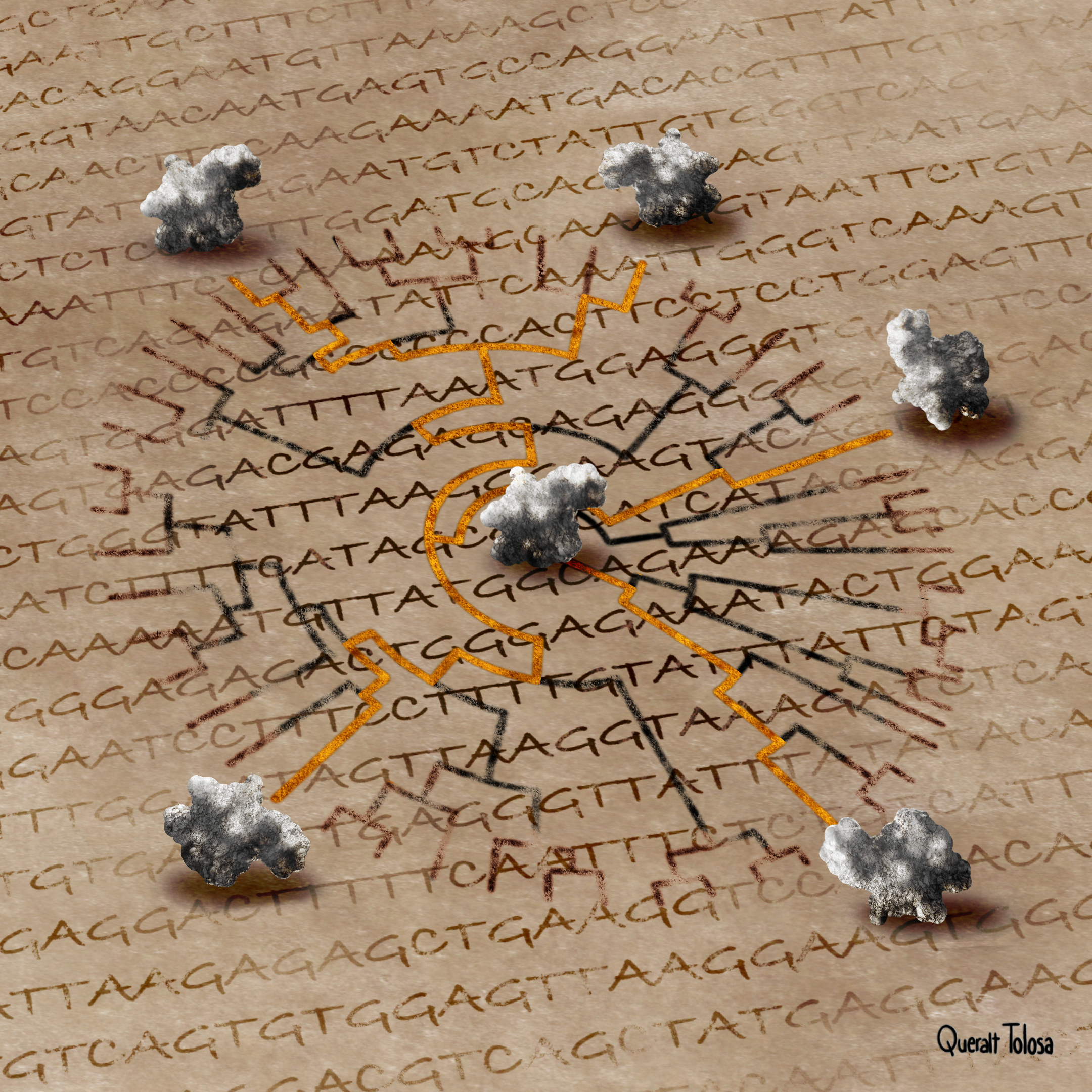
Tracing Evolution Beyond the Erosion of Time – Digital illustration created for the Centre for Genomic Regulation (CRG) press release accompanying the study “multistrap: Boosting Phylogenetic Analyses with Structural Information” by Baltzis, A. et al., published in Nature Communications (2025).
What or who are your most important artistic influences?
My artistic influences have changed a lot over time and are still evolving. They span all sorts of things, from painters and sculptors to cinema and other visual arts.
When it comes to scientific illustration, my early influences were a bit limited, but I’ve always admired the work of people like Leonardo da Vinci and Ramón y Cajal, who managed to combine science and art so well. It’s a pretty basic answer, I’ll admit, but they were huge for me. Growing up, most of the scientific visuals I came across were focused on wildlife and plants, which I didn’t really connect with. It wasn’t until my PhD that I discovered David Goodsell’s work in molecular biology, and that completely opened my eyes to the world of scientific illustration in that area. These days, I’m lucky enough to be influenced by a lot of my colleagues in the field, and being able to interact with them is pretty amazing.
When it comes to broader influences, I’d say my inspirations are eclectic. The first drawings I loved and tried to replicate in order to learn how to draw were the works of Hayao Miyazaki, Tim Burton, and Disney classics. I’ve also been a lifelong fan of manga and anime, which have greatly influenced both my aesthetic and storytelling instincts.
I’m also a huge admirer of classical art, some of my favorite pieces of artwork are from artists like Toulouse-Lautrec, Francisco Goya, Sandro Botticelli, or Artemisia Gentileschi. Nowadays, I follow closely artist such as Laura H. Rubin, Miles Johnston, Zipcy, Lucas David, Kildren, or Guillermo Lorca García.
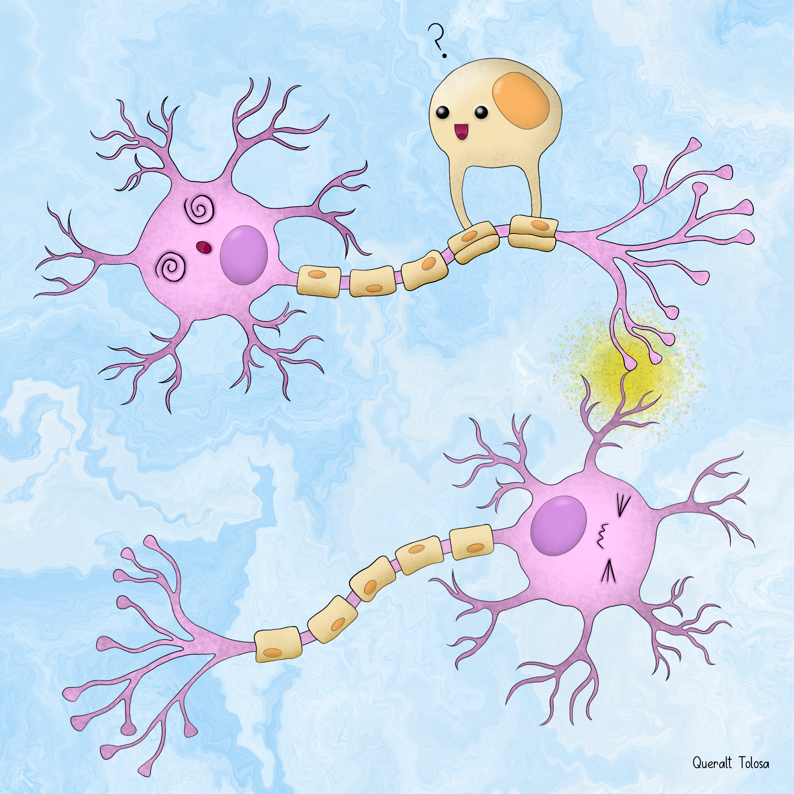
Fun Synapse – Playful digital illustration of a synapse between two neurons, featuring an interacting oligodendrocyte, rendered in a cozy, cartoon-inspired style.
How do you make your art?
For my personal work, I’m drawn to traditional media, particularly graphite and ink. There’s something about the tactile nature of these materials that make the process feel more intimate and fulfilling. I often incorporate a touch of color, but it’s the slower pace and hands-on approach that allow me to truly reconnect with the creative process on a deeper level.
When working professionally, though, my approach shifts entirely to digital. I use Procreate and Photoshop for illustration, which give me the flexibility to experiment and refine ideas quickly. For animation and post-production, I rely on Procreate Dreams, After Effects, and Premiere Pro, which provide all the tools I need to bring my work to life in dynamic ways. For 3D projects, I turn to Blender, which offers the versatility to create detailed, immersive environments.
I believe in always evolving as an artist, so I make it a point to stay curious and open to new techniques. Whenever I can, I dive into new tools, software, or courses that expand my skill set and challenge me to approach my work from fresh perspectives.
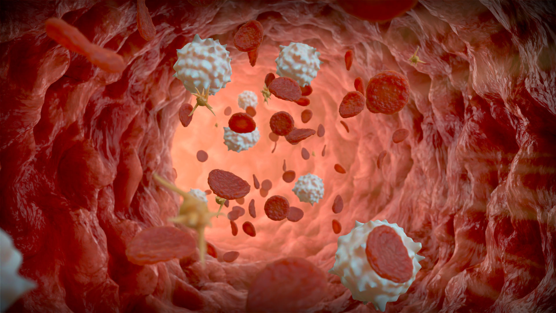
Blood Vessel – 3D digital illustration showing a cross-section of a blood vessel populated with erythrocytes (red blood cells), thrombocytes (platelets), and leukocytes (white blood cells).
Does your science influence your art at all, or vice versa? Or are they separate worlds?
They’re definitely distinct in terms of content, but they absolutely influence each other in surprising ways. My personal art is often more introspective—an outlet for my own thoughts and feelings, where I can step back from the scientific side of things. It’s a space for creativity and self-expression that’s more abstract.
But in my professional work, the two worlds are tightly intertwined. My scientific background gives me the ability to break down complex ideas and understand them in depth. That foundation allows me to create accurate representations of those concepts. On the flip side, my artistic skills help me present these ideas in a way that’s visually engaging and easy to understand, making complex science more approachable and relatable.
It’s all about balance—honoring the rigor of science while using the creativity of art to communicate it in a way that resonates with people. I love how they complement each other, and it’s that intersection that keeps me motivated and inspired.
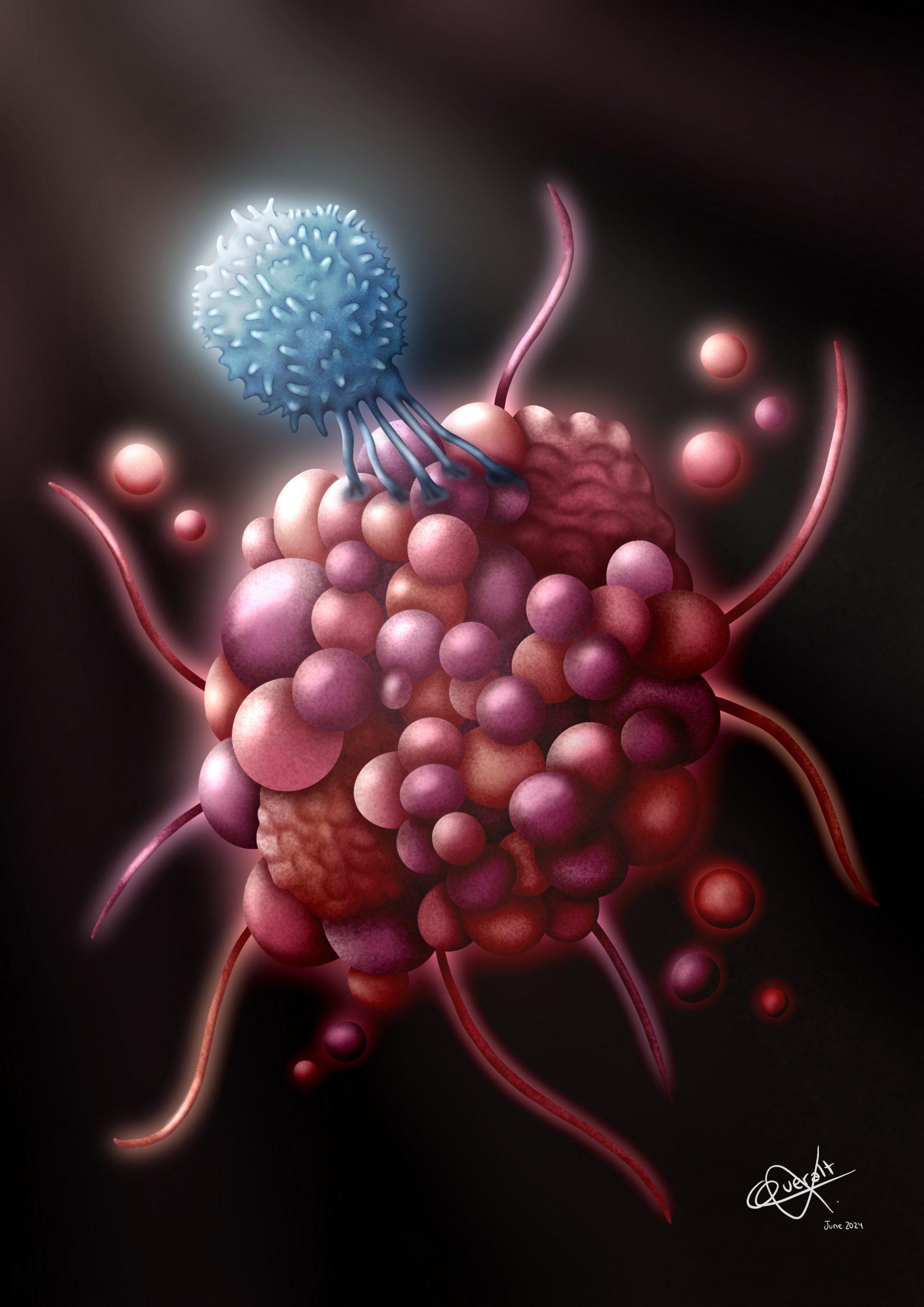
The Beauty of Death – Digital illustration capturing the cellular processes of apoptosis and the subsequent clearance of dying cells.
What are you thinking of working on next?
I’m still in the early stages of my freelance career, so right now I am really focused on building a solid foundation for my scientific illustration and animation business. I am working on reaching more potential clients, building strong relationships, delivering thoughtful work on every project, and fine-tuning my workflow so I can keep getting better and growing.
I am also very committed to continuous learning. I am always looking for ways to improve, whether through online courses or personal projects. I am putting a lot of time into sharpening my techniques and finding the best ways to visually communicate different scientific topics, making sure my work stays accurate, engaging, and high quality.
Looking ahead, I am excited to take on more challenging projects that will push me to grow professionally and help me build a reputation as someone people can trust for beautiful, accurate scientific visuals.
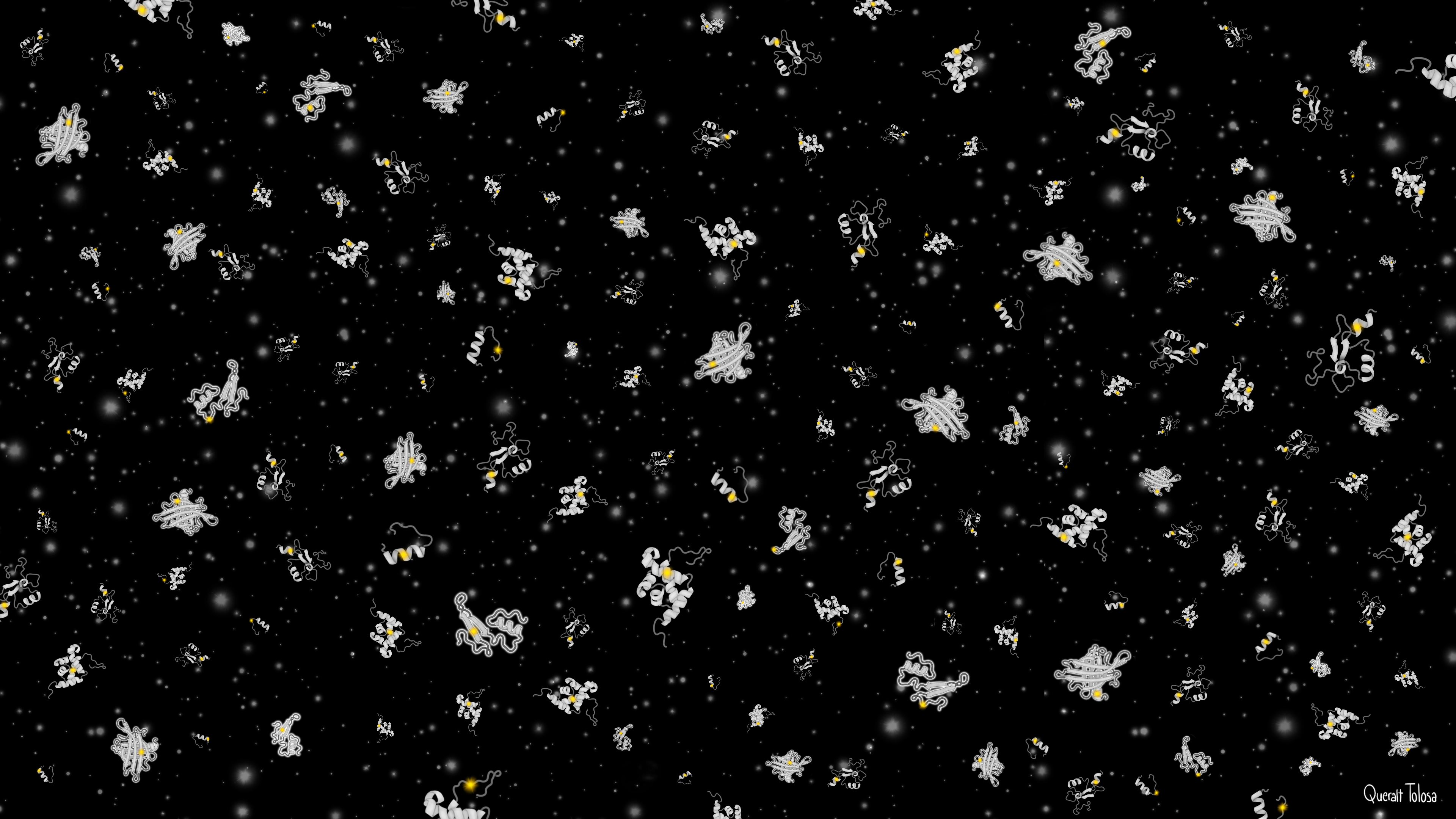
A Galaxy of Protein Domain Variants – Digital illustration developed for the CRG press release highlighting the study “Site-saturation Mutagenesis of 500 Human Protein Domains” by Beltran, A. et al., published in Nature (2025).
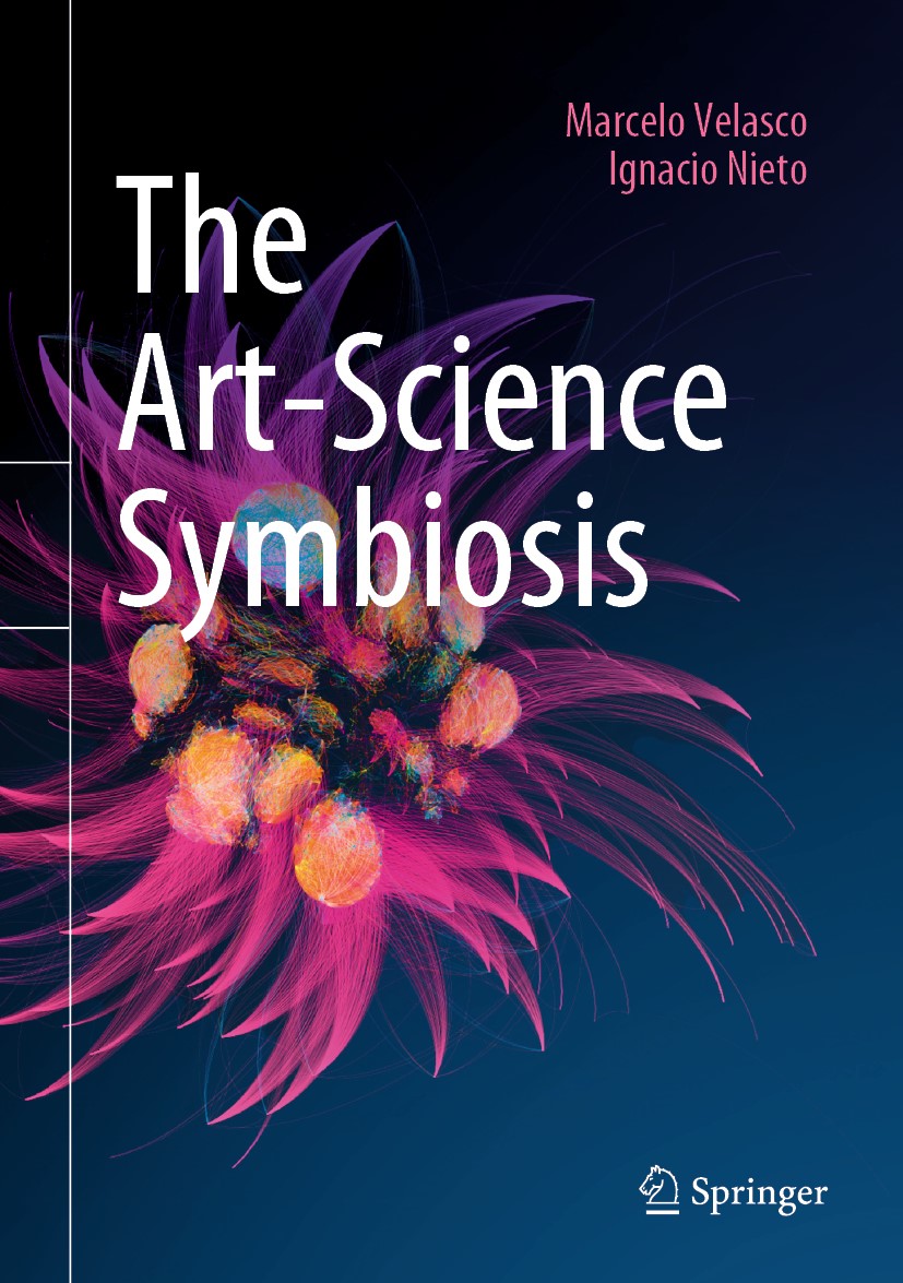
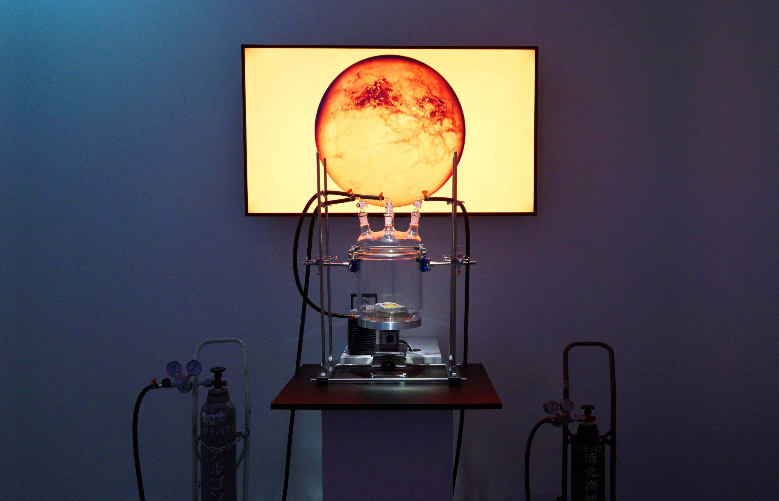
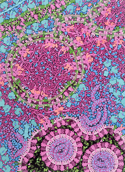
The Art-Science Symbiosis. Book Cover Illustration. Francois-Joseph Lapoint. 2016. From the series Microbiome Selfie. 1000 Handshakes
The increasing influence of science on contemporary art (often referred to as art-science) prompted us to explore a central working question: could contemporary art, in turn, significantly influence scientific activity?
We observed that contemporary art possessed distinctive attributes, absent in more traditional artistic forms, that enabled it to interact fruitfully with the creativity inherent in the scientific process. Our own dual background –Velasco is a Biologist, Nieto a PhD in Aesthetics– provides us with an informed perspective in both fields, and places us in a relatively comfortable position to analyze the inherent strengths and weaknesses of both.
This interest culminated in the publication of a book by the prominent scientific publisher Springer-Verlag, which inevitably leans the work towards a scientific audience. However, precisely because of this orientation, we anticipate that the book will also pique the curiosity of artists and the general public interested in interdisciplinarity. Throughout our research, we discovered notable examples of practicing scientists who have produced significant artistic works, including Manuel Théry (France. PhD. Cytomorpholab Research director @ CEA, EMBO Member); David Goodsell (USA. PhD in DNA X-ray crystallography, illustrator and Professor of Computational Biology at the Scripps Research Institute and a Research Professor at Rutgers University); Jimena Royo-Letelier (Chile. Artist and researcher, PhD in applied mathematics at Université de Versailles Saint-Quentin-en-Yvelines; and François-Joseph Lapointe (Canada. Artist and scientist at the University of Montreal. PhD in evolutionary biology (1992) and a PhD in dance and performance), among many others.
In our approach, we made a conscious effort to avoid the commonplaces that often accompany these discussions. We aimed to integrate theoretical elements to better understand the broad scope of the practice; a curatorial process (reasoned selection) of artworks; relevant historical references; and qualitative research through interviews with the unique artist-scientists included. The curatorial process involved an exhaustive review of the vast art-science-technology landscape, selecting those works that we considered of particular interest to both the scientific and artistic communities.
We are confident that the publication of this book represents a valuable opportunity for establishing collaborative networks, through other platforms for dissemination and debate. The trust placed in our work by Springer allowed us to receive an enthusiastic response from the artist-scientists participating in the project, all developed from Santiago, Chile, and without external funding.
We hope that this exploration will contribute to fostering a deeper dialogue and reveal the symbiotic potential between the analytical rigor of science and the evocative and conceptual capacity of contemporary art.
Kumayl Alloo | Research-Scholar, Columbia University Credit: Emma Asher, 2025
For decades, Parkinson’s disease (PD) research has centered primarily around the disease’s hallmark motor symptoms, including tremors, rigidity, and bradykinesia. However, nonmotor symptoms of the disease—such as anxiety, depression, and cognitive impairments—have remained largely unexplored. Though less visible, these symptoms profoundly affect quality of life, often preceding motor dysfunction and providing critical clues about PD progression.
Kumayl Alloo, a research scholar at the Benson and Huntley Labs at Columbia University and the Icahn School of Medicine at Mount Sinai, is helping address this critical gap in knowledge, delving into the complex interplay between genetic and environmental risk factors in shaping PD’s broader, nonmotor impact on the brain.
“For years, our lab has focused on how the LRRK2-G2019S mutation, a key genetic risk factor for PD, interacts with chronic stress, a prominent environmental risk factor, to drive nonmotor symptoms of the disease,” Alloo explained. “Our earlier studies revealed behavioral and neurological changes in mouse models exposed to both factors, but we lacked clarity on which brain regions or synapses were responsible. Put simply, we knew what was happening, but we didn’t know where.” This mystery formed the foundation of Alloo’s PD research, aimed at pinpointing the specific neural mechanisms driving these observed differences.
In their latest study (Guevara, Alloo, et al., 2025), Alloo and his lab uncovered critical insights into how these risk factors interact in sex-specific ways. Their research demonstrated that the LRRK2 mutation and chronic stress together altered synaptic activity in regions like the medial prefrontal cortex (mPFC), nucleus accumbens (NAc), and basolateral amygdala (BLA)—areas crucial for emotional regulation and cognition. These changes were linked to heightened anxiety-like behaviors in male models, while females showed variable resilience or susceptibility depending on the nature of the stressors.
The findings from this research offer a clearer picture of how risk factors converge to alter brain circuits before motor symptoms emerge, opening doors to earlier intervention strategies and informing future experiments crucial for PD detection, classification, and treatment. “After all, now that we know where the issue is, future therapeutic research would know where to target,” Alloo notes.
A Parkinson’s Foundation Fellow, Saltzman Scholar, Pucker Fellow, and FlexMed Scholar, Alloo’s contributions exemplify the importance of fostering early-career researchers to tackle pressing challenges in neurodegeneration. His contributions are helping to redefine our understanding of PD.
“My long-term goal is to be a physician-scientist—at the intersection of research and treatment,” he says. “This unique overlap between medicine and research allows me to address the clinical needs of patients in the hospital while conducting research tailored to those needs in the lab. The integration of bench-to-bedside approaches—where care and cure inform each other—promises novel cures and therapies built on two different approaches toward disease.”
As Kumayl and his lab continue to advance our understanding of PD, their work exemplifies the power of integrating academic science research with clinical aspirations to address one of the most challenging neurodegenerative disorders of our time.
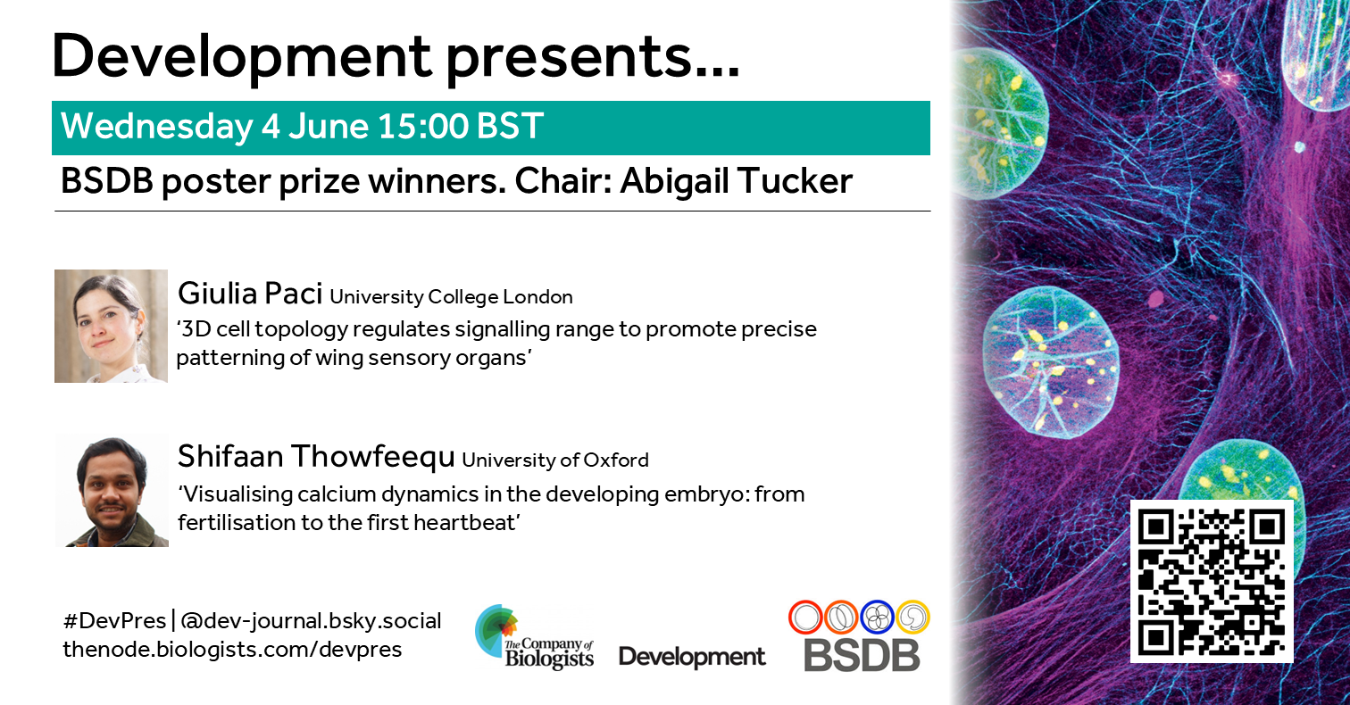
Join us to celebrate the winners of the BSDB postdoc poster prize from the Biologists @ 100 conference. The webinar will be chaired by Abigail Tucker (King’s College London).
Wednesday 4 June – 15:00 BST
Giulia Paci (University College London) ‘3D cell topology regulates signalling range to promote precise patterning of wing sensory organs’
Shifaan Thowfeequ (University of Oxford) ‘Visualising calcium dynamics in the developing embryo : from fertilisation to the first heartbeat’
At the speakers’ discretion, the webinar will be recorded to view on demand. To see the other webinars scheduled in our series, and to catch up on previous talks, please visit: thenode.biologists.com/devpres
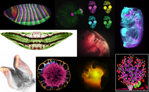
The winners of the Young Embryologist Network (YEN) 2025 Image Competition have been announced at the conference today held at the Francis Crick Institute.
Please check out the top 10 images that have received the most votes from attendees of the YEN 2025 Conference.
We thank those who sent us their images and attendees of YEN 2025 for helping us select these images. If you would like to learn more about YEN visit www.youngembryologists.org
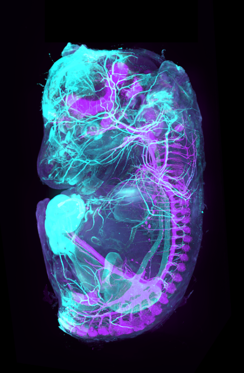
YEN Image Competition winner:
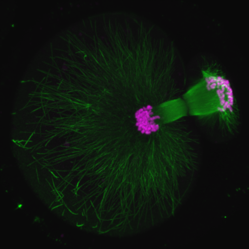
Théo Morel (PARCC Paris, Inserm-UMR970, France) A 3D view beyond the skin What if we could directly visualize the arterial and nervous systems behind the skin of a mouse embryo? To make this possible we used a iDISCO clearing protocol on a 15 dpc mouse embryo, followed by whole mount co-staining with ASMA (light blue, marking arteries) and TH (purple, marking the sympathetic nervous system). The embryos were imaged using light-sheet microscopy and reconstructed in 3D using Imaris software.
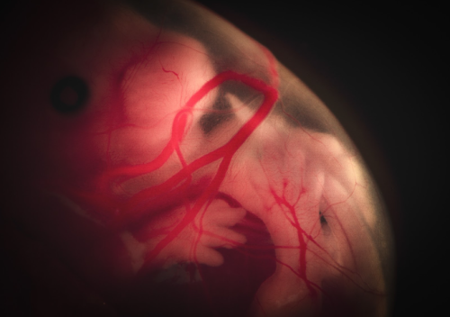
Runner-up:
Lisa Leinhos (University of Oxford, UK) Cosmic life Drifting in the womb like an astronaut in space, the embryo floats peacefully, a moment of cosmic calm in the universe of early life. This image portrays a dissected mouse embryo at E14.5, captured through a Nikon camera on a binocular microscope system. The focus of this experiment is to investigate gene expression at different stages of embryonic development using bulk RNA sequencing. What makes this image particularly compelling is the contrast between its scientific significance and emotional depth. Although the embryo is no longer alive, the image portrays a moment of serene stillness, almost as if it were still within the womb. It captures the profound beauty of life’s early stages while highlighting the mysteries that science seeks to understand. At the same time, there is an inherent sadness, as this specimen, like many others, has been sacrificed in the name of advancing our knowledge of developmental processes. This delicate balance of scientific discovery and respect for life makes the image not only visually striking but also emotionally resonant.
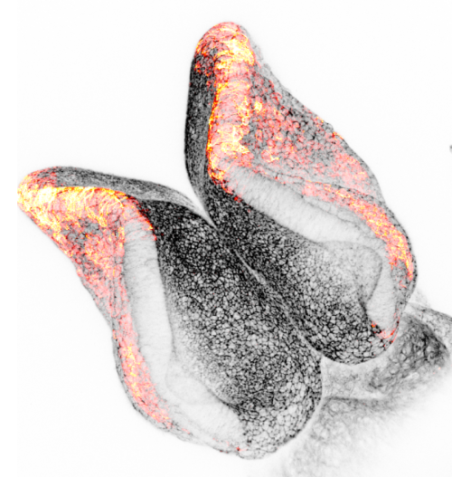
Runner-up:
Andrea Krstevski (Institute of Child Health, UCL, UK) Minnie’s Bow Neural crest cells play a crucial role in neural tube development as they are a population of multipotent cells that emerge from the dorsal neural tube. These cells migrate to various regions of the embryo and differentiate into a diverse array of tissues, including neurons, glial cells, and components of the peripheral nervous system. Their proper development and migration are essential for the correct patterning and function of the nervous system. Any disruption in neural crest cell development can lead to a variety of congenital disorders and malformations. The image shown is a E8.5 mouse embryo displaying actin in black and migrating neural crest cells using marker Pax3 in red. The Zeiss LSM 880 upright confocal multiphoton microscope was used to capture the image.
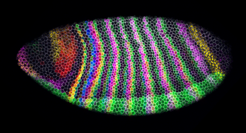
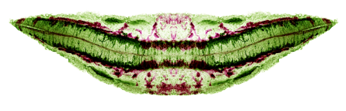
Swanee Douglas and Dr Tom Pettini (Department of Genetics, University of Cambridge, UK) Segment-polarity stripes Drosophila embryo with immunostaining of cell membranes (grey) and 5-plex HCR in situ hybridization to visualize segmentation genes (blue = engrailed mRNA, green = paired + snail mRNA, magenta = odd-skipped mRNA, yellow = wingless mRNA, red = sloppy-paired 1 mRNA) with minimal crosstalk, Microscope: 40X Z-stack on Stellaris 5 confocal microscope (sum projection here), Processing: Python and Fiji
David Grainger (Institute of Developmental and Regenerative Medicine, University of Oxford, UK) SMILE This image showcases a vibratome section of an E9.5 mouse embryo, where KDR immunostaining (magenta) delineates endothelial progenitors and TUBB3 (green) marks developing neurons. Captured using a Zeiss LSM980 confocal microscope, a maximum intensity projection and horizontal mirroring were applied to optimize signal and symmetry.
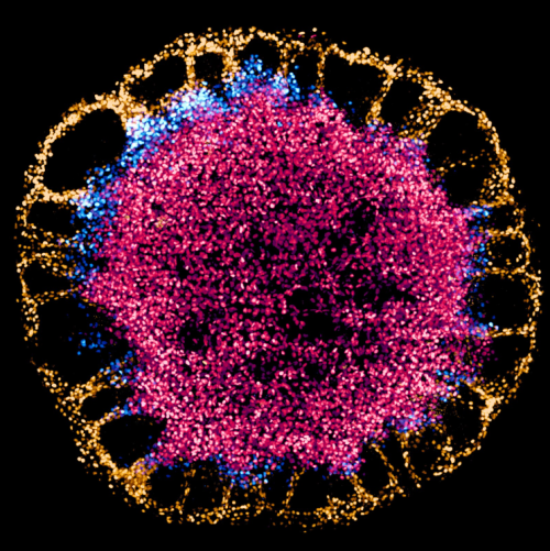
Francesca Montesi (The Francis Crick Institute, UK) A blossom of stem cells Human embryonic stem cells differentiating on a 1000 µm-diameter hydrogel micropattern. Cells differentiate and form hollow cysts at the periphery, while they remain pluripotent at the core of the colony (CDX2, gold; BRA, blue; SOX2, red). Imaged on a spinning disk confocal microscope and processed with Imaris.
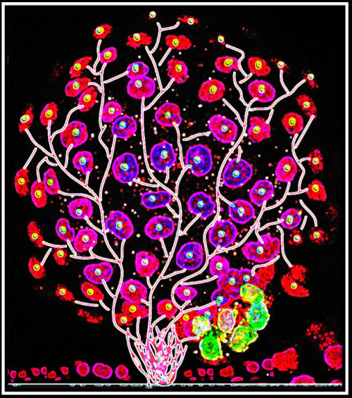
Jinlong Qiu (Hull York Medical School) Blossoming Embryo A fluorescence microscopy representation of a bovine blastocyst, stained with Hoechst 33342, Alexa Fluor 488 conjugated with NANOG and Alexa Fluor 568 conjugated with GATA6. The image was captured using a Zeiss LSM710 confocal microscope and later artistically modified. Tree-like branches were digitally illustrated using Adobe Illustrator, and further contrast adjustments and artistic enhancements, including the ‘Glow Edges’ effect, were applied in PowerPoint. This fusion of science and art transforms the cellular organization of early embryonic development into a vibrant visualization of growth and differentiation, resembling a flourishing tree of life.
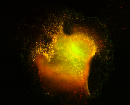
Achira Karunaratna (Institute of Child Health, UCL, UK) Crest Nebula Cranial neural crest explant visualized via immunofluorescence after staining for marker proteins. The image was captured on the Nikon eclipse Ti2 series epifluorescence microscope housed at UCL GOS ICH at 20x followed by gaussian stack focusing of a Z series. Explanting is a widely used ex-vivo approach to study neural crest development in animal models. The neural crest, regarded as the ” fourth germ layer”, gives rise to multiple important cell types within vertebrate bodies, including the craniofacial cartilage, peripheral nerves, and pigment cells among others. This explant experiment is part of a wider attempt to understand the mechanisms of neural crest migration that contributes to neural tube defects in mammalian embryos by understanding cytoplasmic protein methylation in migrating g neural crest cells. Visualised in yellow are neural crest cells (stained for neural crest marker sox10 ) emerging out of the mouse cranial neural folds. Tinges of green at the outer edges represent phalloidin staining marking F-actin localization in cells. The faint red hue marks another membrane-bound protein SETD2, a methyltransferase, within the explant and in migrating cells. The circular looping where neural crest cells emerge is reminiscent of areas of clouds of gas and dust that form when stars are born in nebulae in the pitch-dark corners of our universe. Hence, aptly named a crest nebula, a factory for neural crest cell production.
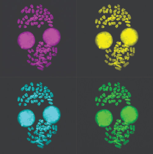
Malgorzata Borkowska (MRC Laboratory of Medical Sciences, Imperial College London, UK) Metaphase Monroe Metaphase spread of 2i/LIF grown mouse embryonic stem cells image obtained with Leica SP5 confocal and processed in Fiji for chromosome counting.
All the world’s a metabolic dance, early career scientists are leading the way!
Emerging perspectives in metabolism
Dr. Holly Thorpe
This week we’ll meet Dr. Holly Thorpe, newly minted PhD from the Chow lab at the University of Utah, who is now continuing her research there as a postdoctoral fellow. Holly’s path into rare disease research began as an undergraduate when she studied multiple sclerosis through computational genetics. A paper from the Chow lab showing how a simple sugar rescued a rare metabolic disorder in flies sparked her fascination towards studying metabolism and rare diseases. Now a freshly minted PhD continuing as a postdoc, Holly models rare disorders like Phosphatidylinositol glycan biosynthesis class A congenital disorder in Drosophila, using the power of fly genetics to uncover disease mechanisms and therapeutic targets. The Chow lab specializes in precision medicine for rare diseases, using advanced genetic tools – demonstrating how basic science is actively curing diseases and impacting human health. Driven by curiosity and compassion, Holly’s research shows how foundational discoveries can become lifelines for patients with no other options. Check out more work from the Chow lab here!
What was your first introduction to the field of metabolism – what’s your first memory? Could you share your journey into studying metabolism using Drosophila and what inspired you to specialize in the field of rare diseases?
For my undergraduate research, I worked in a lab that used computational genetics to study Multiple Sclerosis. I knew from this experience that I wanted to work in a human disease genetics lab for graduate school, but I wanted to have a mix of dry and wet lab in my research. When I found the Chow lab, they had recently published a paper showing that supplementation of N-acetyl glucosamine rescued a Drosophila model of another rare glycosylation disorder. The idea that something as simple as adding a specific sugar to the diet could have an effect was so exciting to me. I knew I wanted to study rare metabolic disorders.
Walk us through the process of studying rare diseases and creating personalized therapies.
Many of the patients reach out to Dr. Chow for help. The rare disease world is interesting because oftentimes the parents of these patients have found each other and started their own communities and foundations. We have had multiple different foundations reach out to Dr. Chow about the running screens for their gene of interest. To screen for phenotypes, we typically start with an RNAi model and knock down the gene ubiquitously and in multiple different tissues in the fly such as the eye, neurons, and muscle cells. Then we look for any phenotypes that might arise. We have successfully used the Drosophila Genetic Reference Panel (DGRP), a group of wild-derived, inbred, fully sequenced flies, to look at the effects of natural genetic variation on the phenotypes. From that we are able to run statistical analyses, such as a genome wide association study (GWAS) to identify potential candidate modifiers.
Tell us how Drosophila serves as an extremely advantageous model for conducting studies on rare genetic disorders.
I think Drosophila are such a good model organism. Roughly 70% of human disease genes have a human orthologue, so we are able to study a lot of different disorders. Most of the disorders we focus on have neuronal phenotypes, and we are able to take advantage of the ability to mimic these phenotypes such as neuromuscular issues and seizures.
Tell us about Phosphatidylinositol glycan biosynthesis class A congenital disorder of glycosylation (PIGA-CDG). How did you work towards characterizing and establishing Drosophila model of PIGA-CDG and how do you think it will be a help in understanding of the pathogenesis of this understudied rare disorder?
PIGA-CDG is an ultra-rare neurodevelopmental disorder. It is caused by loss of function mutations in the gene PIGA which encodes a necessary protein in the glycosylphosphatidylinositol (GPI) anchor synthesis pathway. Patients typically present with seizures, hypotonia, and neurodevelopmental delay. In developing a PIGA model, we found that ubiquitous loss of PIGA in Drosophila was lethal, so we decided to look at more cell-type specific loss. Because of the neurological phenotypes seen in patients, a previous graduate student in the lab performed neuronal- and glial–specific knockdown of PIGA and identified a climbing and seizure defect, respectively. We also had a heterozygous knockout model created to see if ~50% loss of PIGA would give any phenotypes since homozygous knockout flies are lethal. We again found a seizure phenotype. Using these models, and other cell specific models, we can start to tease apart which tissues PIGA is important in and we can run modifier and drug screens to identify other interacting pathways and novel therapeutic targets.
In one of your works, you used pedigree analysis to identify potential protective modifier genes, including a null variant in CNTN2 – walk us through the process and tell us how you genetically validated CNTN2 as a target which could rescue most of the PIGA-associated phenotypes. What were your key findings and what are the future metabolic mechanisms remaining to be uncovered in this regard?
In our study, we used pedigree analysis in a family with variable expression of PIGA-CDG to identify potential protective genetic modifiers. Whole-genome sequencing revealed a null variant in CNTN2 that was present in asymptomatic carriers but absent in the probands. To test the interaction between PIGA and CNTN2, we used tissue specific Drosophila models where knockdown of the CNTN2 ortholog rescued key PIGA-related phenotypes like eye size, seizures, and motor defects. This showed that CNTN2 is a genetic modifier of PIGA, but the mechanism of interaction is still unclear. CNTN2 is a GPI-anchored protein, so it is possible the interaction could be broadly found across many GPI-anchored proteins. The interaction could also be CNTN2 specific and more related to its specific function in the nervous system.
Tell us about your work on evolutionary rates of glycosylation genes. What factors contribute to evolutionary rate differences among glycosylation genes – what are the consequences? How will identifying genetic modifiers of CDG enable our understanding of broad clinical spectrum seen in the patients? What tools have you used for these studies and what future studies should be done to expand these studies to develop targets for CDGs and other rare metabolic diseases?
We used evolutionary rate covariation (ERC) analysis to identify potential genetic modifiers of glycosylation genes. ERC is a computational method that identifies functionally related genes by measuring how similarly their evolutionary rates have changed across species over time. The more similar the evolutionary pattern, the more likely there is a genetic interaction. We discovered that glycosylation genes, particularly those involved in GPI anchor synthesis and N-linked glycosylation, exhibit high ERC values, indicating shared evolutionary pressures and functional interdependence. By identifying genes with high ERC to known glycosylation genes, we pinpointed potential modifiers that may contribute to the clinical variability observed in CDG patients. To validate these findings, we employed Drosophila models, confirming that several candidate genes modulate CDG phenotypes. Glycosylation affects many different genes and biological pathways. Modifier genes can help us to narrow down which pathways may be more important for CDG pathophysiology. Similar pipelines could be applied to other rare metabolic disorders in order to identify modifier genes and potential therapeutic targets.
Tell us about the unique experimental approaches you have taken throughout your work – what tools are you using, how difficult some of these experiments are – did you have to deal with midnight timepoints or require an army of undergrads/ long hours, had to use some un-conventional/creative tools to overcome experimental challenges?
Luckily using Drosophila there are a lot of readily developed tools. Most of the genetic constructs we needed had already been developed, and the different assays we ran are pretty common in the Drosophila world. While there were definitely quite a few weekends and long days, I managed to design my experiments so there were no midnight timepoints.
What are your upcoming plans? What metabolic pathways or signals do you aim to investigate further?
I just defended my PhD, so I will continue to work in the Chow lab as postdoc focusing on a more therapeutic targeted look at a new CDG. I’ll still be using natural genetic variation as an exploratory method, but with the hope of contextualizing and identifying therapeutic targets.
Your work intersects metabolism and genetic variation. How do these fields overlap and how do you integrate these disciplines in your research, and what unique insights have emerged from this approach?
Genetic variation plays a significant role in shaping metabolic function, as variants in metabolic genes can impact numerous interconnected pathways. In my research, I investigate how these genetic differences influence disease risk and severity, particularly by identifying modifiers that alter metabolic outcomes. This approach highlights the importance of studying disease within the context of diverse genetic backgrounds to better understand variability in clinical presentation and therapeutic response.
What role does curiosity play in your life, both within and outside of science? How important it is for you to answer basic science questions on metabolic signaling and how do you see its impact on animal health/relevance on human health?
I was definitely one of those kids that always asked a million questions, so I think my curiosity has really driven my work as a scientist. I think metabolism has such a huge impact on human health. Understanding these basic mechanisms is crucial, as they have direct relevance to human conditions like the rare diseases I study, and more common diseases such as diabetes, obesity, and cancer. Both in and out of the lab, curiosity keeps me asking meaningful questions and pushing for insights that can lead to real-world impact.
What changes have you seen in the scientific community in regard to studying these unique aspects of metabolic signaling in flies? Are we moving toward a more nuanced understanding, or do you see potential pitfalls?
Drosophila offers powerful tools to dissect conserved metabolic pathways in vivo, allowing for high-throughput and genetically precise studies. However, a potential pitfall is oversimplifying or overgeneralizing findings without considering species-specific differences—while flies are incredibly informative, translating insights to human biology still requires careful validation.
Tell us about how you see the future of metabolism evolve with the new upcoming tools – what techniques have you used and which tools are you most excited about?
I think ERC is an incredibly powerful and versatile tool—it can be applied to virtually any gene of interest to uncover new functional relationships and reveal previously unknown aspects of its biology.
Were there any pivotal moments that shaped your career path? What’s an unexpected place you’ve found inspiration for your work?
I’ve always had a passion for science, but I realized I wanted to pursue a PhD in genetics after a conversation with one of my undergraduate professors about her career path. She invited me to join her research lab—an opportunity I hadn’t previously considered—which ultimately opened the door to an entirely new trajectory for me. Rare disease remains an understudied area with immense potential for discovery. In particular, many metabolic rare disorders present rich opportunities for investigation through both computational and experimental approaches.
Tell us about what experiences/results/training motivated you to push forward in grad school?
I have been so lucky in joining the lab that I did. We are all great friends who help encourage each other to keep going. I definitely would not have made it through without the people in my lab.
How do you maintain a balance between your rigorous research activities and personal life? Are there hobbies or practices you find particularly rejuvenating?
The work-life balance is one of my favorite parts of doing grad school in Utah. I work right by the mountains, so all year long, I’m able to go hiking, rock climbing, and paddle boarding. And we frequently take weekend trips to one of the many national parks in the state. It’s always refreshing to get out in nature after a long day, and in Utah it’s so accessible.
If you hadn’t embarked on a career in biological research, what other profession might you have pursued, and why?
If I hadn’t studied science, I would have loved to open up a bakery. The method of baking is so therapeutic to me. I have always loved tinkering with recipes to try and find the best one.
Last week we learnt about the impact of environmental toxins on animal development and their resilient coping mechanisms from “genotypic” to “phenotypic” perspective. Check out the article –From shifting Skies to Toxic Tides (Lautaro Gandara)
 (No Ratings Yet)
 (2 votes)