Last week, I attended the Keystone Symposium “Molecular and cellular basis of growth and regeneration”. This outstanding meeting was the second incarnation of a Keystone meeting on regeneration; the first having been held in 2012 – Development reviewed it here. The expansion in scope of this year’s meeting to encompass both growth and regeneration was, in my opinion, an excellent move on the part of the organisers Alejandro Sanchez Alvarado, Valentina Greco and Duojia Pan. Bringing together these two related fields highlighted the common themes, as well as the differences, between developmental and regenerative programs, and made for a diverse yet coherent meeting. With 115 attendees, who all threw themselves into the interactive spirit of the meeting, the relatively small size was conducive to excellent discussion sessions at the end of each talk and lively poster sessions each evening. All this, along with some excellent snowboarding during the afternoon breaks, made for a highly informative and thoroughly enjoyable conference.

Overall, the standard of talks was fantastic, and I won’t be able to summarise them all here (not least because the majority of the work presented was unpublished), but I will pick out some of my personal highlights and some of the common themes that emerged from the meeting. Many apologies to those whose work I haven’t mentioned: I can assure that it’s not because I didn’t enjoy your talk!
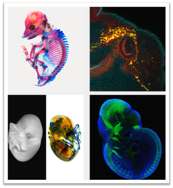
The evo-devo field was well represented at this meeting, kicking off with an excellent Keynote presentation from Hopi Hoekstra – who told an unpublished story on the developmental and molecular basis of pigmentation patterns in rodents. Her talk, like several others, highlighted the value of genomic approaches in non-traditional model organisms to uncover key regulators of developmental processes. Arkhat Abzhanov and Rich Schneider both presented beautiful work on craniofacial morphology in birds. Abzhanov studies Darwin’s finches and other songbirds, which show significant diversity in beak size and shape. Through an impressive combination of genomics, mathematical modelling and comparative embryology, his lab has uncovered mechanisms and regulators of beak growth (he’s written about this on the Node). Schneider, meanwhile, uses quail-duck chimeras (‘qucks’ and ‘duails’) to explore the mechanisms governing jaw size – which differs significantly between these two species. These experiments have uncovered a remarkable degree of autonomy in the behaviour of cranial neural crest cells – donor cells from one species transplanted into a host of the other continue the developmental program of the donor, both spatially and temporally (you can also read more about Schneider’s work on the Node). In what one attendee described as ‘the best talk ever’, David Kingsley presented some of his lab’s recent data on the genetic basis of diversity in 3-spined sticklebacks, as well as a spectacular example of the power of comparative genomics and mouse genetics (McLean et al., 2011) to uncover the molecular basis of human-specific traits – in this case, neocortical expansion. These speakers all focussed on inter-specific variation, while Elaine Ostrander’s interests lie in intra-specific variation, namely in the remarkable diversity of morphological features of dog breeds. With the genome sequences of many breeds to hand, her lab has been able to identify key regulators of phenotypes such as size, leg length, skull shape and fur growth (this review provides a great overview of the field).
An impressive diversity of model species was apparent not only in the evo-devo talks and posters but also in those addressing regeneration. The varying regenerative powers of Stentor coeruleus (a single celled ciliate), Arabidopsis, Nematostella, planarians, Drosophila, sea urchins, sea cucumbers, zebrafish, Xenopus, axolotl, 2 species of mouse (Mus musculus and Acomys cahirinus) and human were all discussed (apologies to any species I’ve left off this list!). Among the talks in this area, I particularly enjoyed Ken Poss’ presentation on clonal heterogeneity in the regenerating zebrafish fin, Alejandro Sanchez Alvarado’s exploration of the properties of planarian neoblasts and their developmental origins, and Valentina Greco’s beautiful live imaging of skin regeneration and homeostasis in mouse.
As well as Greco, Aaron Mertz also showed impressive movies of mouse skin, this time during development, revealing some of the cellular mechanisms underlying stratification of the embryonic epidermis (the paper – Ouspenskaia et al., 2016 – came out on the day he gave his talk). Other stunning examples of the power of live imaging to follow and understand developmental processes came from Yohanns Bellaiche’s analysis of cell division and rearrangement in the Drosophila thorax (Guirao et al., 2015), Marcos Gonzalez-Gaitan’s high resolution imaging to reveal mechanisms underlying asymmetric cell division in Drosophila sensory organ precursors (Derivery et al., 2015), Matt Gibson’s work on early embryogenesis of Nematostella, and Jochen Wittbrodt’s beautiful movies of medaka eye morphogenesis (or ‘retinal gastrulation’ as he referred to it) (Heermann et al., 2015). In several of these cases, analysis of the data required sophisticated processing and analysis workflows and integration with computational modelling – the power of which is becoming ever clearer, and which was beautifully illustrated by L Mahadevan’s talk (which covered systems as diverse as the chick gut, the human brain, and plant pollen tubes!) (Tallinen et al., 2014; Shyer et al., 2013; Campàs & Mahadevan, 2009).
Wittbrodt’s talk touched on another recurring theme of the meeting: the potential and plasticity of adult tissue stem cells (in his case, those that mediate the life-long growth of the fish retina) (Centanin et al., 2014). Jayaraj Rajagopal presented data on the in vivo and in vitro plasticity of mammalian airway epithelial cells – in both regenerative and cancer contexts (Pardo-Saganta et al., 2015). Norbert Perrimon and Bruce Edgar both discussed the Drosophila intestinal stem cell model: Perrimon focussing on mechanical regulation of in homeostasis of the gut epithelium, and Edgar looking at the signalling pathways regulating regeneration following bacterial infection (Jin et al., 2015). Joseph Rodgers, meanwhile, has been investigating stem cell quiescence and its regulation in the mouse muscle system (Rodgers et al., 2014); his data suggest that systemic mechanisms exist to prepare quiescent cells for likely re-entry into the cell cycle. In all these cases, it is clear that a simple model of resident, quiescent tissue stem cells becoming activated upon injury to replenish lost cells through proliferation and differentiation does not account for the complexity of regulatory mechanisms and cell states that exist in adult tissues.
But what happens when tissues can’t rely on proliferation to repair damage? Vicki Losick showed a striking example of non-proliferative tissue repair in the Drosophila adult epidermis. These cells are post-mitotic, and wounds are healed by a combination of dramatic polyploidisation as well as cell fusion (Losick et al., 2013). This was not the only talk showing an important role for endoreplication: the presentations from Bruce Edgar, Mary Baylies (Drosophila muscle development) and Pranidhi Sood (Stentor regeneration) all touched on the idea that control of cell ploidy can play a critical role in development, homeostasis and regeneration.
Several talks highlighted functions for the immune system, or immunity signalling pathways, in developmental or regenerative processes. In adjacent, highly complementary talks, Duojia Pan and Laura Johnston presented data on the Toll pathway in Drosophila; you can read more about Johnston’s work on cell competition here (Meyer et al., 2014), while Pan’s intriguing talk on Hippo-Toll crosstalk is hot off the press at Cell (Liu et al., 2016). Also in Drosophila, Andreas Bergmann is looking at the role of hemocytes in the regulation of apoptosis-induced proliferation. Moving into vertebrates, Nadia Rosenthal and colleagues focus on the roles of immune cells in regeneration – in both the context of the axolotl limb and the mammalian heart. Her work demonstrates the importance of minimising fibrotic responses to improve regenerative capacity, as she discussed in a recent Development review (Furtado et al., 2016). The role of immune cells and pathways in development and regeneration seems to me an up-and-coming topic, and I expect many more exciting developments in this field in the coming months and years.
Finally, one of the real highlights of the meeting for me was the talk from Antonio Giraldez, who is interested in the molecular mechanisms underlying the maternal-zygotic transition in zebrafish. Giraldez’ work has revealed a hitherto unappreciated layer of regulation of post-transcriptional gene expression that involves codon optimality. The study is still unpublished, so I can’t go into further details, but I would urge you to look out for this in the coming months – it’s an fascinating story with far-reaching implications!
In summary then, this was a truly inspiring meeting for me, and – I hope – for all the speakers and attendees. Huge thanks to all for putting together such a great event, and for participating in it so fully, and I look forward to the next instalment!
 (6 votes)
 Loading...


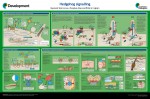 The Hedgehog (Hh) signalling pathway is one of the key regulators of metazoan development. Here,
 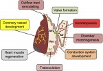 Endocardial cells are cardiac endothelial cells that line the interior of the heart tube. Here,
 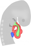 Nadia Rosenthal and co-workers discuss the role of cardiac fibroblasts in scarring and regeneration, as well as how these cells are specified during development and the unique characteristics that define them. See the Review on p.
  (No Ratings Yet)
 The award is named after
  The BSDB is proud to announce the first winner of the Cheryll Tickle Medal, which will be awarded at the 2016 BSDB-BSCB Spring meeting in Warwick to
 –
–
 -We featured this