The community site for and by
developmental and stem cell biologists

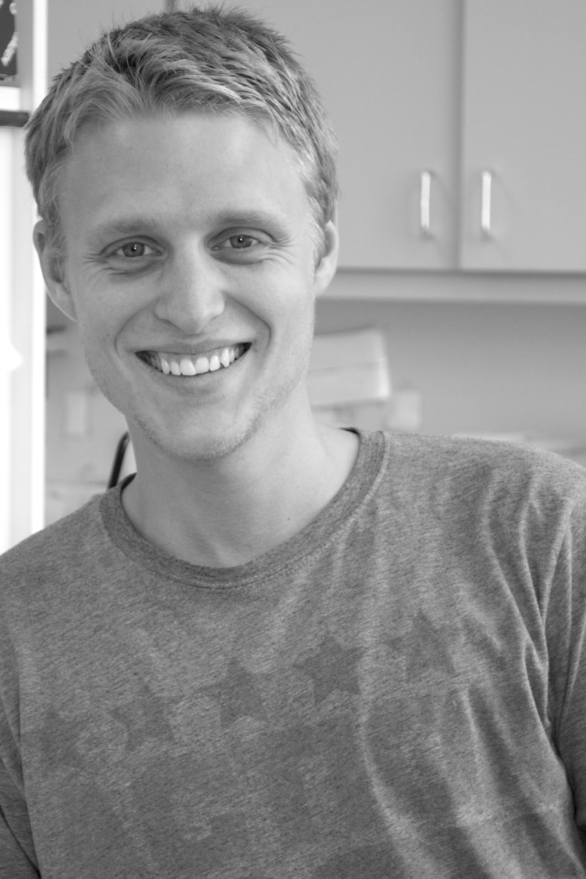
Interview with Beddington Medal winner William Razzell

Posted by , on 14 April 2014

BeddingtonMedalEach year, the British Society for Developmental Biology (BSDB) awards the Beddington Medal to the best PhD thesis in developmental biology. This year’s award went to William Razzell, who completed his PhD in Paul Martin’s lab at the University of Bristol. At the BSDB Spring Meeting last month, Will presented his thesis studies of wound healing, in which he’d used Drosophila as a model. We caught up with Will after his talk to ask him more about his work, to find out what he’s doing now, and to ask him if he has any advice for PhD students.
Congratulations on winning the Beddington Medal. Can you tell us a bit more about your thesis work?
Thank you. I used the Drosophila embryo to model two aspects of wound healing. First, I looked at how innate immune cells are rapidly recruited to wounds in response to a hydrogen peroxide signal, which is made by an enzyme called DUOX. I found that calcium signals in the wounded epidermis, that it can kick start the wound inflammatory process by activating DUOX, and that removing DUOX’s ability to sense calcium prevents hydrogen peroxide release and recruitment of innate immune cells. The second aspect I studied is how epithelial wounds in the Drosophila embryo repair, as they close extremely rapidly and efficiently compared to mammalian adult skin wounds. This is because they close by assembling an actomyosin cable that contracts the wound edges together until actin protrusions extending from the wound margin interdigitate to seal the epithelial hole. We know that these wound edge actin machineries are important for wound closure, but we know less about how cells surrounding the wound contribute to wound repair. I looked at cell shape changes associated with wound closure in the Drosophila embryo and saw that the cells have to stretch towards the wound but also shrink their junctions with neighbours in a myosin-dependent manner. This is important for remodelling the epithelium surrounding the wound edge, allowing for cell intercalation events, so that the actin cable can efficiently pull the wound edges together. The same events occur during an earlier developmental event in the embryo to drive tissue elongation, consistent with the idea that cells at the wound edge can reactivate developmental pathways in order to close the wound.
Obviously there are lots of different models you could have used, but you chose Drosophila as a model for studying wound repair. Why?
Drosophila have two really big advantages. One is that they are genetically-tractable, which means that we can do very rapid genetic studies on the embryo, such as genetic screening or efficiently mutating genes with ever advancing tools including CRISPR technology. The other huge advantage is that you can live image embryos. For example, we can simultaneously label and follow different populations of cells during wound closure, such as the damaged epithelium as it closes and the innate immune cells as they migrate to wounds. This gives us the opportunity to observe cell behaviours that contribute to wound closure live in the embryo and analyse how these very dynamic events contribute to wound repair.
What can your studies tells us about wound healing in humans?
Through these sorts of studies we can identify genes or signalling pathways that are involved in wound closure that might be conserved during mammalian wound healing. We can also use live imaging studies in Drosophila to get clues about how cell behaviours may contribute to closure of human wounds, which we cannot yet live image. For example, the junctional rearrangements I observed in Drosophila wound repair may give us clues into the junctional changes that could be involved during the repair of human epithelial wounds.
And I guess, in the long term, the aim is to improve that process?
Of course! We would really like to improve and speed up the wound healing process. At the moment, there are a lot of people who have chronic wounds or suffer from impaired wound healing or excessive scarring – we want to be able to enhance wound repair in these patients.
You recently moved to New York. Can you tell us about the work that you are doing there now?
I joined Jennifer Zallen’s lab at the Sloan Kettering Institute in New York. I’m using the Drosophila embryo again, but I’m now looking at the earlier developmental event of germband extension, in which cells undergo intercalation events driven by polarised recruitment of myosin to cell junctions. I want to understand the signalling pathways leading to this myosin behaviour, and how this contributes to global tissue architecture.
Do you have any advice for PhD students?
One thing I found particularly helpful during my PhD is talking about my science to absolutely everyone. Visiting PIs, other PhD students…just anyone! I have shown my confocal movies to my parents, and although they are not scientists they can still look at them and see things that I can’t. You focus on the same things every day, and just showing your results to someone else can really open up your view. Giving presentations is also a great way to meet people and discuss your science with others. I didn’t do enough of this during my PhD but I think it is definitely something that can be a huge advantage to any PhD student.
William Razzel photo


Razzell, W., Wood, W., & Martin, P. (2014). Recapitulation of morphogenetic cell shape changes enables wound re-epithelialisation Development DOI: 10.1242/dev.107045


Thumbs up (2 votes)
Loading...

Tags: , , ,
Categories: Interview

Navigate the archive

Use our Advanced Search tool to search and filter posts by date, category, tags and authors.

(Developmental) Biology around the internet- April 2014

Posted by , on 13 April 2014

Here is our monthly round-up of some of the interesting content that we spotted around the internet:


News & Research:

– This year’s Waddington Medal was awarded by the British Society for Developmental Biology to Prof Phil Ingham!

– Are you a budding science writer? The Wellcome Trust launched their 2014 Science Writing Competition, and are also posting writing tips on their blog.

– The British Library (London, UK) has launched a great exhibition about the beauty behind science visualisation. You can visit ‘Beautiful Science’ until the 26th of May.

– The Francis Crick Institute (London, UK) is running a free symposium on advances in optical microscopy.

– The Wellcome Trust’s Human Induced Pluripotent Stem Cells Initiative is calling for clinicians and scientists to provide samples from patients with inherited genetic diseases for the project.

– The Knoepfler lab Stem Cell blog is running a stem cell image competition.

– And the winner of the 2014 March of Dimes prize for developmental biology is neuroscientist Dr Huda Zoghbi.


Weird & Wonderful:

Foldscope is a  clever origami, paper-based microscope that can be made for less than a dollar. Also watch their TED talk.

– If you work on C. elegans you will find ‘Nematode news in brief‘ highly amusing

– A company names all of their fluorescent proteins after Santa’s reindeers and other christmassy things. Next lab Christmas gift?

– And if you are a developmental biologist working on axolotls, you can now buy your own axolotl necklace here.


Beautiful & Interesting images:

– Science cakes continue to make appearances on Twitter- check out this great cellular cake, and some biocupcakes.

– The Wellcome Trust announced the winners of their 2014 Image Competition. Check their website for some stunning images.

– These cute and informative posters are for the young cell biologist-to-be in your life.

– This diagram explains why we are all born scientists.

– And we spotted this colourful image of a waterbear:


Videos worth watching:

– Remember the fantastic ‘Inner Life of the Cell‘ animation? Harvard University and XVIVO got together again to create an updated version of the animation that reflects the chaos of a real cell.

– 24 hours of zebrafish embryonic development in 1 minute.

– And is the Royal Society Charter Book the world’s greatest autograph book?


Keep up with this and other content, including all Node posts and deadlines of coming meetings, by following the Node on Twitter.


Thumbs up (1 votes)
Loading...

Tags:
Categories: News, Video

What’s your favourite gene?

Posted by , on 11 April 2014

Which gene tops them all?

I recently took part in the ‘I’m a scientist, get me out of here!’ outreach event. As soon as the school children found out I was a developmental geneticist and worked out what I did, one question I was repeatedly asked was: “what’s your favourite gene and why?” so for a bit of fun, I thought I’d share my thoughts and see what everyone else’s are too.


Now, I could have gone into detail about a gene of utmost importance in my work or one we literally couldn’t live without (although picking either of those would be tricky as that hardly narrows the list down). However, my first thought when picking my ‘favourite gene’ is always listing the funny-named ones that stuck out from my university lectures. That’s not to say these genes don’t also fit into the important and essential-to-life categories, but they have that added ‘pazzaz’ of an ear-catching name that would wake you from your university slumber, thinking “did he actually just say what I think he said…?”. So here is my shortlist, the top 5 genes based almost entirely on their names:


5. Tinman


Tinman is an absolute gem in the history of creative gene naming. As with all the best gene names, tinman is named after the phenotype seen in Drosophilia when it is suppressed. In case you haven’t guessed, mutating tinman in Drosophilia embryos results in flies without a heart, despite the rest of the embryo continuing to develop. The vertebrate relatives of this gene have been named under a much less creative process with NK2 homeobox genes making up the homologous family. Just in case the origin of the name is still unclear, think back to the magical land of Oz, where Dorothy meets the tinman whose only wish is to have a heart. A sweet tale; however, while the tinman in The Wizard of Oz was always sentimental anyway, tinman mutant Drosophilia don’t have the advantages of a fairytale ending…


4. Swiss cheese


You may find a theme with these genes as yet another gene with a brilliant name comes from the phenotypic description in flies. Think about the classic cartoon cheese and you’ve got a fairly good description of the adult fly brain seen in Drosophilia with a faulty version of the gene: full of holes. To be more precise, the swiss cheese mutant results in brain degeneration through apoptosis spreading from the CNS. Notably, the swiss cheese gene paved the way for the blue cheese gene, another gene which, when mutated, affects the fly brain in a way that may resemble the mouldy, marbled appearance of blue cheeses.


3. INDY


This gene has made the cut through its clever acronym name. INDY simply stands for ‘I’m not dead yet!’. In addition, the reference to the ‘Monty Python and the Holy Grail’ scene where a corpse being collected shouts “I’m not dead yet!” means this gene gets a strong haul of bonus points! This gene is shown to greatly extend the lifespan of fruit flies when mutated, so I guess the name makes sense as well. INDY is a nice example of scientists trying to hide jokes in their work; as of course we are a hilarious bunch of people.


2. Clark kent, superman and kryptonite (I know that’s cheating a bit!)


Finally stepping outside the realms of fruit flies, Arabidopsis thaliana has a lovely offering in a trio of genes that are named together. The clark kent and superman mutant genes make these plants extra macho – increasing the number of stamens found on their flowers. Superman mutants have an even greater increase than clark kent but if the kryptonite gene gains a mutation… uh oh! The mutation in the kryptonite gene causes both the clark kent and superman genes not to be expressed and makes the plant impotent. I think the naming of these genes greatly reflects the link many scientists share to the characters on ‘The Big Bang Theory’ and their passion for comic books!


1. Sonic hedgehog


Sonic hedgehog is possibly the most famous funny gene name and a personal favourite for sentimental reasons as well as the obvious brilliance of its name. Often shortened to SHH (although who would miss an opportunity to say ‘sonic hedgehog’ in full?), this gene is part of a gene family of hedgehogs with other mammalian relatives named desert hedgehog and indian hedgehog. I can vividly remember the development lecture I was sitting in when sonic hedgehog was first mentioned and how it took us all a good half an hour before someone bravely put their hand up to say what we were all thinking: “I’m sorry, are you actually saying this gene is named after a blue, speedy, cartoon hedgehog?!”. Well to put it simply, yes it is. Sonic hedgehog is heavily involved in vertebrate development and in flies the hedgehog mutant phenotype covers the surface of the Drosophilia in tiny pointy projections – similar to a hedgehog! In Zebrafish, hedgehog genes are also named tiggywinkle hedgehog and echidna hedgehog (although the latter is no longer commonly used) making full use of the creative possibilities stemming from naming a gene family ‘hedgehogs’.


The issue of gene naming has however come up in the news, and public opinion is divided. Whilst some of these creative gene names are amusing, memorable and sometimes quite clever, we have to remember that for every gene there is the potential for it to be implicated in a real disease or disorder. That means that somewhere, someone could be told that their child will not survive into adulthood because of mutations in their sonic hedgehog gene. I wouldn’t want to be the bearer of that bad news, let alone have to stand there and use a gene name like that in the explanation. Phenotypic description gene names have been useful in the past, especially as we began our exploration and discovery of genes. However, in a world now dominated by computers and bioinformatics, a more standard method for naming genes has to be implemented. Hence why many mammalian genes (whose functions were discovered after the original Drosophilia outbursts) are named based on structure and function, an attempt at moving gene naming into the same realms as taxonomy.


There are still plenty of genes around with somewhat hysterical names. Thankfully we now have alternatives for most, if not all, of these options, meaning the ‘in-jokes’ of science can be put in the past when necessary! For now though, peruse the options out there and be prepared for the inevitable question, ‘what’s your favourite gene…?’


Notable others (to google in your own free time!):

–       Barbie and Ken

–       Grim reaper

–       Pikachurin

–       Casanova

–       Rolling stones

–       Spock

–       Van Gogh

–       Callipyge

–       Dracula

–       Dumpy

–       Cheap date

–       Braniac

–       Cabernet

–       Chardonnay

–       Riesling

–       Cleopatra

–       Maggie

–       Tigger

–       Cyclops

–       Dreadlocks

–       Lava lamp

–       Hamlet

–       Gooseberry

–       Bagpipe

–       One-eyed pinhead

–       Half stoned

–       Lunatic fringe, Radical fringe, Maniac fringe

Thumbs up (3 votes)
Loading...

Tags: , , , ,
Categories: Discussion, Lab Life

Outreach competition- the winners

Posted by , on 10 April 2014

Science outreach often involves using metaphors, where a real life object or situation is used to explain a complex scientific concept. Last December we launched an outreach competition, and we asked the Node readers to submit pairs of images: a photograph of a real life scene and an image of the scientific concept that it helps explaining.

We can now announce the winners of this competition. Many congratulations to Ewart Kuijk, a post-doc at the Hubrecht Institute and author of the post Cellular Reincarnations, and Roel Hermsen, who is about to complete his PhD at the Hubrecht Institute ‘after which he will return to the “real world”’. They won a £50 Amazon voucher and a copy of Benny Shilo’s book on the outreach project that inspired this competition.


Here is their winning entry:

“The basis for life is encrypted in the DNA. Cells can activate or inactivate certain regions of their DNA by making it more or less accessible. Consequently, cells can be very different (e.g. heart cells versus neurons), while share the same genetic content. DNA accessibility and concomitant gene activity depends on the binding of specific proteins. The photograph of the “real world” shows an office building attached to a spiral staircase. The staircase represents the double-helix of the DNA and the building represents a DNA binding factor that modulates DNA accessibility. The scientific image is a picture of an embryonic ovary stained for H3K27me3 (green), DNA (blue), and a germ cell marker (red). Foci of H3K27me3 mark the inactivated X chromosome, of which the DNA is not accessible resulting in suppression of gene activity from this chromosome.”

 Spiral Repression outreach competition


We hope that this competition will inspire you to find a real life metaphor that can help you explain your work to the wider public! And our thanks to everyone who entered this competition.


Outreach logo new squareThis post is part of a series on science outreach. You can read the introduction to the series here and read other posts in this series here.


Thumbs up (1 votes)
Loading...

Tags: ,
Categories: Images, News, Outreach

Cellular Architectures at the Nuit Blanche 2013 in Paris

Posted by , on 9 April 2014

The “Nuit Blanche” in Paris. A city wide exposition of contemporary arts from dusk till dawn. Performances, light shows, dance, installations. Along the Canal Saint-Martin the visitors stroll from one exhibition to the next or sit down and take a break, chatting and drinking. A bit further up Boulevard Avenue Richerand the south-west entrance to the Hospital Saint Louis gives entry to a backyard. On the facade of the 17th century building, retinal pigmented epithelial cells perform their dance. Nothing but their cytoskeleton exposed in white before the dark building, magnified 10000-fold, accelerated 100-fold. One cell gets hold of a support beam, gradually spreading out over all its length. Loosing grip, rounding up and going into mitosis. Each daughter cell spreading out again. Other cells remain at one place, yet their cytoskeleton still moves, searching an exit from the confined space of a window.

When Manuel Thery came to me with the idea that we have to project cells onto a building – and cells which had been grown on micro-patterns in the shape of that same building – it was obvious that this idea simply had to be done. First, the general public has little access to all those beautiful images one takes when working with cells. Second, curiosity and “wanting to understand” needs to be promoted as a value by itself. Physics is not engineering, and biology is not R&D for pharmaceuticals. And the cytoskeleton with its appearing and disappearing regularities has the graphical power to draw you in and want you to grasp its function, without necessarily thinking about the benefit. Third, we try to find cues of how cells react to extracellular geometrical constraints in our day-to-day science, and we were curious ourselves, what we would see on these complex shapes. Fourth, as a structural scaffold for the cell (amongst other functions), the cytoskeleton shares some properties and constraints of engineering and architecture, and the metaphor has been made before. Fifth, simply, because we can.

The plan was simple:

1) take a photo of a public building
2) extract some interesting shapes from the building
3) produce micro-patterns of these shapes, the size of a single or several cells – micro-patterns are protein patterns for cell attachment on a substrate that otherwise repels cells
4) culture cells with fluorescently labeled cytoskeletal elements on these micro-patterns
5) do some live cell video microscopy
6) select the nicest videos and project the cells back on the original place of the pattern on the building, the size of the building, accelerated, for everyone to see.

Although that project may sound simple, it is by no means a one person, one weekend prep. At least, if you are a newbie to micro-patterning, live cell imaging, and your cell culture experience dates back more than a PhD’s time. Keep in mind that photo toxicity of the stain will kill the cells pretty quickly if you illuminate too strongly and frequently. On the other hand, if you want to play your time-lapse movies at a speed that people do not recognize it as a sequence of images, you need hundreds of frames for tens of seconds of footage, so you require days with an image every 5-10 min. And have cells survive under a microscope for 48h, on previously untested patterns of adhesive regions… is performing an experiment. And as such may just not work the first few times you try. Add the little pressure of a deadline (the day of the event that cannot be shifted).

But it gets even more involved with the work of the artists from the amazing Groupe Laps:

cut of the movie (the choreographies for the cells); giving the cells music to move to; correcting the image of the house for spherical aberration, so that the projection would align with the building; organizing the equipment for the projection; finding, in all those hundreds of movies, those that would be used;  organizing the permissions to project onto the building; sticking non-reflecting foil over the windows on the day of the event; aligning the images with the features of the building; …


For someone having been involved in any sort of larger scale artistic event, all this may sound evident, but I was amazed by just how complex the basic idea would get.

Another change is the mindset between me and the artist. For them a cool sequence of cells would do. For me it also had to reflect a typical cell on these patterns – while still yielding footage with good enough contrasted structures to allow the projection.

Only on the evening itself did I see how well the composition by Groupe Laps had worked. It was very different from what I had imagined on the outset, and much richer for the fact that it did not look like all the images you would see in a publication – if you cared to project them on a house, that is.


Since we could not assume that the public would grasp that the images showed live cells, we also provided two information boards to describe the project in a short text as you might expect in an exhibition. However, we were surprised by the number of people who were at the event, and because a 10m high projection of something moving draws more attention than 1m high panel of written text, only a minority took advantage. Therefore, the level of understanding ranged from fellow scientists “yay, z-stack of microtubules”, over raising curiosity with many, to some complete ignorance that this had anything to do with biology. On the other hand, many spectators were clearly drawn in by the videos, watching the installation several times over. And I like to believe there was a lot wider spectrum of interpretation and thought, specifically because there was no classroom-ready explanation available.

In the case that you plan an outreach activity with collaborators foreign to your field or even science, my main advise would be to make sure you take the time to collaborate closely; to get to know each other and mutually showing additional possibilities and clarifying impossibilities on either side takes some iterations. This may apply less to smaller events, or repeating ones, where you can learn on the job and grow the event slowly. But for larger scale one-offs this is paramount. In fact, the sole regret I have about my experience with the nuit blanche project is that – because of distance and safety regulations – I could never show the involved artists our laboratories, so my work still remains abstract to them, and that we did not have regular “lab”-meetings, as is the case with my research.

If you ask yourself whether you should get involved in some outreach activity of the sort, involving people foreign to science, and specifically if you are not familiar with their line of work: Do it! It broadens the horizon and experiencing some recognition from someone outside of ones usual line of work has a special quality for both sides.


Outreach logo new squareThis post is part of a series on science outreach. You can read the introduction to the series here and read other posts in this series here.


Thumbs up (2 votes)
Loading...

Tags: , ,
Categories: Outreach

Regenerating the aged thymus

Posted by , on 9 April 2014

thymus regenerationThe latest issue of Development includes a paper by Clare Blackburn and colleagues at the Medical Research Council Centre for Regenerative Medicine, University of Edinburgh, showing that the aged mouse thymus can be regenerated in vivo by the upregulation of a single transcription factor, FOXN1. This work has generated quite a lot of interest in the media, so we reproduce below the press release by the Medical Research Council. You can read the research article here.


Caroline Hendry, PhD
Stem Cells & Regeneration Reviews Editor at Development

Scientists regenerate immune organ in mice
Scientists have for the first time used regenerative medicine to fully restore a degenerated organ in a living animal, a discovery that could pave the way for future human therapies.
The team from the Medical Research Council (MRC) Centre for Regenerative Medicine, at the University of Edinburgh, rebuilt the thymus of very old mice by reactivating a natural mechanism that gets shut down with age.
The regenerated thymus was very similar to one in a young mouse in terms of structure and the genes expressed. The function of the organ was also restored, and mice receiving the treatment began making more T cells – a type of white blood cell important in fighting infection. However, the researchers do not yet know if the immune system of the older mice was strengthened. The research is published today in the journal Development.
Professor Clare Blackburn from the MRC Centre for Regenerative Medicine, at the University of Edinburgh, who led the research, said:
“By targeting a single protein, we have been able to almost completely reverse age-related shrinking of the thymus. Our results suggest that targeting the same pathway in humans may improve thymus function and therefore boost immunity in elderly patients, or those with a suppressed immune system. However, before we test this in humans we need to carry out more work to make sure the process can be tightly controlled.”
The thymus, located in front of the heart, is the first organ to deteriorate as we age. This shrinking is one of the main reasons our immune system becomes less effective and we lose the ability to fight off new infections, such as flu, as we get older.
Researchers targeted a key part of this process – a protein called FOXN1, which helps to control how important genes in the thymus are switched on. They used genetically modified mice to enable them to increase levels of this protein using chemical signals. By doing so they managed to instruct immature cells in the thymus – similar to stem cells – to rebuild the organ in the older mice. The regenerated thymus was more than twice the size than in the untreated mice.
Dr Rob Buckle, Head of Regenerative Medicine at the MRC, said:
“One of the key goals in regenerative medicine is harnessing the body’s own repair mechanisms and manipulating these in a controlled way to treat disease. This interesting study suggests that organ regeneration in a mammal can be directed by manipulation of a single protein, which is likely to have broad implications for other areas of regenerative biology.”
Previous attempts to provoke thymus regeneration have involved using sex hormones, but these have resulted in only temporary recovery of size and function of the organ. In this study, the recovery of the thymus was sustainable, but more work is needed to ensure there are no unintended consequences of increasing FOXN1.
The research was funded by Leukaemia and Lymphoma Research, the Darwin Trust of Edinburgh, the MRC and the European Union.

ResearchBlogging.org
Bredenkamp, N., Nowell, C., & Blackburn, C. (2014). Regeneration of the aged thymus by a single transcription factor Development, 141 (8), 1627-1637 DOI: 10.1242/dev.103614

Thumbs up (1 votes)
Loading...

Tags: ,
Categories: Research

Identified a new possible target to combat muscle wasting

Posted by , on 9 April 2014

The pathological atrophy of skeletal muscle is a serious biomedical problem for which no effective treatment is currently available. Those most affected populations are the elderly diagnosed with sarcopenia and patients with cancer, AIDS, and other infectious diseases that develop cachexia.

A study by scientists at the Institute for Research in Biomedicine (IRB), headed by Antonio Zorzano, also full professor of the University of Barcelona, reveals a potential therapeutic target to tackle muscle wasting in these risk populations.

In the study published today in the Journal of Clinical Investigation (JCI), one of the journals with highest impact in experimental medicine, the researchers associate the activity of the DOR protein with muscle atrophy and point to DOR as a plausible target against which to develop a drug to prevent muscle deterioration in certain diseases.

DOR (Diabetes- and Obesity-regulated gene), also known as TP53INP2, is a protein involved in autophagy, a quality control process that ensures cells stay healthy. The researchers have found that increased DOR expression in the muscle of diabetic mice leads to enhanced autophagy, which in turn favours the loss of muscle mass in these animals.

The advantage of developing a DOR inhibitor is that autophagy, a process necessary to keep cells healthy, would not be completely blocked in the absence of this protein. DOR is not essential for autophagy, but acts more as an accelerator. Thus, the inhibition of DOR would only partially reduce autophagy as other molecules involved would exert their activity normally, thus maintaining the levels of autophagy in a beneficial range for cells.

“If we could treat patients with sarcopenia and cachexia, or people at risk of these conditions, using a drug to inhibitor DOR then we would be able to stop or prevent muscle wasting,” explains the expert in diabetes and obesity Zorzano, head of the “Heterogenic and Polygenic Diseases” lab at IRB.

“We are showing pharmaceutical researchers a new possible therapeutic target for two diseases that seriously impair the quality of lives of those who suffer from them,” says the scientist.

An answer to why type 2 diabetic patients lose less muscle than those with type 1

The study also solves a biomedical enigma related to diabetes. Physicians did not understand why patients with type 2 diabetes—who become resistance to insulin or have very low levels of this hormone—are able to maintain muscle mass or minimize muscle wasting compared to patients with type 1 diabetes—who do not produce insulin—who show a clear loss of muscle mass. The IRB researchers demonstrate that the repression of DOR in muscle cells of type 2 diabetic animals allows the maintenance of muscle mass.

“We interpret DOR repression, which occurs naturally, as an adaptation mechanism to preserve muscle mass and to maintain greater muscular strength in type 2 diabetics,” explains David Sala, first author of the study, who has recently started a post-doctoral training period at Sanford-Burnham Medical Research Institute, in La Jolla, California.

Besides working with mice, the scientists have performed experiments on biopsies from skeletal muscle of patients with diabetes and patients resistant to insulin, thanks to collaboration with clinicians from the Université Lyon 1, in France, and from the Medical University of Byalistok, Poland, also included among the authors.

The project developed in Dr. Zorzano’s lab at IRB has been funded by the Centro de Investigación Biomédica en Red de Diabetes y Enfermedades Metabólicas (CIBERDEM), the Spanish Ministry of Economy and Knowledge, and the European project DIOMED, part of the Interreg-SUDOE programme.

Reference article:
ResearchBlogging.org
Sala D, Ivanova S, Plana N, Ribas V, Duran J, Bach D, Turkseven S, Laville M, Vidal H, Karczewska-Kupczewska M, Kowalska I, Straczkowski M, Testar X, Palacín M, Sandri M, Serrano AL, & Zorzano A (2014). Autophagy-regulating TP53INP2 mediates muscle wasting and is repressed in diabetes. The Journal of clinical investigation PMID: 24713655


This article was first published on the 9th of April 2014 in the news section of the IRBBarcelona website.

Thumbs up (No Ratings Yet)
Loading...

Tags: , , , , , ,
Categories: Research

Scheduled Node maintenance- 9th of April

Posted by , on 8 April 2014

We will be doing some maintenance work on the Node on Wednesday the 9th of April, and unfortunately there will be no access to the site during that period. You can expect the Node to be down from 7 p.m. (British Summer time) for approximately 4 hours. We are sorry for the disruption, especially for those Node readers in America and the early risers in the Pacific. We will be up and running again as soon as we can!

If you spot any problems and would like to get in touch our email address is the node [at] biologists.com


construction Node2

Thumbs up (1 votes)
Loading...

Tags:
Categories: News

6th Young Embryologist Annual Meeting

Posted by , on 8 April 2014

ImageJ=1.48a

6th Young Embryologist Annual Meeting
Friday
27th June 2014
JZ Young LT, Anatomy Building, UniversityYEN2014_small College London

Registation and abstract submission now open

The 6th Young Embryologist Meeting aims to bring together developmental biologists from across the UK and abroad to discuss their work at a leading UK institution by presenting a talk or a poster. This year, we are pleased to include The Sammy Lee Memorial Lecture presented by Professor Bill Harris (University of Cambridge). We will also have a Q&A session about ‘Careers in Science’. In addition, prizes will be awarded to the best talks and posters, including a medal in memory of Sammy Lee

This unique meeting is run by PhD students and Post Docs at UCL, KCL, NIMR and  Imperial and aims to create a more interactive research community. Everyone with an interest in developmental biology is welcome to attend this free meeting!
 
We hope the meeting will provide a platform for young researchers (preference is given to PhD and Post Docs) to present their work in a series of 20 minute talks and in the poster session. We will favour diversity of topics and models in the field of embryology.
 
Our main goal is to generate a space where students, postdocs and PIs can discuss a wide variety of topics in developmental biology in a relaxed setting. Ultimately, we would like to strengthen  the Young Embryologist Network to enhance the research environment for embryologist, so please come!
 
Registration is free and can be done here: http://yen2014.eventbrite.co.uk
If you are interested in giving a 20 minute talk or presenting a poster please submit your abstract by clicking here
Deadline for submission is May 19th 2014
 
More information can be found on our website: http://www.youngembryologist.org/
If you want to get more out of your research, meet fellow researchers and get new ideas then join the Young Embryologist Network (YEN) by emailing: youngembryologistnetwork [at] gmail.com
Thumbs up (1 votes)
Loading...

Tags: , , , , , , , ,
Categories: Events

In Development this week (Vol. 141, Issue 8)

Posted by , on 8 April 2014

Here are the highlights from the current issue of Development:


Spine-tingling new role for Sall4

FigureWnt, Fgf and retinoic acid signalling play a key role in patterning the posterior neural plate to form the midbrain, hindbrain and spinal cord. Despite intense study of Wnt signalling and neural patterning, only a few target transcription factors that mediate spinal cord development have been identified and the mechanism remains unclear. In this issue (p. 1683), Richard Harland and colleagues reveal a role for Spalt-like 4 (Sall4) in promoting the differentiation of neural progenitor cells in Xenopus via the repression ofpou5f3 (oct4). Morpholino-induced knockdown of Sall4 results in defects in neural tube closure and neural differentiation in the embryo, while morpholino injection at the 4-cell stage reduces expression of spinal cord markers hoxb9hoxc10 and hoxd10 without affecting pan-neuronal identity. The authors find that when Sall4 activity is disrupted, expression of pou5f3increases, while overexpression of pou5f3 disrupts the expression of key spinal cord identity genes. These data uncover a novel role for Sall4 in neural patterning, with a specific role in spinal cord differentiation.

Ageing thymus out-FOXed

FigureThe thymus is central to the adaptive immune system, but it is one of the first organs to undergo an age-related decline in function. Reduced expression of the thymic epithelial cell (TEC)-specific transcription factor FOXN1 has been associated with thymus degeneration, but whether restoration of FOXN1 expression can regenerate an aged thymus is unknown. Now, on p. 1627, Clare Blackburn and colleagues show that provision of FOXN1 in the thymus can reverse fully established age-related thymic degeneration. The authors use an elegant transgenic mouse model to induce the expression of FOXN1 exclusively in the TECs of aged mice, and show that the resulting rejuvenated thymus displays tissue architecture and gene expression similar to that of a much younger mouse. Importantly, the regenerated thymus can generate and export new T cells: a function that is crucial for its role in the adaptive immune system. This is the first report of the regeneration of a whole, aged organ by a single factor and has exciting implications for regenerative medicine.

Muscling in on stem cell hierarchy

FigureMuscle stem cells, called satellite cells, are responsible for muscle growth and repair throughout life. Different subsets of satellite cells have varying degrees of self-renewal and differentiation potential, but how and when these different subsets arise has not been addressed in vivo. Now, on p. 1649, Andrew Brack and colleagues analyse the precise timing of phenotypic and functional divergence of different satellite cell subpopulations in mouse muscle. The authors use a genetic approach to label satellite cells with an inducible reporter, which becomes diluted with every round of cell division. In this way, the authors identify label-retaining cells (LRCs) that possess greater self-renewal potential than non-LRCs, which are prone to differentiation. The LRCs emerge shortly after birth, become functionally distinct at later stages of postnatal muscle maturation, and are re-established after injury. By comparing slow and fast dividing cells, the authors identify the cell cycle inhibitor p27kip1 as a novel regulator of LRCs, required to maintain their self-renewal potential.

Nucleolus precursor body makeover

FigureUnlike somatic cells, the nucleus of the oocyte and very early embryo contains a morphologically distinct nucleolus called the nucleolus precursor body (NPB). Although this enigmatic structure has been shown to be essential for normal mammalian development, its precise function remains unclear. In this issue, Helena Fulka and Alena Langerova now demonstrate (p. 1694) a crucial role for the NPB in regulating major and minor satellite DNA sequences and chromosome dynamics in the mouse. Absence of the NPB during the first embryonic cell cycle causes a significant reduction in satellite DNA sequences, and the authors also observe extensive chromosome bridging of these sequences during the first embryonic mitosis. The authors further demonstrate that the NPB is unlikely to be involved in ribosomal gene activation and processing as previously believed, since this process can still occur in NPB-depleted early embryos. This study uncovers an interesting and novel role for the NPB in early embryogenesis.

Moss stem cells do it differently

FigureThe WUSCHEL (WUS) family of transcription factors is well known for its role in stem cell maintenance in seed plants. There are two paralogues of the WUS-RELATED HOMEOBOX 13 (WOX13) gene in the moss Physcomitrella patens, but their function is unknown. Now, on p. 1660, Mitsuyasu Hasebe, Thomas Laux and colleagues investigate the role of theWUX13L paralogues in moss and find that the two genes act redundantly to promote stem cell formation, but via a mechanism that differs from that of seed plants. Using a double knockout of the WOX13Lparalogues, the authors show that WOX13L activity is required to initiate the cell growth that is necessary for stem cell formation from detached leaves. Further transcriptome analysis of the double mutant compared with wild-type moss reveals that the WOX13L genes are required for the upregulation of cell wall-loosening genes, revealing a novel function of the WOX gene family.

Change of heart for RA signalling

FigureRetinoic acid (RA) is essential for many developmental processes, but signalling levels must be tightly regulated since too much RA signalling can cause developmental defects. Cyp26 enzymes help to control this balance, metabolising RA and ensuring the correct specification of multiple different organs. Loss of Cyp26 activity can affect heart formation, and now (see p. 1638) Ariel Rydeen and Joshua Waxman reveal a mechanism that may underpin this. The authors show that Cyp26 activity in the zebrafish anterior lateral plate mesoderm (ALPM) is required for the correct specification of cardiac versus vascular lineages. Specifically, loss of Cyp26 activity in zebrafish embryos results in an accumulation of RA and a subsequent increase in the specification of atrial cells at the expense of endothelial progenitors. The authors propose that the Cyp26 enzymes can have non-cell-autonomous consequences through regulating the amount of RA in the local environment to promote vascular specification by defining the boundary between atrial and endothelial progenitor fields in the ALPM.


PLUS…

The evolution and conservation of left-right patterning mechanisms

1603Morphological asymmetry is a common feature of animal body plans, from shell coiling in snails to organ placement in humans. Many vertebrates use cilia for breaking symmetry during development: rotating cilia produce a leftward flow of extracellular fluids that induces asymmetric expression of the signaling protein Nodal. By contrast, Nodal asymmetry can be induced flow-independently in invertebrates. Here, Martin Blum et al ask when and why flow evolved, and propose that flow was present at the base of the deuterostomes and that it is required to maintain organ asymmetry in otherwise perfectly bilaterally symmetrical vertebrates. See the Hypothesis on p. 1603


The Hippo pathway effectors TAZ and YAP in development, homeostasis and disease

1614Over the past 20 years, diverse roles for the Hippo pathway have emerged, the majority of which in vertebrates are determined by the transcriptional regulators TAZ and YAP.  Accurate control of the levels and localization of these factors is thus essential for early developmental events, as well as for tissue homeostasis, repair and regeneration. Here, Bob Varelas provides an overview of the processes and pathways modulated by TAZ and YAP and outlines how TAZ and YAP contribute to organ homeostasis and regeneration. See the Review on p. 1614


Thumbs up (No Ratings Yet)
Loading...

Categories: Research