 British researchers working with human embryos for IVF have been wondering about the effects of components of the culture medium they use.
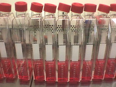
In the UK, culture media used for IVF and fertility research are regulated by the Medicines and Healthcare Regulatory Agency (MHRA) and at European level. Regulation ensures that all researchers and clinicians in the field are using industry standard medium, but manufacturers can change the composition of the media at any time without input from its end users.
When IVF researchers became curious about the role of the various components in the culture medium, and whether the manufacturers could justify changes to the medium, they contacted the HFEA (Human Fertilization & Embryology Authority) – an independent regulatory body in the UK that oversees IVF and related research.
Among many of its other tasks, the HFEA has been trying to make sure that culture medium used for in vitro maturation and preimplantation cultures is safe and optimized for use with human embryos. For example, in 2001, they surveyed IVF clinics about the culture medium they use, mainly to investigate any potential infection risks.
In December 2011, the authority’s Scientific and Clinical Advances Advisory Committee (SCAAC) published a report outlining the current knowledge about the effect of culture media components on embryo viability and development. It’s available on their website.
This most recent report notes that “Although generally considered to be safe based on past and current experience, there are still uncertainties about the long term effects of the culture media used for in vitro fertilisation, and questions remain about the effect of varying components in media. Varying concentrations of components such as growth factors, amino acids, energy substrates, and antibiotics may potentially impact on early embryo development and may have longer term health implications.”
The main conclusion of the report is that, at the moment, the research is inconclusive. They recommend more research into effects of growth medium on early and late development, as well as further investigation into long-term health effects.
The report summarizes some of the studies that have been done to date, which suggest that it’s crucial to keep an eye on what manufacturers add to the media. In one example, an article by Harper et al in Human Reproduction points out that culture medium used in human embryo development was designed based on studies in animal development. It’s not optimized for human development, because for many conditions we simply don’t know what the optimal human condition is. What we do know is that the conditions used in embryo cultures may have long-term effects. They cite a study by Dumoulin and colleagues that showed that different preimplantation media result in differences in birth weight after successful IVF pregnancies. Since birth weight may be associated with disease risk later in life, this research in particular highlights the importance of studying the effect of the various components of culture media.
Studies such as these suggest that any changes in culture media for human embryos need to be assessed very closely. However, the manufacturers of culture media are not obliged to disclose the exact composition of the media they sell. That makes it very difficult to understand the effect of various factors on development.
In an ongoing effort, the HFEA is collecting reports and studies such as these, and is gathering feedback from IVF clinics and researchers. They hope to find out what can be done to ensure that the effects of the culture media used in this field are well-regulated and understood, and plan to communicate their findings to the MHRA.
These studies may take years to complete, especially those assessing long-term effects, but a stronger scientific understanding of the role of in vitro culture medium in human development will eventually translate to a higher success rate and lower risk of IVF procedures.
 Harper J, Magli MC, Lundin K, Barratt CL, & Brison D (2012). When and how should new technology be introduced into the IVF laboratory? Human reproduction (Oxford, England), 27 (2), 303-13 PMID: 22166806
Dumoulin JC, Land JA, Van Montfoort AP, Nelissen EC, Coonen E, Derhaag JG, Schreurs IL, Dunselman GA, Kester AD, Geraedts JP, & Evers JL (2010). Effect of in vitro culture of human embryos on birthweight of newborns. Human reproduction (Oxford, England), 25 (3), 605-12 PMID: 20085915
 (3 votes)
 Loading...
 Gene expression is translationally regulated during many developmental processes. Translation is mainly controlled at the initiation step, which involves recognition of the mRNA 5′ cap structure by the eukaryotic initiation factor 4E (eIF4E). Eukaryotic genomes often encode several eIF4E paralogues but their biological relevance is largely unknown. Here (
 Mammary epithelial cells undergo structural and functional differentiation at late pregnancy and parturition to initiate milk secretion. TGF-β and prolactin signalling act antagonistically to regulate this process but what coordinates these pathways? On
  During the development of the central nervous system, neurons and/or neuronal precursors travel along diverse routes from the ventricular zones of the developing brain and integrate into specific brain circuits. Neuronal migration has been extensively studied in the forebrain but little is known about this key developmental event in the embryonic midbrain (mesencephalon). On
  Anterior-posterior patterning of both the primary embryonic axis and the secondary body axis (limbs and digits) in mammals requires regulated Hox expression. Polycomb-mediated changes in chromatin structure control Hox expression during the first patterning event but are they also involved in the second? Here (
 Mutations in the human Shwachman-Bodian-Diamond syndrome (SBDS) gene, which functions during maturation of the large 60S ribosomal subunit, cause a disorder characterised by exocrine pancreatic insufficiency, chronic neutropenia and skeletal defects. Steven Leach and colleagues have now refined a zebrafish model of this ‘ribosomopathy’ (see
  The sequence and location of every gene in the human genome is now known but our understanding of the relationships between human genotypes and phenotypes is in its infancy. To better understand the role of every gene in the development of an individual, the International Mouse Phenotyping Consortium aims to phenotype targeted gene knockout mice throughout the genome (∼23,000 genes). Because many of these mice will be embryonic lethal, methods for phenotyping mouse embryos are needed. Michael Wong and colleagues are developing such an approach and, on
  The tenth annual RIKEN Center for Developmental Biology symposium ‘Quantitative Developmental Biology’ held in March 2012 covered a range of topics. As reviewed by Davidson and Baum, the studies presented at the meeting shared a common theme in which a combination of physical theory, quantitative analysis and experiment was used to understand a specific cellular process in development. See the Meeting Review on p.
  Meyerowitz and colleagues present a glossary of image analysis terms to aid biologists and discuss the importance of robust image analysis in developmental studies.
 Members of the MADS-box transcription factor family play essential roles in almost every developmental process in plants. Kaufmann and colleagues review recent findings on MADS-box gene functions in Arabidopsis and discuss the evolutionary history and functional diversification of this gene family in plants.
 (No Ratings Yet)
