Meeting report: Fossils, Phylogenies, Genomes, Embryos & the Evolution of the Deuterostomes
Posted by Helen Robertson, on 30 May 2022
The Fossils, Phylogenies, Genomes, Embryos & the Evolution of the Deuterostomes symposium took place at the Natural History Museum in London to honour the work and contributions of the late palaeontologist R.P.S. ‘Dick’ Jefferies. In a field that is often looking to the future, it can be easy to take for granted the work that has come before or to overlook the early iterations of hypotheses we investigate today. This brilliant symposium, featuring speakers working across the scope of deuterostome evolution, was a fitting celebration of the pioneering work carried out by Dick Jefferies.
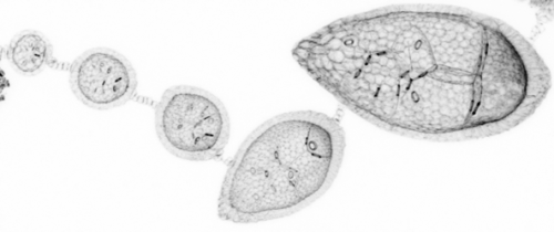
Joining the British Museum in the 1960s, Dick’s research focused on deuterostome fossils. Although many of his predictions have subsequently been disproved, some of his then-radical ideas have in fact been confirmed by more modern techniques. For example, his perhaps unexpected hypothesis of a clade comprising the tunicates and vertebrates (the Olfactores), has been subsequently supported by molecular data. Modern imaging approaches have also been key to confirming the validity of some of his ideas. A brilliant talk by Imran Rahman showed beautiful high-resolution X-ray scans supporting Jefferies’ idea of gill slits in early echinoderm fossils called the Stylophora. A particularly entertaining talk by Bertrand Lefebvre to round out the day detailed the painstakingly slow fossil sectioning-and-tracing technique employed by Jefferies’ to draw the same hypotheses now reached with very rapid microscopy approaches.
A particular highlight of the symposium was the breadth of research represented in the talks. Trends in developmental biology might sometimes lean towards developmental genetics and -omics, but the ‘evo’ side of evo-devo holds valuable contributions for the field. An excellent talk by Elizabeth Clark detailed the modelling of locomotion from fossil traces, and several talks (Paschalia Kapli, Graham Budd, Rachel Warnock) discussed persistent problems in phylogenetics. It was great to see the representation of such diverse research, and clear that there are still many open questions in the field – some of them pondered by Jefferies himself – remaining to be answered.
The Fossils, Phylogenies, Embryos & the Evolution of the Deuterostomes meeting was held on 12 May. It was supported by The Company of Biologists and The Palaeontological Association and organised by Max Telford, Jeffrey Thompson, Tim Ewin, Tim Littlewood, Greg Edgecombe, and Paul Barrett.
 (No Ratings Yet)
 (87 votes)