A short-term post-doc position (1st September until 20th July 2020, with potential extension) is available in Professor Kate Storey’s group to develop ongoing work with our human iPS cell derived neural differentiation assay to investigate cell biological mechanisms that regulate neurogenesis. This will include generating and characterising reporter hiPS cell lines and their use in live cell imaging experiments to monitor cell behaviour. Candidates require a PhD in a relevant biological subject and expertise in molecular and cell biology as well as imaging – although training in our specific imaging approaches can be provided. Please feel free to contact Professor Kate Storey k.g.storey@dundee.ac.uk for further information. Apply online here: https://www.lifesci.dundee.ac.uk/vacancies/2019/jul/31/postdoctoral-research-assistant The Dundee and School of Life Sciences are amazing places to live and work – world class life sciences research in a friendly and affordable city with a rich heritage in design and technology and the sea and mountains on your doorstep.
Welcome to our monthly trawl for developmental biology (and related) preprints. The preprints were hosted on bioRxiv, PeerJ, andarXiv. Let us know if we missed anything, and use these links to get to the section you want:
Blastula stage specification of avian neural crest
Maneeshi S. Prasad, Eileen Uribe-Querol, Jonathan Marquez, Stephanie Vadasz, Nathan Yardley, Patrick B. Shelar, Rebekah M. Charney, Martin I. Garcia-Castro
Mesenchymal Igf2 is a major paracrine regulator of pancreatic growth and function
Constanze M. Hammerle, Ionel Sandovici, Gemma V. Brierley, Nicola M. Smith, Warren E. Zimmer, Ilona Zvetkova, Haydn M. Prosser, Yoichi Sekita, Brian Y.H. Lam, Marcella Ma, Wendy N. Cooper, Antonio Vidal-Puig, Susan E. Ozanne, Gema Medina-Gómez, Miguel Constância
Molecular and genetic regulation of pig pancreatic islet cell development
Seokho Kim, Robert L. Whitener, Heshan Peiris, Xueying Gu, Charles A. Chang, Jonathan Y. Lam, Joan Camunas-Soler, Insung Park, Romina J. Bevacqua, Krissie Tellez, Stephen R. Quake, Jonathan R. T. Lakey, Rita Bottino, Pablo J. Ross, Seung K. Kim
HIPK4 is essential for murine spermiogenesis
J. Aaron Crapster, Paul G. Rack, Zane J. Hellmann, Joshua E. Elias, John J. Perrino, Barry Behr, Yanfeng Li, Jennifer Lin, Hong Zeng, James K. Chen
Gclc deletion in surface-ectoderm tissues induces microphthalmia
Brian Thompson, Ying Chen, Julien Philippe, David Anderson, Jaya Prakash Golla, Emily Davidson, Nicholas Apostolopoulos, Kevin Schey, Nicholas Katsanis, David J. Orlicky, David Thompson, Vasilis Vasiliou
Cell types of the human retina and its organoids at single-cell resolution: developmental convergence, transcriptomic identity, and disease map
Cameron S. Cowan, Magdalena Renner, Brigitte Gross-Scherf, David Goldblum, Martin Munz, Jacek Krol, Tamas Szikra, Panagiotis Papasaikas, Rachel Cuttat, Annick Waldt, Roland Diggelmann, Claudia P. Patino-Alvarez, Nadine Gerber-Hollbach, Sven Schuierer, Yanyan Hou, Aldin Srdanovic, Marton Balogh, Riccardo Panero, Pascal W. Hasler, Akos Kusnyerik, Arnold Szabo, Michael B. Stadler, Selim Orgül, Andreas Hierlemann, Hendrik P. N. Scholl, Guglielmo Roma, Florian Nigsch, Botond Roska
Longitudinal single cell transcriptomics reveals Krt8+ alveolar epithelial progenitors in lung regeneration
Maximilian Strunz, Lukas M. Simon, Meshal Ansari, Laura F. Mattner, Ilias Angelidis, Christoph H. Mayr, Jaymin Kathiriya, Min Yee, Paulina Ogar, Arunima Sengupta, Igor Kukhtevich, Robert Schneider, Zhongming Zhao, Jens H.L. Neumann, Jürgen Behr, Carola Voss, Tobias Stöger, Mareike Lehmann, Melanie Königshoff, Gerald Burgstaller, Michael O’Reilly, Harold A. Chapman, Fabian J. Theis, Herbert B. Schiller
Transcription factor NF-κB in a basal metazoan, the sponge, has conserved and unique sequences, activities, and regulation
Leah M. Williams, Melissa M. Inge, Katelyn M. Mansfield, Anna Rasmussen, Jamie Afghani, Mikhail Agrba, Colleen Albert, Cecilia Andersson, Milad Babaei, Mohammad Babaei, Abigail Bagdasaryants, Arianna Bonilla, Amanda Browne, Sheldon Carpenter, Tiffany Chen, Blake Christie, Andrew Cyr, Katie Dam, Nicholas Dulock, Galbadrakh Erdene, Lindsie Esau, Stephanie Esonwune, Anvita Hanchate, Xinli Huang, Timothy Jennings, Aarti Kasabwala, Leanne Kehoe, Ryan Kobayashi, Migi Lee, Andre LeVan, Yuekun Liu, Emily Murphy, Avanti Nambiar, Meagan Olive, Devansh Patel, Flaminio Pavesi, Christopher A. Petty, Yelena Samofalova, Selma Sanchez, Camilla Stejskal, Yinian Tang, Alia Yapo, John P. Cleary, Sarah A. Yunes, Trevor Siggers, Thomas D. Gilmore
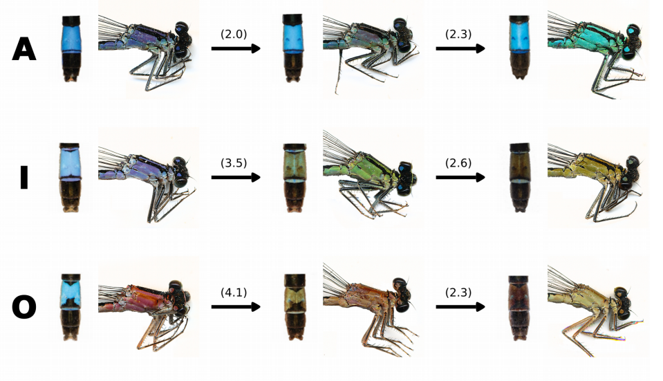
Common Bluetail damselflies from Willink, et al.’s preprint
Ins2 gene bursting activity defines a mature β-cell state
Honey Modi, Søs Skovsø, Cara Ellis, Nicole A.J. Krentz, Yiwei Bernie Zhao, Haoning Cen, N Noursadeghi, Evgeniy Panzhinskiy, Xiaoke Hu, Derek A. Dionne, Shouhong Xuan, Mark O. Huising, Timothy J. Kieffer, Francis C. Lynn, James D. Johnson
A Cre-amplifier to generate and detect genetic mosaics in vivo
Francesco Trovato, Riccardo Parra, Enrico Pracucci, Silvia Landi, Olga Cozzolino, Gabriele Nardi, Federica Cruciani, Laura Mosti, Andrzej Cwetsch, Laura Cancedda, Laura Gritti, Carlo Sala, Chiara Verpelli, Andrea Maset, Claudia Lodovichi, Gian Michele Ratto
Differential expansion microscopy
Sebastian P. Pernal, Asiri Liyanaarachchi, Domenico L. Gatti, Brent Formosa, Rishika Pulvender, Eric R. Kuhn, Rafael Ramos, Akshata R. Naik, Kathleen George, Suzan Arslanturk, Douglas J. Taatjes, Bhanu P. Jena
School of Life Sciences, University of Warwick, UK
A full-time, twelve-month Senior Research Technician position is available for an immediate start in the laboratory of Dr Kristen Panfilio, for molecular genetics and lab management tasks to support investigations of insect embryology for both biomedical and pest management research. Our lab uses a range of imaging, sequencing, transgenic, comparative genomic, and other approaches to examine the function of essential developmental genes. The primary focus is on cell and tissue structure in protective epithelial tissues, and how the tissues dynamically remodel during development, visualised with live imaging microscopy.
The technician’s principle role will be to conduct molecular biology and related research tasks in support of the PI and other members of the research team, and to do stock maintenance for our research organisms: the flour beetle, Tribolium castaneum, and the milkweed bug, Oncopeltus fasciatus. Tasks will support ongoing BBSRC- and MRC-aligned research on (a) the functional relevance of polyploidy in protective, barrier epithelial tissues, (b) tissue structure and integrity in relation to birth defects, and (c) testing the role of novel genes in lineages of pest insect species.
The candidate needs to have prior practical experience in standard molecular biology and developmental genetics techniques, and experience with insect cultures is desired. However, full training will be given in new techniques. The successful candidate is expected to be an active participant in a growing research group, so a record of successful time and project management is essential.
To view further particulars of the position and to apply, visit the University of Warwick Human Resources posting for this position. The closing date is 19th August.
Please submit your application including a CV and covering letter stating why you are interested in the topic and what you would bring to the project through the on-line application system. For informal enquires or further information about the project, please contact Kristen Panfilio (K.Panfilio@warwick.ac.uk).
In the interests of promoting transparency around the editorial process, Development will now be publishing a ‘Peer review history’ file alongside published papers, where the author has opted-in to such a file being published. All research papers submitted on or after 1 August 2019 are eligible. The file can be found on the ‘Info & metrics’ tab on the article page, and will include decision letters, referee reports and author point-by-point responses, along with a timeline of the submission and revision process, and the name of the handling editor. Reviewer reports will be published under a CC-BY license.
Authors will be able to opt out of having their peer review history files published, but we hope that most authors will be happy to include these as part of the published record. Referees, by accepting to review a paper for Development, will be indicating their willingness to have their comments published, although we will maintain anonymity unless a referee chooses to reveal their identity. Confidential comments to the editor will remain confidential (although we encourage referees to use these only under exceptional circumstances and would prefer all information to be included in the report to the authors), as will comments made through the cross-referee commenting process.
We hope that these files will provide additional insights into the published paper, by way of commentary from experts in the field, as well as opening up the process of peer review to interested readers.
Please note that Development reserves the right not to publish a ‘peer review history’ file relating to a paper in special cases, for example, if the tone or language used is inappropriate or defamatory or due to an ethical consideration.
For any queries or feedback about this policy, please contact the editorial office.
Development partners with Publons for peer reviewer recognition
Development is also pleased to announce a new partnership with Publons, part of the Web of Science Group (a Clarivate Analytics company). Publons gives reviewers formal recognition of their peer review contributions using the Reviewer Recognition Service.
Our partnership with Publons allows reviewers to easily track and verify every review by choosing to add the review to their Publons profile when completing the review submission form. Publons makes it simple for reviewers to showcase their peer review contributions in a format that can be included in job and funding applications (without breaking reviewer anonymity). Reviews completed previously can also be added by forwarding review receipts (i.e. ‘Review received’ emails) to reviews@publons.com.
We are genuinely grateful for the time and expertise volunteered by our reviewers each year. We regularly acknowledge their contributions and support by publishing a list of reviewers who have provided reports in the previous year.
The Kidney Regenerative Medicine Laboratory at the Rogosin Institute is searching for a talented and highly motivated individual interested in understanding how we can apply our understanding of kidney developmental biology to generating new tissue from stem- and primary cells. The goal of the lab is to generate kidney tissue that faithfully reproduces adult kidney functions when engrafted into experimental animals. Projects are flexible, and will take advantage of the applicant’s expertise and interests. Skills in stem cell culture, organ culture, physiological measurement, microscopy, and rodent surgery would be great assets. Strong cell culture skills are a requirement. The Rogosin Institute Kidney Regenerative Medicine Laboratory headed by Dr. Leif Oxburgh is located on the Upper East Side of Manhattan in the large biomedical campus consisting of New York-Presbyterian Hospital, Memorial Sloan-Kettering Cancer Center, New York Blood Center, Weill-Cornell Medical College and Rockefeller University. Facilities are outstanding and the local academic environment will provide top-notch training and networking opportunities. The lab works very collaboratively within the field of kidney development and regenerative medicine, both locally and nationally/internationally. The Rogosin Institute offers competitive employment packages adjusted for the cost of living in New York.
Please fill in our short listener survey, and you’ll be entered into a prize draw to win a signed copy of Kat Arney’s book, Herding Hemingway’s Cats.
The falling cost and rising speed of DNA technology has meant that genetic tests like those offered by 23andMe are now cheap enough to be advertised as fun family Christmas presents. But these tests only look at snapshots across the genome, rather than the whole thing.
The next frontier is direct to consumer whole genome sequencing, and the lower cost now makes it feasible for people with an interest in digging deeper into their genome and a bit of cash to spend. But is it a good idea?
If you enjoy the show, please do rate and review and spread the word. And you can always send feedback and suggestions for future episodes and guests to podcast@geneticsunzipped.com
The collaboration will study evolution of Wnt signalling and alternative transcript expression, splicing and function of TCF/LEF genes. This will be a close collaboration between the research group of David Ferrier at the Scottish Oceans Institute in St. Andrews and Hoppler group at the Institute of Medical Sciences in Aberdeen and will also involve Seb Shimeld and Nanopore in Oxford and Nori Satoh in Okinawa, Japan.
POSTDOCTORAL POSITION is available to study different aspects of the lymphatic vasculature in health and disease. Some of the projects include the characterization of the role of lymphatics in mammalian organogenesis in health and disease. Highly motivated individuals and have a strong background in vascular and molecular tools and are familiar with the use of mouse models are encouraged to apply. Interested individuals should send their curriculum vitae, a brief description of their research interests, and the names of three references to:
Guillermo Oliver, Ph.D
Thomas D Spies Professor of Lymphatic Metabolism
Director Center for Vascular and Developmental Biology
Northwestern University Feinberg School of Medicine
The term “embryonic development” was originally proposed some 2,400 years ago by Aristotle, however, many aspects of how the genome regulates development remain unclear. One major challenge of the modern Genomics Era is to better understand how transcriptional factors, transcriptional activators and repressors, regulate gene expression in order to support spatiotemporal outputs that change over the course of development. Recently, Dr. Theodora Koromila – senior postdoc in the Stathopoulos lab, together with Dr. Angelike Stathopoulos, at the California Institute of Technology, took a different approach to gene regulation by focusing mostly on transcriptional repressors. Using the quantitative MS2-MCP live imaging technology, they have provided insight into the mechanisms of action of the broadly-expressed transcriptional factors Runt (Run; Runx ortholog) and Suppressor of Hairless [Su(H)], throughout the early Drosophila embryo.
Specifically, based on the predicted transcriptional factors binding sites, both of these proteins were identified in an enhancer of the BMP antagonist gene short-gastrulation (sog) called the sog_Distal enhancer. Upon mutagenesis of these binding sites, the reporter-driven expression patterns exhibit dynamic spatiotemporal width changes. To assess these phenotypes systematically, Koromila and Stathopoulos devised a MS2-MCP live imaging approach to measure the spatiotemporal outputs across the entire embryo (see Movie). In this context, when the Run binding site was mutated, active expression was detected earlier than in the control. This suggests that Run normally acts to regulate the timing of transcription (see Graphical Abstract). Furthermore, their data demonstrated that Su(H) regulates expression levels, and that both factors control spatial expression. On the other hand, whereas Su(H) functions as a dedicated repressor, Run temporally switches its activity from repressor to activator in the context of the sog_Distal enhancer (see Graphical Abstract).
This research has just been published in the scientific journal Cell Reports (https://doi.org/10.1016/j.celrep.2019.06.063) and demonstrates that broadly-expressed repressors not only contribute to dynamic gene expression, but also play temporally-distinct roles. These findings provide an important basis for future research on how broadly-acting transcription factors control gene expression, which will likely be applicable to higher organisms, including vertebrates, as cis-regulatory mechanisms are generally conserved in metazoans.
The full study, “Distinct roles of broadly-expressed repressors support dynamic enhancer action and change in time” appears in Cell Reports (July 2019).
The Department of the Director Prof Miltos Tsiantis is looking for early stage researchers to employ at the Ph.D. and Post Doc level in the areas of plant development, morphogenesis and evolution (http://www.mpipz.mpg.de/226344/tsiantis-dpt).
Ph. D. candidates should have an M. Sc in appropriate discipline (Plant biology, Developmental biology, Computer Science, Genetics, Biochemistry, Statistical, Evolutionary or Population Genetics). Post Doc candidates typically will have obtained a Ph.D. degree no more than two years ago.
Ideal candidates will have shown evidence for working towards academic excellence, creativity, strong communication skills both orally and on paper and ability to work independently and harmoniously as part of a team. The Department also has a number of active international collaborations in which students will be encouraged to participate.
The positions are available at either PhD and Post Doc level and appointments will depend on the merit of interested candidates. Payment and benefits will depend on age and experience and will be according to the German TVöD scale.
Areas of research:
The interplay between growth and patterning in shaping organ form
Morphogenesis, pattern formation, growth and form, leaf development, live imaging, computational modelling
Natural variation of developmental traits in Cardamine hirsuta
Genetics, bioinformatics, local adaptation, ecology and evolution
Genetic Networks shaping leaf development in angiosperm plants
Transcriptional enhancers in development and diversity, plant homeobox genes, genome editing
Interested candidates should provide a motivation letter explaining how their skills and interests fit the areas above. Please, apply by sending a combined pdf document (your_name_PhD or PostDoc.pdf) including your motivation letter and CV, to Dr. Ismene Karakasilioti (applications.tsiantis@mpipz.mpg.de), until 17 September 2019. Applications received after the deadline will not be considered. Only shortlisted candidates will be contacted.
 (No Ratings Yet)


 (1 votes)