What did Watson and Crick discover? Rosalind Franklin’s notes…
In this episode from our centenary series exploring 100 ideas in genetics, we’re unravelling the story of the double helix, cracking the triplet code, and sketching out a Punnett square.
If you enjoy the show, please do rate and review and spread the word. And you can always send feedback and suggestions for future episodes and guests to podcast@geneticsunzipped.com
We are looking for a lab technician to assist in research on muscle stem cells, development, regeneration, disease, and evolution. More details about our research can be found at http://www.kardonlab.org/. Technician will assist in management of a mouse colony and conduct supervised research (leading to publications). Technician must be reliable, well organized, detail-oriented, excited about research and committed to working in our lab for at least two years. Prior lab experience is preferred (although not necessarily required), and class work in biology and enthusiasm for science is essential. Lab is located at the University of Utah in Salt Lake City, affording amazing opportunities for science and outdoor recreation. Looking for someone to start July-August 2019.
Contact Gabrielle Kardon (gkardon@genetics.utah.edu) with CV, list of references, and a brief statement about why you are interested in the position. BS or BA required.
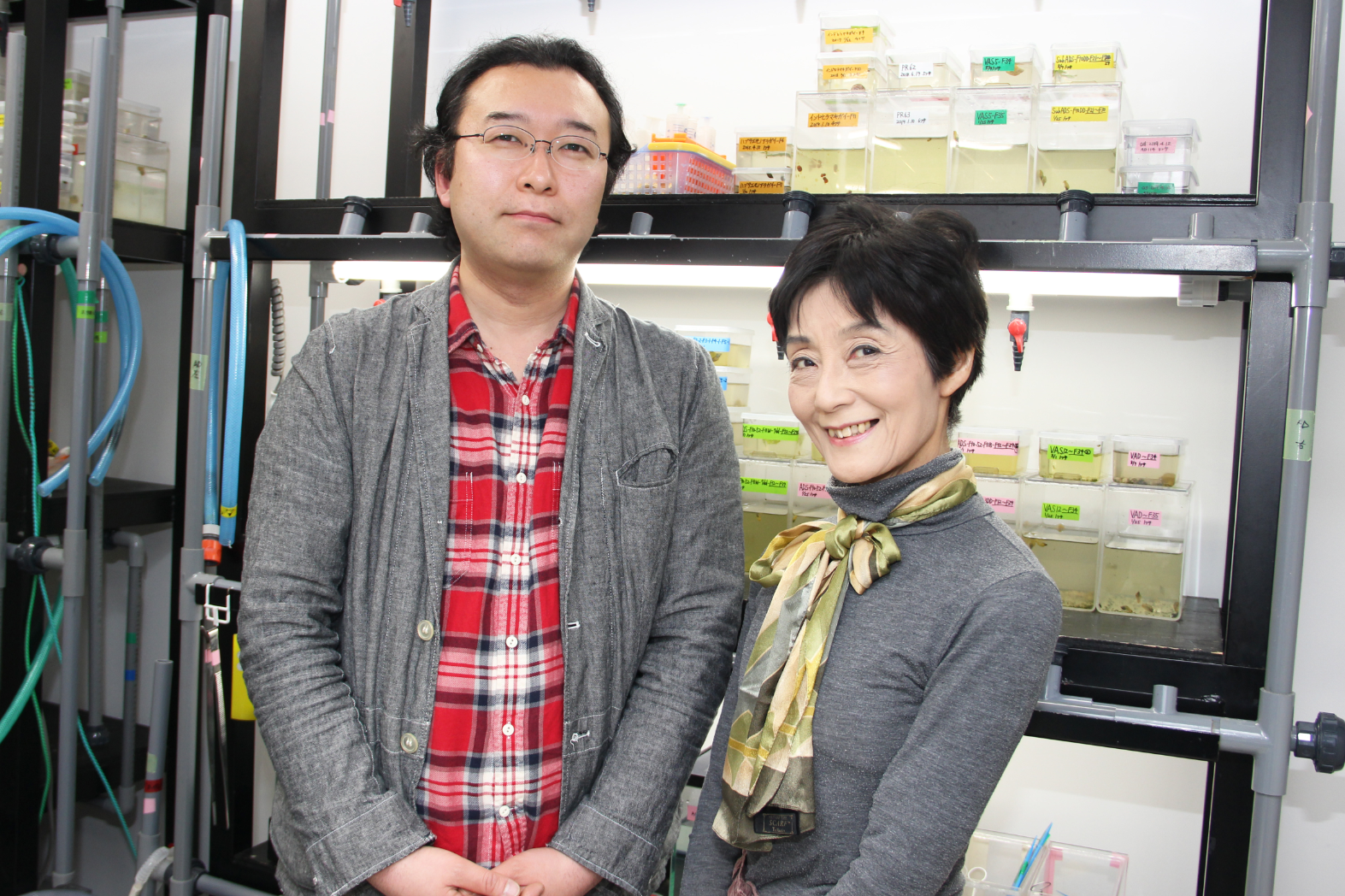
One of the most obvious examples of left-right asymmetry in animal bodies comes from snails: in most species or strains, the shells coil dextrally, but some coil sinistrally. The control of coiling is genetic and begins in the early embryo. Previous work has implicated the formin diaphanous in the regulation of snail shell chirality, and a new paper in Development now decisively proves its involvement, thanks to the first application of CRISPR/Cas9 gene knockouts in molluscs. We caught up with the author team behind the paper: Masanori Abe and his supervisor Reiko Kuroda, Professor at Chubu University in Japan (recently moved from Tokyo University of Science), to find out more.
Masanori and Reiko
Reiko, can you give us your scientific biography and the questions your lab is trying to answer?
RK During my PhD at the University of Tokyo and in my first postdoc period under Professor Stephen Mason at King’s College London, I studied chemistry and carried out research on X-ray crystallography and spectroscopy of chiral transition-metal complexes. I then moved to the Biophysics Department and worked on DNA-carcinogen/anti-cancer drug interactions using X-ray crystallography, computer graphics and molecular biology. I was appointed Associate Professor and then Professor in the Department of Chemistry, Graduate School of Arts and Sciences of the University of Tokyo, and then recently, Professor at Tokyo University of Science, and very recently Professor at Chubu University. As I am interested in chirality, my attention was inevitably drawn to the molecular mechanisms of snail coiling. I was a late starter in developmental biology, but I have now been working in the field for the last 20 years.
In 1999, I coined the term ‘chiromorphology’, which expresses the concept of my research to link macroscopic and microscopic morphological phenomena in both biology and chemistry domains through chirality. Eventually, I hope to understand the impact and the origin of the homochiral biological world [in which almost all proteins contain only L-amino acids and RNA/DNA contain D-(deoxy)ribose]. In the area of crystallography, I try to understand how billions of molecules gather to form either chiral or non-chiral crystals, and how chirality is generated, recognized, transferred and/or amplified in the solid state. In the area of spectroscopy, I have developed chiroptical spectrophotometers, which can analyze samples in their condensed phases, such as the crystalline, gel or membrane state, without the contamination of artefact signals that can arise from intrinsic macroscopic anisotropy. We have used them to explore the dynamics of secondary-structure transitions of β-amyloids and hornet silk, in addition to the crystals of organic and inorganic compounds.
Masanori: how did you come to work with Reiko, and what drives your research?
MA When I joined the Kuroda lab at the Graduate School of the University of Tokyo, I was initially not interested in snails at all, but in research on DNA recognition compounds and proteins. However, when this research got stuck, I decided to participate in the snail work. And so far, I have been very excited about the elucidation of the mechanism of left-right asymmetry determination in snails. I think it suits me to look for interesting things that people don’t normally pay attention to, and this research is just that.
How was Lsdia1 first implicated as a candidate chirality gene, and why did you need to turn to CRISPR/Cas9 to prove its role decisively?
RK We reached the conclusion that Lsdia1 is the strongest candidate for the handedness-determining gene based on positional cloning and on various experiments using pure dextral, pure sinistral and F10backcrossed lines that we had established. We found a sinistral strain that carries a frameshift mutation that abrogates full-length LsDia1 protein expression, already at the one-cell stage (published in 2016). Although we were confident about the results, we could not ignore the possibility that Lsdia1 is simply genetically linked to a true handedness-determining gene. We envisaged that directly knocking out the Lsdia1 gene by CRISPR/Cas9 would provide proof. Although only MA and RK are involved in the current work, we are grateful to all the scientists/technicians/students who worked in Kuroda lab in the past for their contributions in establishing pure and backcrossed strains, rearing the snails, and enabling positional cloning, and to the Japan Science and Technology Agency (JST) for the funding of ERATO and SORST Kuroda Chiromorphology projects (to RK, 1999-2004 and 2004-2009, respectively).
Can you give us the key results of the paper in a paragraph?
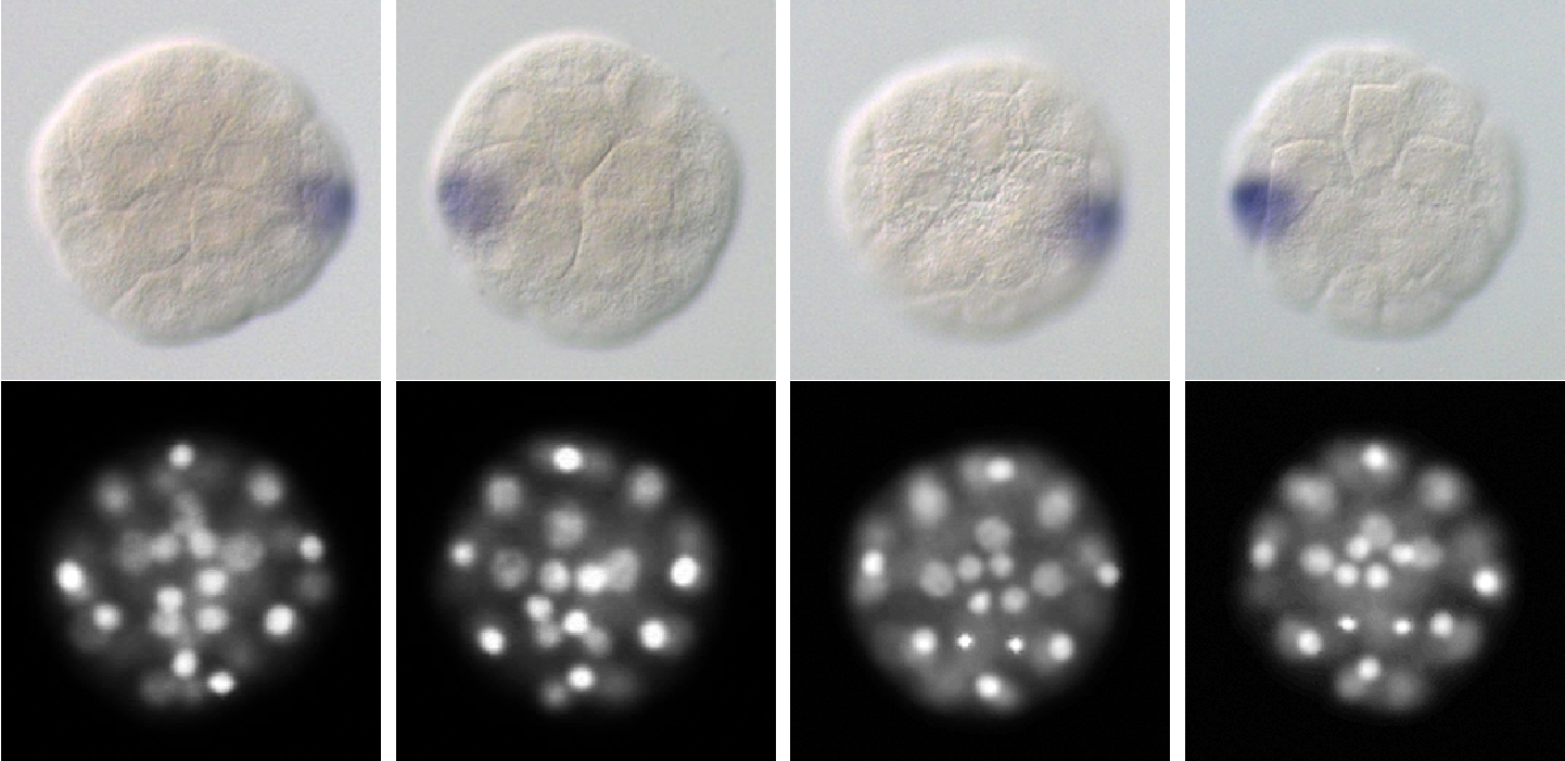
RK We have decisively identified that Lsdia1 is the long-sought handedness-determining gene of Lymnaea stagnalis by developing CRISPR/Cas9 gene editing techniques. Biallelic frameshift mutations introduced into the gene produced sinistrally coiled offspring generation after generation, in the otherwise totally dextral genetic background. We could also show that the gene sets the chirality already at the one-cell stage, the earliest observed symmetry-breaking event linked directly to body-handedness in the animal kingdom. The early intracellular chirality is superseded by the intercellular chirality during the third cleavage, leading to asymmetric expression of nodal and Pitx (two genes known to regulate chirality in vertebrates) and then to organismal body handedness.
Gene-edited F1 snails (49-cell stage), with in situ hybridisation for nodal, DAPI staining and selected cell types labelled.
How do you think chirality is transmitted through development?
RK This is the project we are currently working on. It is interesting that intracellular chirality within a cell is superseded by the intercellular chirality during the third cleavage (as proven by the fate of mechanically manipulated embryos at this stage), and then to the 24-cell stage when the cell fates are determined. We hope to report the results in the near future.
Left-right (LR) asymmetry is a widespread feature of animals – how much conservation do you think there is at a genetic and cytoskeletal level?
RK Although diverse mechanisms have been proposed for different phyla, we think a unified mechanism and the involvement of cellular chirality are probable. We believe that there are conserved mechanisms, given that diaphanous is a protein present in many phyla. We plan to elucidate the mechanisms of chirality establishment by LsDia1 at the molecular level, which may give insight into the conservation of the genetic control of LR asymmetry.
Do you have any advice for people wanting to do CRISPR/Cas9 mutagenesis in snails?
RK Gene editing of snails by CRISPR/Cas9 is not particularly difficult, as the method itself has already been well established in model organisms, although one must check for possible off-target effects. The most difficult aspects are raising the gene-edited eggs to adult snails, and successfully breeding them to produce the next generation.
When doing the research, did you have any particular result or eureka moment that has stuck with you?
MA Since it was not guaranteed that Lsdia1 was a handedness-determining gene, I sometimes felt anxious that the gene-edited individuals created over a long period would be wasted. At that time, the F0 individuals were starting to lay eggs, so I decided to count the number of hatched juveniles, and then felt a strong sense of discomfort. At that moment, I realized that the direction of shell coiling had changed into sinistral! The juvenile snails did not show any abnormalities except the shell-coiling inversion and moved around in the Petri dish vigorously. Then I was finally convinced that Lsdia1 was the snail handedness-determining gene. I was also surprised when I found one-cell-stage chirality.
And what about the flipside: any moments of frustration or despair?
MA It can sometimes get frustrating spending a long time caring for snails. Containers for breeding individuals should be cleaned weekly, thus I can’t take a long vacation leaving the lab. In particular, we are doing experiments with eggs just laid, but the quality of the eggs varies greatly depending on the health and age of the parent snail. It can’t be avoided in order to obtain correct results with good reproducibility.
So what next for you after this paper?
MA It was found that LsDia1 and LsDia2 give LR asymmetry to the one-cell-stage egg. Next, I would like to investigate the origin of organismal LR asymmetry by delving into the molecular level, focusing on the analysis of cytoskeleton dynamics in snails. In addition, as we have established gene editing technique for this snail, which is often used in the study of perception, learning and memory, I hope that our work will help this snail become a model organism for human disease research.
Where will this work take the Kuroda lab?
RK When one question is solved, more questions arise. We have so many interesting themes to work on: the roles of LsDia1, LsDia2 and other proteins in explaining the Dia1-dependent chirality within one cell, how the intracellular chirality is superseded by intercellular chirality (cytoskeletal dynamics), and how information on the geometrical arrangement of blastomeres at the eight-cell stage is transferred to the 24-cell stage, and then to nodal/Pitx expression, at the molecular level. My long-term research objective is to link macroscopic and microscopic phenomena, i.e. across the biological hierarchy, using chirality.
When one question is solved, more questions arise
Finally, let’s move outside the lab – what do you like to do in your spare time in Tokyo?
MA I like taking a walk doing Pokemon Go as it is good exercise. There are many interesting spots in Tokyo, historic buildings and monuments, beautiful parks and cityscapes, etc. Also I enjoy making small discoveries and surprises even in the back alleys that I had never visited before.
RK We both moved this April from Tokyo to Chubu University, Kasugai, near Nagoya. When in Tokyo, I often went to special exhibitions at the National Science Museum (such as ‘Lascaux – Cave Paintings of the Ice Age’, ‘Hunters of the Ocean’, ‘Wine – the Exhibition’ and ‘Deep Sea’) or other museums and galleries in Tokyo (e.g. ‘Winne-the Pooh – the original drawings from the V&A Museum London’ and ‘Vermeer and Dutch Art’). Once we have settled down in Kasugai, I hope to explore the area.
We are seeking a proactive and highly motivated individual to deliver gene targeting capabilities at the Babraham Institute.
The successful applicant will work closely with scientific staff and will be embedded in the Epigenetics Programme in order to foster a strong scientific working environment, whilst providing expert support in gene targeting and delivering transgenic mice to researchers across the Institute. Opportunities for training and to participate in significant high impact research leading to publications will be available.
The ideal candidate will have enthusiasm and proven experience with developing and applying molecular biology technologies in genome engineering. This includes being able to independently design, generate and use gene targeting reagents and recently developed editing technologies such as CRISPR/Cas9. The ideal candidate will possess the skills required to independently undertake all aspects of the generation of transgenic mouse embryos. Prior experience with embryo collection and electroporation is highly desirable.
The job holder will be expected to compile, maintain, interpret and present data. Strong communication and interpersonal skills will be essential in order to clearly present technical and conceptual information verbally and in writing to a wide variety of people with varying levels of knowledge.
Closing date for applications is Thursday, 4th July 2019.
NO AGENCIES PLEASE
The Babraham Institute holds a silver Athena SWAN award and is committed to promoting and developing a culture of excellence, diversity and mutual respect that supports the Institute’s ambitions and attracts highly motivated and talented people. The Babraham Institute is a Disability Confident Employer and has a positive approach to employing disabled people.
Rita Levi-Montalcini Postdoctoral Fellowship in Regenerative Medicine
The Center of Regenerative Medicine at Washington University in St. Louis invites applications for the Rita Levi-Montalcini Postdoctoral Fellows in Regenerative Medicine program. These fellowships honor Rita Levi-Montalcini, whose Nobel-winning discovery of Nerve Growth Factor was among the first regenerative biology research to be done at WUSTL.
The Center of Regenerative Medicine (CRM) seeks individuals of outstanding talent with a doctoral degree to provide them with the opportunity to pursue research within a CRM lab. As Rita Levi-Montalcini was herself an international scholar working at WUSTL, we strongly encourage international applicants to apply. Additional information about the Center of Regenerative Medicine can be found at: https://regenerativemedicine.wustl.edu/. Full program details can be found at: https://regenerativemedicine.wustl.edu/about/rlm_fellowship/rlm_programdetails/
Qualifications
RLM Fellowships are intended for exceptional scientists of great promise who have recently been awarded, or who are about to be awarded, the doctoral degree. Fellows are required to work in the lab of a CRM faculty member on a project that directly focuses on regenerative medicine. Current employees, fellows, and students of Washington University in St. Louis are not eligible. Applicants currently on H-1B visas are not eligible. We anticipate awarding two fellowships in 2019.
Terms of Appointment
RLM Fellowships will be granted for a period of two years.
A Ph.D./D.Sc./M.D. must be awarded and proof furnished to the CRM before the start of the Fellowship.
The RLM Fellowship provides annual compensation of $55,000, as well as fringes and health insurance, research funds, and relocation and travel funds.
Review of applications will begin on August 30, 2019.
Please provide a full current CV, two letters of reference, indicate a potential CRM faculty host (https://regenerativemedicine.wustl.edu/people-page/), and a brief description of scientific accomplishments and long-term goals.
Washington University is an Equal Opportunity Employer. All qualified applicants will receive consideration for employment without regard to race, origin, religion, age, sex, sexual orientation, gender identity or expression, national origin, genetic information, disability, or protected veteran status.
The Wellcome Trust – Medical Research Council Cambridge Stem Cell Institute is an international centre of excellence for stem cell research and regenerative medicine. Scientists in the Institute collaborate to advance our knowledge of various stem cell types and to perform pioneering work in translational research areas, providing the foundation for new medical treatments (https://www.stemcells.cam.ac.uk/).
Research position is open in the laboratory of Professor Austin Smith to join investigations into the fundamental biology of pluripotency and pluripotent stem cells in different mammals. Candidates should have a publication record from their previous research, but prior experience with pluripotent stem cells is not essential. Homepage: www.stemcells.cam.ac.uk/research/pis/smith
We are looking for a Research Assistant/Associate – Bioinformatics. Research Associate candidates should have a PhD or equivalent research doctorate in computational biology or bioinformatics. Research Assistant candidates should have a Bachelor’s degree or higher in a relevant subject. The successful candidate will have experience in next generation sequencing pipelines, and good understanding of molecular cell biology. Under the guidance of a senior bioinformatician, you will develop and implement algorithms, analysis methods and visualisation tools for cross-species comparisons using scRNAseq, ChIPseq and ATAC-seq data.
Candidates will have proven capacity to design, execute, and interpret experiments or data analyses. Good communication skills and the ability to work effectively in a team are essential. Our lab is international in character and this project is collaborative with groups in China and Japan.
Fixed-term: The funds for this post are available for 2 years in the first instance.
Click the ‘Apply’ button below to register an account with our recruitment system (if you have not already) and apply online at http://www.jobs.cam.ac.uk/job/21931/
The closing date is 10 July 2019, with interviews to be confirmed.
Please ensure that you upload a covering letter and CV in the Upload section of the online application. If you upload any additional documents which have not been requested, we will not be able to consider these as part of your application.
Please include details of your referees, including email address and phone number, one of which must be your most recent line manager.
Please quote reference PS19502 on your application and in any correspondence about this vacancy.
The University actively supports equality, diversity and inclusion and encourages applications from all sections of society.
The University has a responsibility to ensure that all employees are eligible to live and work in the UK.
The Wellcome Trust – Medical Research Council Cambridge Stem Cell Institute is an international centre of excellence for stem cell research and regenerative medicine. Scientists in the Institute collaborate to advance our knowledge of various stem cell types and to perform pioneering work in translational research areas, providing the foundation for new medical treatments (https://www.stemcells.cam.ac.uk/).
Research positions are open in the laboratory of Professor Austin Smith to join investigations into the fundamental biology of pluripotency and pluripotent stem cells in different mammals. Candidates should have a publication record from their previous research, but prior experience with pluripotent stem cells is not essential. Homepage: www.stemcells.cam.ac.uk/research/pis/smith
Two positions are available, each for 2 years in the first instance:
Post 1: Molecular embryologist with specialist skills in culture and manipulation of pre-implantation embryos and post-doctoral experience studying blastocyst development and lineage segregation.
Post 2: Molecular biologist/biochemist with PhD training in the molecular genetic analysis of signalling pathways and downstream transcriptional regulation.
Candidates will have proven capacity to design, execute, and interpret experiments or data analyses. Good communication skills and the ability to work effectively in a team are essential.
Our lab is international in character and this project is collaborative with groups in China and Japan.
Fixed-term: The funds for this post are available for 2 years in the first instance.
Click the ‘Apply’ button below to register an account with our recruitment system (if you have not already) and apply online at http://www.jobs.cam.ac.uk/job/20694/
The closing date is 10 July 2019, with interviews to be confirmed.
Please ensure that you upload a covering letter and CV in the Upload section of the online application. If you upload any additional documents which have not been requested, we will not be able to consider these as part of your application.
Please include details of your referees, including email address and phone number, one of which must be your most recent line manager.
Please quote reference PS18395 on your application and in any correspondence about this vacancy.
The University actively supports equality, diversity and inclusion and encourages applications from all sections of society.
The University has a responsibility to ensure that all employees are eligible to live and work in the UK.
Keywords: Actin cytoskeleton; Yeast genetics; Cell imaging and Single molecule imaging.
The actin cytoskeleton is a complex and dynamic system involved in several force-generating processes such as cell motility or cell division [1]. Depending on their function in cells, actin filaments can be organized into different structures. For example, crawling cells form a lamellipodium made of branched actin filaments at the front and filopodia which are composed of parallel actin filaments (see Fig. 1). In these structures, new actin filaments are generated and elongated by various families of nucleation factors. These structures are also constantly remodeled by multiple families of actin binding proteins (ABPs).
Importantly, the different actin structures present in cells are differently regulated because they interact with distinct sets of ABPs. Many conserved families of ABPs are found only associated with specific actin structures, while excluded from others [2]. How cells organize their interior is one of the central questions in biology. While it applies also to actin networks, the community largely ignores how ABPs are sorted efficiently to distinct sub-structures of actin filaments.Two non-exclusive hypotheses may explain how actin filaments within actin networks may acquire a specific identity to recruit ABPs selectively [3]. First, actin filaments may be assembled from distinct actin isoforms. Second, actin filaments may be decorated by some specific additional factors while they are being assembled. The aim of this PhD will be to verify these hypotheses and to determine whether the mechanisms by which cells generate a diversity of actin substrates. The candidate will use yeast genetics to test his/her hypotheses in cells and top-end single molecule imaging to understand these mechanisms in vitro.The host team is interdisciplinary and composed of physicists, chemists and biologists. The candidate should be interacting actively with the team members and be driven by his/her curiosity. The team is also international, so speaking and presenting in English is expected.
CONTACT
Please send your CV, a letter of motivation and con- tacts for references.
Michelot lab Institut de Biologie du Développement de Marseille (IBDM), Aix-Marseille Université
E-mail: alphee.michelot@univ-amu.fr
REFERENCE
[1] Blanchoin et al., Actin dynamics, architecture, and mechanics in cell motility, Physiol Rev, 2014
[2] Kovar et al., Three’s company: the fission yeast actin cytoskeleton, Trends Cell Biol, 2010
[3] Michelot and Drubin, Building distinct actin filament networks in a common cytoplasm, Curr Biol, 2011
¹Center for Chronobiology,¹The Visual Systems Group, Abrahamson Pediatric Eye Institute, Division of Pediatric Ophthalmology, ²Division of Developmental Biology, Cincinnati Children’s Hospital Medical Center, Cincinnati, OH 45229, USA. ²Department of Ophthalmology, University of Cincinnati, College of Medicine, Cincinnati, OH 45229, USA
We live on a planet that is close to a yellow dwarf star. This means that throughout the evolution of life, photons have been available. Living systems have exploited photon availability for adaptive advantage using pathways like photosynthesis, the circadian clock and the visual system. However, in addition to these obvious examples, we are learning that metazoans can decode light information in other ways. Here we comment on recent work from the Lang lab that describes how light signals can provide developmental timing cues.
The embryonic eye has an elaborate vessel network, called the hyaloid vasculature, that supplies nutrients and oxygen to the developing retina. This network is transient and undergoes programmed regression concomitantly with the development of retinal vasculature. Regression of the hyaloid vessels is an adaptation for high acuity vision: Since the structure lies in the optical path and would scatter light, its regression is necessary for optical function of the eye. The clinical conditions persistent fetal vasculature (PFV) and persistent hyperplastic primary vitreous (PHPV) are a consequence of abnormal hyaloid vessel regression.
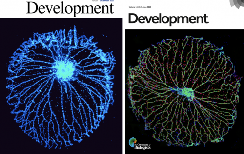
Research in the Lang lab has focused on development of vasculature in the eye since 1993 when we showed that myeloid cells play an important role in scheduledvascular regression(Lang and Bishop, 1993). The molecular pathways requiredfor hyaloid vessel regression (the Wnt, Angiopoietin and VEGF pathways) have been defined by our group and by several others(Gale et al., 2002; Glass II et al., 2005; Kurihara et al.; Liu and Nathans, 2008; Lobov et al., 2002; Nayak et al., 2018; Rao et al., 2007; Yoshikawa et al., 2016). The hyaloid vessel system is an interesting subject of study in part because it can be dissected from the eye as a whole mount and the vessels easily counted. The hyaloid vasculaturemay seem like an obscure structure (it is) but has featured on the front cover of Development twice (Nayak et al., 2018; Rao et al., 2007)(Fig. 1).
Fig 1. Hyaloid vessels featured on the cover of Development
Despite all that had been learned, the trigger that initiatedhyaloid regression remained to be elucidated. When the Lang lab considered this question, we thought about the possibility that light stimulation could be the trigger. After all, this was an eye, a structure designed to respond to light, and hyaloid vessel regression began soon after birth when light exposure levels would presumably be elevated. To investigate this initially, we (that is, Lang Lab alumnus Sujata Rao) dark-reared mice and found hyaloid vessel persistence, an indication that light stimulation could be involved.
What unfurled next was a nice illustration of how science investigations sometimes work. Our colleague Maureen McCall was visiting from Louisville and, after she heard about our work, recommended we contact David Copenhagen (UCSF) because he was also working on light response pathways in neonatal mice. Excited by the possibilities, we contacted David and learned that newborn mice have a negative phototaxis (a light aversion response) mediated by melanopsin (OPN4), one of the non-canonical retinal opsins (Johnson et al., 2010). This raised the possibility that melanopsin might also regulate hyaloid regression and sure enough, when Sujata assessed Opn4 null mice, we found hyaloid vessel persistence (thanks for the hot tip Maureen). Subsequently, the Lang and Copenhagen labs worked together to understand how melanopsin controlled the timing of vascular regression.
Key findings included the observation that dark reared and Opn4 null mice developed an over-abundance of retinal neurons, showed elevated oxygen demand and elevated levels of vascular endothelial growth factor A (VEGFA), a crucial promoter of angiogenesis and vascular endothelial cell survival (Rao et al., 2013). Elevated levels of VEGFA provided a good explanation for the promiscuous retinal angiogenesis and hyaloid vessel persistence characteristic of the Opn4 null and dark-reared mice (Rao et al., 2013). In a little bit of a surprise, the analysis suggested that the light-OPN4 pathway functioned before birth in a fetal eye that was directly light responsive. While some of our colleagues have struggled with the idea that deep tissue photoreceptors can receive sufficient light to function, there is evidence throughout the animal kingdom that this is one mode of light detection (Miyashita et al., 1996; Nakane et al., 2010; Okano et al., 1994) and recent analysis from the Lang and Copenhagen labs confirms that this is the way this pathway works (stay tuned….).
Prompted by this work, the Lang lab become much more curious about the possibility that other light response pathways could play an important role in development.Genome sequencing nearly two decades earlierhad identified two other non-canonical opsins, neuropsin (OPN5,(Tarttelin et al., 2003)) and encephalopsin (OPN3,(Blackshaw and Snyder, 1999)). Both of these were expressed in the retina and so as a first assessment, the Lang lab determined whether a null mouse for either opsin has a vascular phenotype in the eye: In Opn5 null mice, we found one (Nguyen et al., 2019).
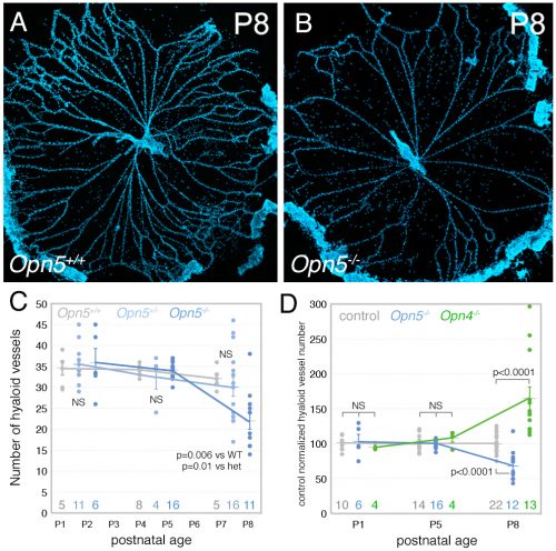
Fig 2. Precocious hyaloid vessel regression in the Opn5 null mouse. (A, B) Hoechst 33258 (blue) labelled hyaloid preparations from P8 Opn5 control and Opn5 null mice. (C) Quantification of hyaloid vessel number in control (grey), Opn5 heterozygote (light blue) and Opn5 null (dark blue) mice over a P1-P8 time-course. (D), as in (C) but relative hyaloid vessel numbers for control (grey), Opn5 null (blue), and Opn4 null (green) mice. First published in Nguyen et al., Nature Cell Biology, 2019.
The Lang lab has trained a small cadre of researchers to perform hyaloid vessel dissections (the dissection requires steady hands, patience, specialized technique, and freshly harvested eyes). On the day we first analyzed the Opn5 mouse hyaloid vessels, we had three litters all at the same stage of development. This meant a long day of dissections for Postdoc Shruti Vemaraju(50 eyes and about 6 hours of dissection) in which she was blinded to the genotype and so had no idea of the outcome as she worked. After all the staining and counting was complete, we were excited to learn that loss of OPN5 function resulted in precocious hyaloid vessel regression(Nguyen et al., 2019). This wasunusual because all hyaloid phenotypes described to date showed vessel persistence (Fig. 2). OPN5 is known to be stimulated by violet light with peak absorbance at 380nm(Kojima et al., 2011). Withdrawing this wavelength during the neonatal period mimicked the precocious hyaloid vessel regression seen with loss of OPN5 function(Nguyen et al., 2019). This made a strong argument that OPN5 was functioning as an opsin to regulate hyaloid regression.
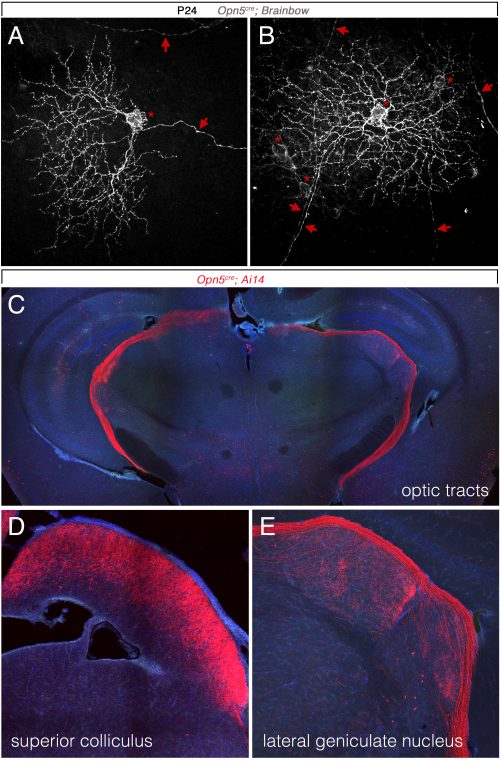
So, to summarize, we now knew that two distinct opsins, OPN4 and OPN5 each regulated hyaloid vessel regression, but because the phenotypes were opposite, probably used distinct mechanisms. We knew from prior work (a collaboration with the labs of Russell Van Gelder, King-Wai Yau and Ethan Buhr) that Opn5 was expressed in retinal ganglion cells (RGCs)(Buhr et al., 2015). Since we had not been able to identify any antibodies that detected OPN5, we generated an Opn5cre allele as a means of targeting and characterizing Opn5 RGCs. Conversion of the Ai14 tdTomato cre reporter confirmed that positive cells had all the characteristics of RGCs(Fig. 3)including projections to the lateral geniculate nucleus and superior colliculus.
Fig 3.Opn5 is expressed in retinal ganglion cells. (A, B) Flat mount retinae with labeling of cell bodies (asterisks), dendritic fields and axons (arrows) for retinal ganglion cells labeled by the Brainbow3.2 reporter in P24 Opn5cre mice. (A-C) tdTomato signal (red) in Opn5cre; Ai14 P28 mouse brain cryosections in the optic tracts (C), superior colliculus (D) and lateral geniculate nucleus (E). These projections are characteristic of retinal ganglion cells. First published in Nguyen et al., Nature Cell Biology, 2019.
When Lang lab Research Associate Minh-Thanh Nguyen was characterizing the Opn5 null retina, she noticed an unusual pattern of expression of tyrosine hydroxylase, the rate limiting enzyme in the pathway that synthesizes dopamine. This suggested that dopamine might be changed and, according to an ELISA assessment, it was low in retinal neurons and high in the extracellular vitreous (where the hyaloid vessels reside). This was interesting because dopamine can have an anti-vascular function: via the dopamine receptor DRD2, dopamine can activate the phosphatase Shp2 and promote dephosphorylation of VEGF receptor 2(Sinha et al., 2009). This suggested the hypothesis that an OPN5 light response pathway regulated hyaloid vessel regression using dopamine as a signaling intermediate.
Aided by dopamine expert and collaborator Mike Iuvone at Emory University, we investigated this idea further and showed that hyaloid regression could be both positively (precocious regression with receptor agonists) and negatively (vessel persistence with receptor antagonists) regulated by dopamine signaling. We further showed that a key regulator of dopamine uptake, the dopamine transporter (DAT) was activated (via phosphorylation(Foster et al., 2012) by 380 nm light in an Opn5-dependent manner. Phospho-DAT was detected in a network of neuronal processes throughout the inner retina and so was well placed to globally regulate dopamine levels. Using a pharmacological inhibitor of DAT, we could revert hyaloid vessel persistence resulting from dark-rearing to normal numbers and also reproduce an Opn5 null precocious hyaloid vessel phenotype in Opn5 heterozygote mice. These findings led to the hypothesis that hyaloid vessel regression is regulated by an intricate balance between dopamine and VEGFA signaling.
The hypothesis that dopamine was a signaling intermediate also required that hyaloid vascular endothelial cells (VECs) expressed a dopamine receptor. We detected DRD2 in the hyaloid vessels by immunolabeling and further showed that conditional deletion of a Drd2floxed allele in VECs resulted in hyaloid persistence. When combined with Opn5 loss-of-function (elevated dopamine levels in vitreous, precocious regression), deletion of Drd2 from VECs abrogates the effects of dopamine signaling and restores hyaloid vessel numbers to normal. This was consistent with a direct effect of dopamine promoting hyaloid vessel regression.
A further prediction of the hypothesis was that VEGFR2 signaling in the hyaloid vessels would be influenced by dopamine. To test this, Lang lab Postdoc Yoshinobu Odaka performed immunoblotting on the vanishingly small quantities of cell lysates available from hyaloid vessel preparations. He assessed the levels of total and phospho–tyrosine 1173-VEGFR2 (an activating modification) in control and Drd2 VEC conditional deletion hyaloids. Consistent with the hypothesis, pY1173-VEGFR2 was elevated when Drd2 was deleted. This supported the idea that normally DA signaling suppresses VEGFR2 and provided a mechanistic explanation for how the high levels of vitreal dopamine in the Opn5 null could promote precocious hyaloid vessel regression.
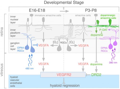
During review of the manuscript describing this work, we were asked quite a few questions about how the OPN4 and OPN5 vascular pathways were integrated. The schematic (Fig. 4) describes our current model.We propose that blue light photons activate OPN4 before birth. Through a mechanism not currently understood, this suppresses the number of retinal neurons that develop. When mice are dark–reared or have OPN4 loss-of-function, cellularity and oxygen demand are higher, and this stimulates production of VEGFA. Under those abnormal conditions, VEGFA within the retina produces promiscuous angiogenesis while vitreal VEGFA suppresses regression of the hyaloid vessels. This means that normally, blue light stimulation of fetal retina suppresses the levels of VEGFA as a prerequisite for regression of the hyaloid vessels.
Fig 4. Schematic describing integration of the OPN4-VEGFA and OPN5-dopamine hyaloid regression pathways. The schematic identifies two phases of development, E16-E18 and P3-P8 when OPN4 and OPN5 are each required. In late gestation, blue light stimulation of OPN4 RGCs suppresses retinal cellularity. In dark-reared or in Opn4 null mice, elevated cellularity increases oxygen demand ([O2]) and, via the hypoxia response pathway, increases VEGFA expression in amacrine cells and RGCs. Elevated VEGFA causes promiscuous retinal angiogenesis and suppresses hyaloid vessel regression. According to the present analysis, violet light stimulation of OPN5 RGCs postnatally suppresses dopamine in the vitreous by upregulating T53 phosphorylation of the dopamine transporter (P-DAT/SLC6A3) in neurons in the inner plexiform layer. Normally, OPN5-dependent phosphorylation of DAT results in elevated dopamine uptake and a reduced flux of dopamine from dopaminergic amacrine cells to the vitreous. In the absence of OPN5, or the violet light that stimulates OPN5, vitreous dopamine is precociously elevated. This results in premature activation of dopamine receptor DRD2 in hyaloid VECs, suppression of VEGFR2 survival signalling and precocious regression. These data indicate that both 480 nm blue light via OPN4, and 380 nm violet light via OPN5, function as developmental timing cues. First published in Nguyen et al., Nature Cell Biology, 2019.
Current analysis suggests that OPN5 functions after birth to suppress the level of vitreal dopamine. This dopamine suppresses VEGFR2 signaling and promotes hyaloid vessel regression. However, this occurs when dopamine levels in the eye are generally rising because the TH+ amacrine cells that are the source of dopamine are differentiating and becoming active. This series of positive and negative influences means that the net effect of violet light and OPN5 activity is to sustain the hyaloid vessels. This mechanism likely evolved to ensure that hyaloid vessel regression does not occur before the first layer of retinal vasculature is complete: this would prevent hypoxia by ensuring that one of the vascular networks was always functional.One way to think about OPN4 and OPN5 function in the developing eye is that they are the detectors for developmental timing cues. A reminder that in development, timing is everything.
References
Blackshaw, S. and Snyder, S. H. (1999). Encephalopsin: a novel mammalian extraretinal opsin discretely localized in the brain. J Neurosci19, 3681–3690.
Buhr, E. D., Yue, W. W., Ren, X., Jiang, Z., Liao, H. W., Mei, X., Vemaraju, S., Nguyen, M. T., Reed, R. R., Lang, R. A., et al. (2015). Neuropsin (OPN5)-mediated photoentrainment of local circadian oscillators in mammalian retina and cornea. Proc Natl Acad Sci U S A112, 13093–13098.
Foster, J. D., Yang, J. W., Moritz, A. E., ChallaSivaKanaka, S., Smith, M. A., Holy, M., Wilebski, K., Sitte, H. H. and Vaughan, R. A. (2012). Dopamine transporter phosphorylation site threonine 53 regulates substrate reuptake and amphetamine-stimulated efflux. J. Biol. Chem.287, 29702–29712.
Gale, N. W., Thurston, G., Hackett, S. F., Renard, R., Wang, Q., McClain, J., Martin, C., Witte, L., Witte, M. H., Jackson, D., et al. (2002). Angiopoietin-2 is Required for Postnatal Angiogenesis and Lymphatic Patterning, and Only the Latter is Rescued by Angiopoietin-1. Dev Cell3, 411–423.
Glass II, D. A., Bialek, P., Ahn, J. D., Starbuck, M., Patel, M. S., Clevers, H., Taketo, M. M., Long, F., McMahon, A. P., Lang, R. A., et al. (2005). Canonical Wnt signaling in differentiated osteoblasts controls osteoclast differentiation. Dev. Cell8,.
Johnson, J., Wu, V., Donovan, M., Majumdar, S., Renteria, R. C., Porco, T., Van Gelder, R. N. and Copenhagen, D. R. (2010). Melanopsin-dependent light avoidance in neonatal mice. Proc Natl Acad Sci U S A107, 17374–17378.
Kojima, D., Mori, S., Torii, M., Wada, A., Morishita, R. and Fukada, Y. (2011). UV-sensitive photoreceptor protein OPN5 in humans and mice. PLoS One6, e26388.
Kurihara, T., Kubota, Y., Ozawa, Y., Takubo, K., Noda, K., Simon, M. C., Johnson, R. S., Suematsu, M., Tsubota, K., Ishida, S., et al. von Hippel-Lindau protein regulates transition from the fetal to the adult circulatory system in retina. Development137, 1563–1571.
Lang, R. A. and Bishop, J. M. (1993). Macrophages are required for cell death and tissue remodeling in the developing mouse eye. Cell74,.
Liu, C. and Nathans, J. (2008). An essential role for frizzled 5 in mammalian ocular development. Development135, 3567–76.
Lobov, I. B., Brooks, P. C. and Lang, R. A. (2002). Angiopoietin-2 displays VEGF-dependent modulation of capillary structure and endothelial cell survival in vivo. PNAS99, 11205–11210.
Miyashita, Y., Moriya, T., Yokosawa, N., Hatta, S., Arai, J., Kusunoki, S., Toratani, S., Yokosawa, H., Fujii, N. and Asami, K. (1996). Light-sensitive response in melanophores of Xenopus laevis: II.Rho is involved in light-induced melanin aggregation. J. Exp. Zool.276, 125–31.
Nakane, Y., Ikegami, K., Ono, H., Yamamoto, N., Yoshida, S., Hirunagi, K., Ebihara, S., Kubo, Y. and Yoshimura, T. (2010). A mammalian neural tissue opsin (Opsin 5) is a deep brain photoreceptor in birds. Proc. Natl. Acad. Sci.107, 15264–15268.
Nayak, G., Odaka, Y., Prasad, V., Solano, A. F., Yeo, E.-J., Vemaraju, S., Molkentin, J. D., Trumpp, A., Williams, B., Rao, S., et al. (2018). Developmental vascular regression is regulated by a Wnt/β-catenin, MYC and CDKN1A pathway that controls cell proliferation and cell death. Development145, dev154898.
Nguyen, M.-T., Vemaraju, S., Nayak, G., Odaka, Y., Buhr, E. D., Alonzo, N., Tran, U., Batie, M., Upton, B. A., Darvas, M., et al. (2019). An Opsin 5-dopamine pathway mediates light-dependent vascular development in the eye. Nat. Cell Biol.In press,.
Okano, T., Yoshizawa, T. and Fukada, Y. (1994). Pinopsin is a chicken pineal photoreceptive molecule. Nature372, 94–7.
Rao, S., Lobov, I. B., Vallance, J. E., Tsujikawa, K., Shiojima, I., Akunuru, S., Walsh, K., Benjamin, L. E. and Lang, R. A. (2007). Obligatory participation of macrophages in an angiopoietin 2-mediated cell death switch. Development134,.
Rao, S., Chun, C., Fan, J., Kofron, J. M., Yang, M. B., Hegde, R. S., Ferrara, N., Copenhagen, D. R. and Lang, R. A. (2013). A direct and melanopsin-dependent fetal light response regulates mouse eye development. Nature494, 243–246.
Sinha, S., Vohra, P. K., Bhattacharya, R., Dutta, S., Sinha, S. and Mukhopadhyay, D. (2009). Dopamine regulates phosphorylation of VEGF receptor 2 by engaging Src-homology-2-domain-containing protein tyrosine phosphatase 2. J Cell Sci122, 3385–3392.
Tarttelin, E. E., Bellingham, J., Hankins, M. W., Foster, R. G. and Lucas, R. J. (2003). Neuropsin (Opn5): a novel opsin identified in mammalian neural tissue. FEBS Lett554, 410–416.
Yoshikawa, Y., Yamada, T., Tai-Nagara, I., Okabe, K., Kitagawa, Y., Ema, M. and Kubota, Y. (2016). Developmental regression of hyaloid vasculature is triggered by neurons. J. Exp. Med.213, 1175–1183.
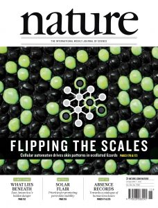
In the context of a multidisciplinary study funded by an ERC advanced grant, the Milinkovitch lab offers one position for an outstanding, highly motivated, and creative experimental wet-lab biologist or biophysicist (at the Post-doc level or, possibly, PhD student level) with strong skills in Cell and Developmental Biology as well as experience in Biophysics. The position is for 3 to 5 years and must start between September and December 2019. The successful candidate will use molecular/cell/developmental biology and biophysics methods to investigate (i) how cell proliferation and tissue differentiation are coupled to mechanical tension during scale development in reptiles and birds, and (ii) how geometry of the skin affects reaction diffusion during the development of skin colour patterns in reptiles. These analyses will be performed on new model species (lizards, snakes, crocodiles, birds) already established in the Milinkovitch lab.
Excellent written and verbal communication skills in
English are mandatory. Other specific requirements are strong expertise in
Cell and Developmental Biology: CRISPR-Cas9 technology, ex-vivo tissue cultures, confocal and light-sheet microscopy, immuno-histochemistry, in-situ hybridisation, transcriptomics, in-vivo assays;
Biophysics: micro-indentation, physical experiments with PDMS/hydrogels, laser ablations, etc.
Candidates must have a PhD in Biology or biochemistry or Biophysics. The position is available at the level of Post-Doc / Research Associate. Exceptional master students can be considered for a PhD student position.
The University of Geneva (UNIGE) is world-renowned for its research and is among the top 1% best universities in the world. Geneva is an international city occupying a privileged geographical situation.
Candidates must send their application — in the form of a single PDF file including a brief letter of interest, a CV, as well as contact information (not support letters) of three persons of reference — to: lane-jobs@unige.ch
Deadline for application: July 15, 2019.
References
: Elastic instability during branchial ectoderm development causes folding of the Chlamydosaurus erectile frill. eLIFE (in press). Locally-curved geometry generates bending cracks in the African elephant skin.Nature Communications 9 (2018); A Living
Mesoscopic Cellular Automaton Made of Skin Scales. Nature 544: 173-179 (2017); The Anatomical Placode in Reptile Scale Morphogenesis Indicates Shared Ancestry Among Skin Appendages in Amniotes. Science Advances 2, 6: e1600708 (2016); Photonic Crystals Cause Active Colour Change in Chameleons.Nature Communications 6: 6368 (2015); The genome sequence of the corn snake (Pantherophis guttatus).Int. J. Dev. Biol. 58 : 881 – 888 (2014); Crocodile Head Scales Are Not Developmental Units But Emerge from Physical Cracking. Science 339, 78-81 (2013). Crocodylians Evolved Scattered Multi-Sensory Micro-Organs. EvoDevo 2013, 4:19.
 What did Watson and Crick discover? Rosalind Franklin’s notes…
 (No Ratings Yet)
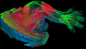
 (3 votes)