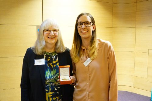
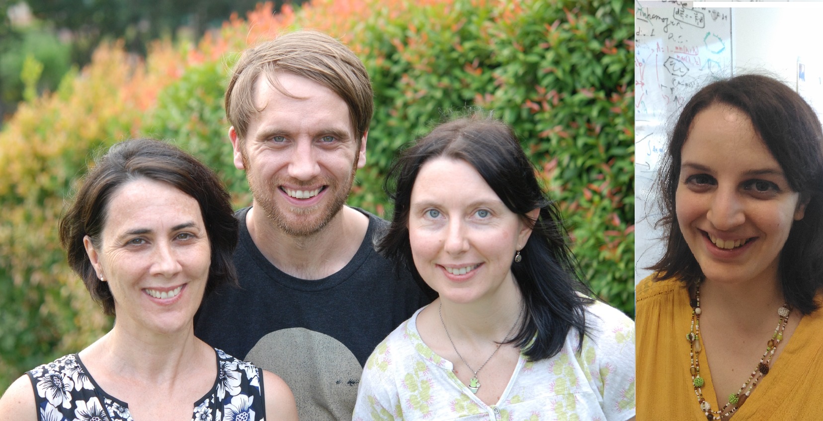
The people behind the papers – Heidi Connahs, Sham Tlili, Timothy Saunders and Antónia Monteiro
Posted by the Node Interviews, on 28 May 2019
This interview, the 63rd in our series, was recently published in Development
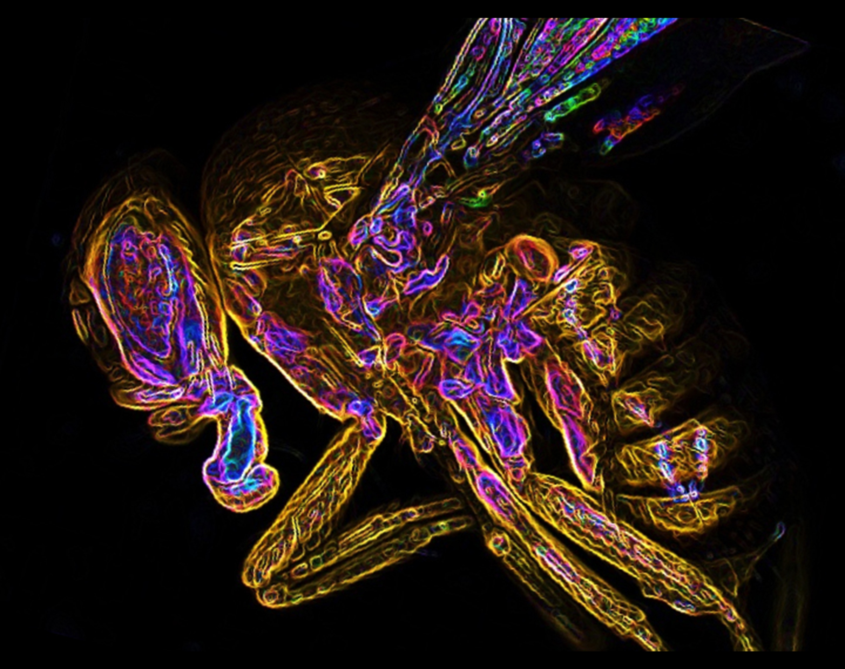
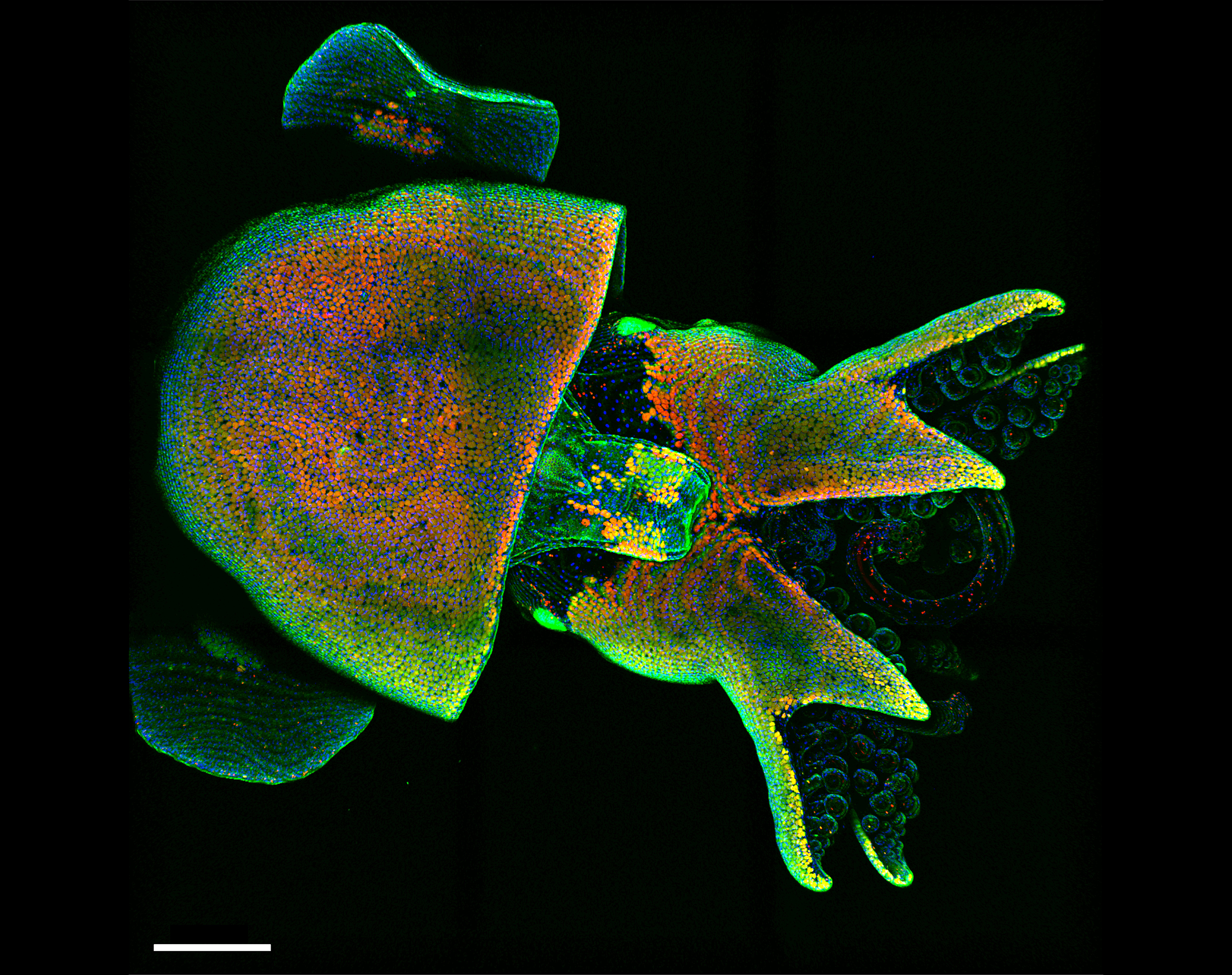
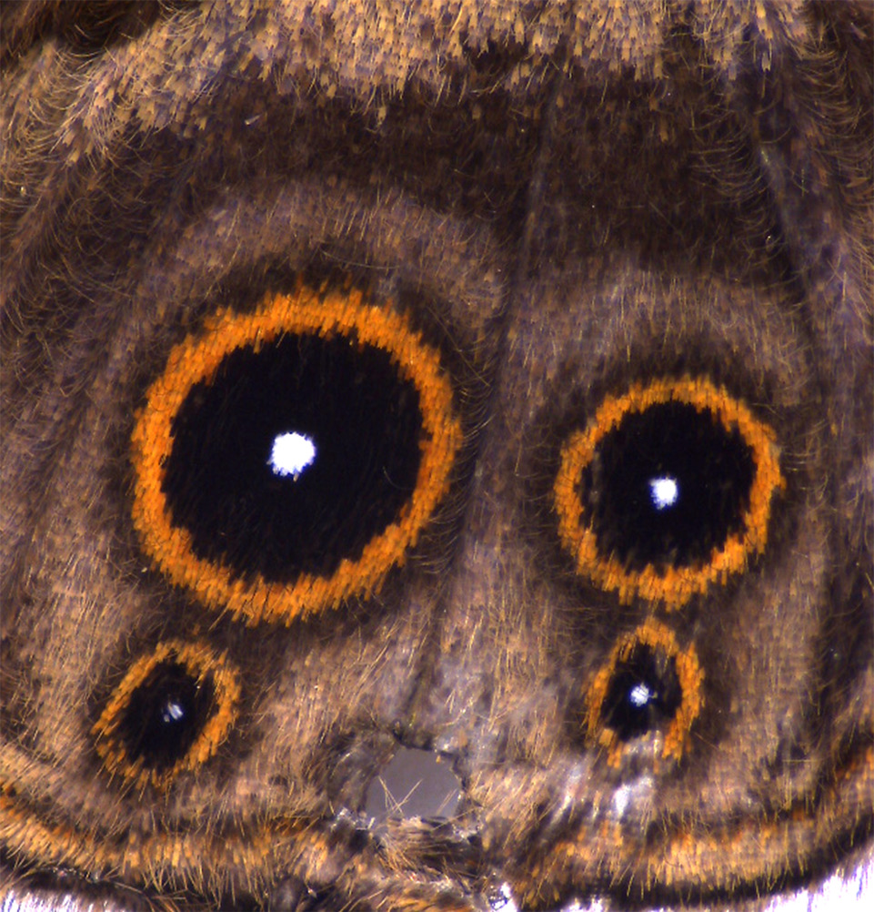
Butterfly eyespots are striking examples of animal patterning, but their developmental origins are still relatively poorly understood. A new paper in Development– the result of a collaboration between two Singapore-based labs – now combines CRISPR-Cas9 gene targeting with theoretical modelling to address the role of the Distal-less transcription factor in eyespot patterning. We caught up with co-first authors Heidi Connahs and Sham Tlili, and their respective supervisors Timothy Saunders (Assistant Professor at the Mechanobiology Institute, National University of Singapore) and Antónia Monteiro (Associate Professor at the Department of Biological Sciences, National University of Singapore and Yale-NUS College) to find out more about the story.
Tim and Antónia, can you give us your scientific biography and the questions your labs are trying to answer?
AM I was trained in population genetics and developmental biology, and have been trying to figure out how eyespot patterns develop on the wings of butterflies for most of my career, including investigating their origin, their evolution in number and also their ability to change in size with changes in environmental cues such as temperature. We have also been addressing how males and females use these eyespots in sexual signalling and in evading predators.
TS I was trained in theoretical physics but moved to developmental biology in 2007. Initially, I was interested in developing mathematical models, particularly of morphogen gradients, to understand how developing organisms reliably form, despite the presence of temperature variations and other natural fluctuations. I realised that access to experimental data was critical, and so I learnt Drosophila genetics and imaging during a post-doc at the European Molecular Biology Laboratory (Heidelberg, Germany). I have had my own lab since 2013 at the Mechanobiology Institute where we primarily focus on how complex organ shape emerges during development. We use both Drosophila and zebrafish embryogenesis to perform quantitative live imaging of organ formation. The lab also incorporates mathematical modelling and image analysis, to build a deeper understanding of how tissues form complex morphological shapes.
And Heidi and Sham: how did you each come to join your respective labs, and what drives your research?
HC During my PhD, I became fascinated with butterfly wing patterns and their eyespots in particular, so I was already reading a lot of Antonia’s work. And about 18 months before the end of my PhD, I attended a careers seminar where the speaker said that if there was someone we really wanted to post-doc with, we should just go ahead and contact them. So, I emailed Antonia right away and sent her my CV. I got lucky, because a semester before I graduated she contacted me and said she had funding for a postdoc and asked if I was still interested, and of course I said yes! Mostly what drives my research is a desire to understand how butterfly wing patterns develop, and specifically to identify the developmental processes that generate eyespot diversity.
ST I was trained as a physicist and enjoy doing both experimental work and modelling. I am especially interested in biological problems where spatial components play a role, and this necessitates the generation of quantitative maps of quantities such as cellular movements. I completed my PhD in Paris working on the mechanical properties of cell aggregates and cell monolayers. During the last year of my PhD, I had started to think about doing a post-doctoral work on more in vivosystems while keeping a quantitative biophysics approach. I discovered Tim through his website: he had just started his group at the Mechanobiology Institute. I was motivated by the highly interdisciplinary component of his group and by the fact that he was trained as a theoretical physicist developing experimental aspects in the lab. I was finally convinced to join his lab after meeting all the members of the lab during my interview.
How did your two labs come to collaborate on this project?
AM I think we started this collaboration via an undergraduate student, Trisha Loo, who joined Tim’s lab with an interest in modelling and was also doing some wet lab work in my lab. But the collaborative work really got going when we started getting very interesting Distal-less (Dll) crispants, such as butterflies with split eyespot centres, that really begged for modelling of potential morphogenetic processes that were differentiating those centres.
TS Antonia and I first met at a department faculty retreat. On the bus journey to the venue, we discussed patterning and complexity. We realised there was potential synergy between our labs work, and so we jointly took on Trisha to explore mathematical modelling of eyespot centre specification. The project then grew from there, with Heidi and Sham joining, who significantly pushed forward the science. Pleasingly, Tricia is now a PhD student in my lab, doing theory and image analysis – a large change from her undergraduate studies in biology!
Can you give us the key results of the paper in a paragraph?
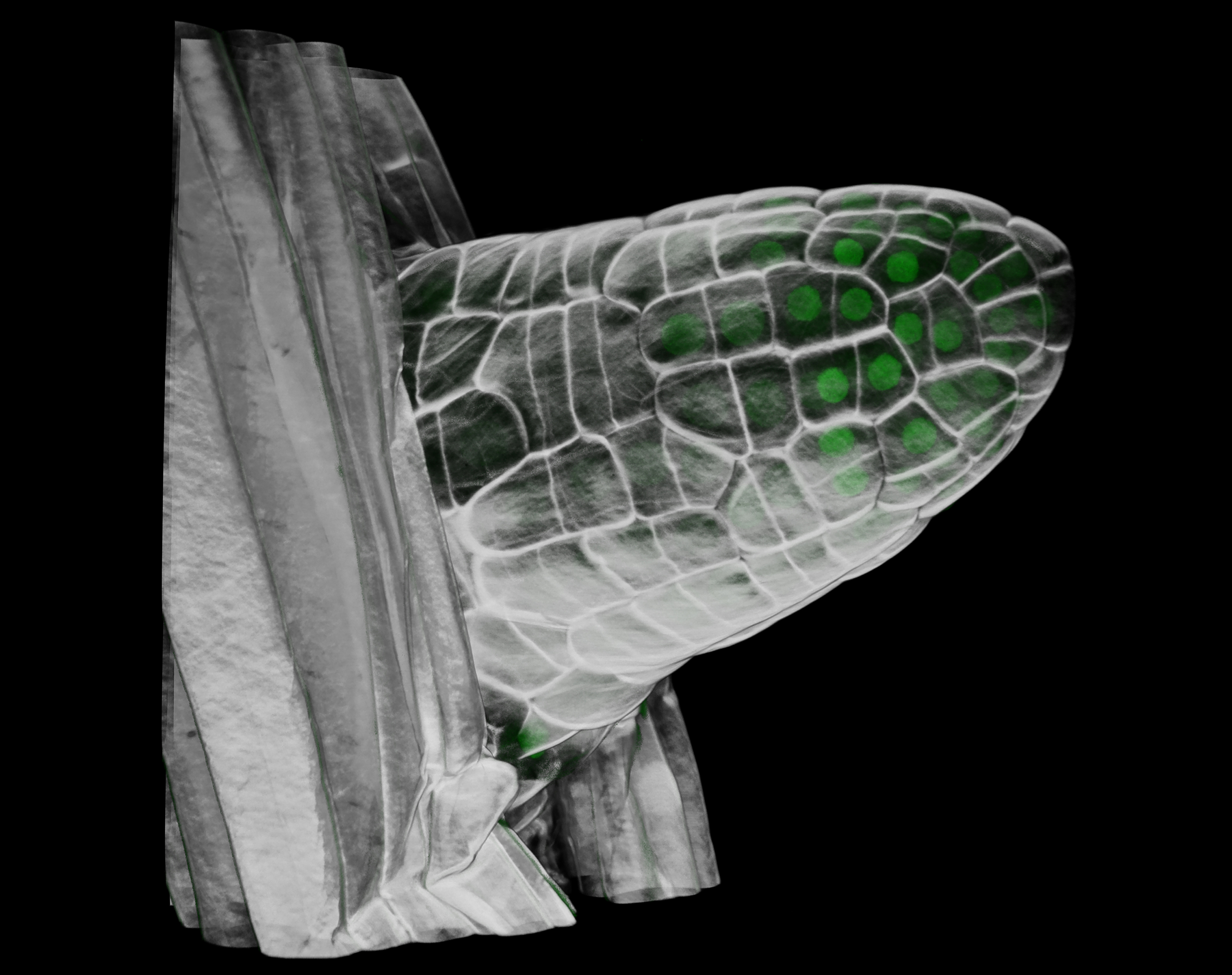
AM The paper shows that mutations in different exons of the gene Dll affect the morphogenetic process that differentiates the cells at the centre of an eyespot pattern. Close examination of these crispants suggests that Dll is involved in a reaction-diffusion process where continuous variation in Dll levels can lead to eyespots appearing on a wing sector, to eyespots splitting into two, to finally acquiring a tear-drop shape.
TS For me, a key result of the paper is the use of modelling to describe complex phenotypes. The crispants created a diverse range of phenotypes and our model was able to explain all these observations with minimal changes.
ST I would add that modelling the spatial component of the problem was critical in this case to make sense of the complex eyespot shapes obtained.
HC Our work shows that, in Bicyclus anynana, Dll is required for eyespot formation and it also appears to have other roles such as in regulating melanin pigmentation and scale development. The experimental and modelling work suggests that different eyespot phenotypes can be explained by variation in levels of Dll expression. Our findings lead us to conclude that, as Dll expression levels decrease, eyespots become smaller or disappear altogether. However, when Dll levels increase, this leads to the duplication of eyespot centres resulting in extra eyespots developing on the wing.
Your experiments demonstrate exon-skipping/gain-of-function phenotypes from certain CRISPR-induced mutations in Dll. Do you think this is a widespread issue in the field?
AM This exact same Dll exon-skipping phenomenon has recently been shown in sepsid flies, leading to the occurrence of ectopic sternite brushes (Rajaratnam et al., 2018), and it has also been documented in other studies using cell lines (Kapahnke et al., 2016; Lalonde et al., 2017; Mou et al., 2017). What is still unclear to us is the extent that natural variation in exon skipping takes place in natural populations to alter gene function from a reduced or loss-of-function to a gain-of-function outcome.
HC There does seem to be a growing awareness now of the potential for CRISPR to induce exon skipping, and also the importance of sequencing not only the genomic DNA but the mRNA from crispants.
Why might Dll lacking exon 2 induce such weird and wonderful phenotypes?
AM: We still don’t know. The Dll truncated protein, lacking exon 2 but still containing a functioning homeobox, might be more stable and resistant to degradation, mimicking a gain-of-function phenotype.
HC Or perhaps the truncated 5′UTR increases the translation efficiency of the protein. More research is needed to understand the properties of this truncated version of Dll, and this will probably require using Drosophila transgenics to express the truncated protein.
Your theoretical model can replicate eyespot formation and Dll mutant phenotypes: do you think it might be extended to a general mechanism for how organisms make spots?
HC, ST, TS & AM Perhaps, but morphogenetic processes in the insect epidermis are largely considered a bit different from those taking place in the skin of mammals such as leopards or fish. In vertebrates, cells containing pigment molecules actually move about to take their place in a field of cells, whereas in insects, the cells are primarily differentiated in situ via interpretations of local morphogen gradients, etc. We are more excited with the idea that the models developed in this paper could help us understand limb specification in the thorax of insects via similar mechanisms.
When doing the research, did you have any particular result or eureka moment that has stuck with you?
HC When I found my first crispant which had the ectopic eyespots, that was a big eureka moment for me. Also, when I got back some sequencing results and realized that exon 2 had been spliced out, that was very exciting. I had to look at the results multiple times before I could believe it.
ST We first investigated how the classical Gierer-Meinhardt activator-inhibitor model could explain the Dllcrispants. Although this model was giving interesting results, we were struggling to find a unique set of parameters that could explain the ensemble of the crispants phenotypes. Then, Antonia motivated us to look for a model where the morphogens are anti-colocalised instead of colocalised – to be in closer agreement with the experimental data. Shortly after implementing the Gray-Scott model, I found that this model easily generated phenotypes strikingly similar to the crispants phenotypes. This was a very satisfying moment.
And what about the flipside: any moments of frustration or despair?
HC Yes, I definitely had a lot of those moments too. Initially when I started doing the CRISPR experiments, we had a lot of trouble getting it to work and so for several months there was this heart-pounding moment each time I went to check if there were any butterflies with interesting wing phenotypes and it was very disheartening to see normal looking butterflies. Eventually we realized it was the cas9 protein, and once we ordered a new one, the experiments finally worked: that was a huge relief!
ST Maybe after sending the first draft versions, when some readers were not reading the modelling part of the story and thinking the crispants phenotypes were just not making any sense. I think that, in this story, the modelling and experimental aspects are tightly entangled. These criticisms made me realise how important it is to make the model as pedagogical as possible in a highly interdisciplinary context.
I had to look at the results multiple times before I could believe it
So what next for you two after this paper?
HC I am now focusing on targeting the enhancers of Dllusing CRISPR so we can try to understand the origins of butterfly eyespots by identifying pleiotropic enhancers. This has been quite a difficult project to work on as using CRISPR to target enhancers comes with its own unique set of challenges, but we are now starting to get some preliminary results that look quite promising!
ST I just moved back to France after three great years in Tim’s group. I started a new post-doc on embryonic stem cell aggregates mimicking early mammalian development. This project will combine tissue mechanics and potentially morphogen reaction-diffusion again!
Where will this work take the Saunders and Monteiro lab? Any plans for further collaborations?
AM Would love to collaborate further with Tim’s lab. The insights they provided into our crazy looking crispant mutants were amazing. We are now mutating other genes that are also expressed in eyespots and we are observing similar eyespot splits, etc. The reaction-diffusion process seems to involve multiple genes and we might need to model the action of these genes as well.
TS This work has nicely gone alongside our lab’s study on complex shape emergence. Both patterning and mechanics play important roles in organ formation. In the future, we are hoping to integrate reaction-diffusion modelling with mechanobiology. Of course, I’d be delighted to work further with Antonia – the butterfly is an awesome system.
Finally, let’s move outside the lab – what do you like to do in your spare time in Singapore?
AM I love to hike through Singapore’s forested parks on the weekends. My husband and I usually do a 10 km trek that ends in VivoCity – a mall with many lunch options!
TS I climb with my wife regularly and much of my weekend is spent exploring Singapore with our 6-year-old daughter.
HC Usually on the weekends I enjoy relaxing with my boyfriend and visiting different science/art/nature-themed attractions or exhibitions in Singapore. I also enjoy watercolour painting and keeping aquarium fish.
ST In Singapore, I really enjoyed walking in the parks along the harbour which gave a really nice view of the sea, Indonesian islands in the horizon and all these coloured container ships coming from all over the world.
 (No Ratings Yet)

 (2 votes)