An NIH-funded postdoctoral position is available in Jianbo Wang’s lab at the Univ. of Alabama at Birmingham. We study how shape forms during embryogenesis, and how aberrant shape formation leads to congenital birth defects. The process of shape formation is known as morphogenesis, and is regulated in part by the planar cell polarity (PCP) pathway. PCP is a unique signaling mechanism that coordinates cellular polarity and regulates dynamic, directional cell behavior. Our studies involve multi-disciplinary approaches including genetic, imaging, cell biology and biochemistry; and use multiple model organisms including the mouse, chick and Xenopus. Our goals are to 1) uncover novel mechanisms and logic of PCP signaling; 2) elucidate the role of PCP in cardiovascular, skeletal and neural development; and 3) investigate how human PCP gene variants contribute to congenital disorders.
We are looking for highly motivated applicants with training in Developmental Biology, Genetics, Cell Biology or a related discipline. To apply, please email your CV, a description of previous research and contact information for three references to j18wang@uab.edu.
This blog post is a reflection on the Company of Biologists-organized workshop on Chromatin-Based Regulation of Development that I recently attended at Wiston House, located in the countryside of Sussex in the UK.
Wiston House, Steyning, UK
To my mind, this workshop was the ideal of what a meeting should be. As a freshly minted assistant professor at the University of California, San Francisco, this workshop came at an excellent time for me to learn, glean inspiration, and extract advice.
A good conference is much more than the sum of its parts – the “parts” being the topics represented by the scientist attendees. By reading publications, you learn what experiments worked and hung together in a narrative. By hearing that work presented in person, you can infer the hunches or obsessions that drove the scientist to ask the question in the first place, as well as see the pieces of data that are forming the nucleus for the next phase of work.
A good conference provides an opportunity to rapidly survey ongoing work in your field. Through discussions and debates, you can learn whether your field has reached a consensus that a prevailing model consistently explains observations, or whether alternative explanations are being fomented. In this frame of mind, a new question that no one else seems to be asking might occur to you. Equally helpfully, you may discern whether a particular area is overpopulated and incorporate that knowledge to adjust your own project strategy. By assimilating all of this, you expand your knowledge base and learn by observing how other scientists think.
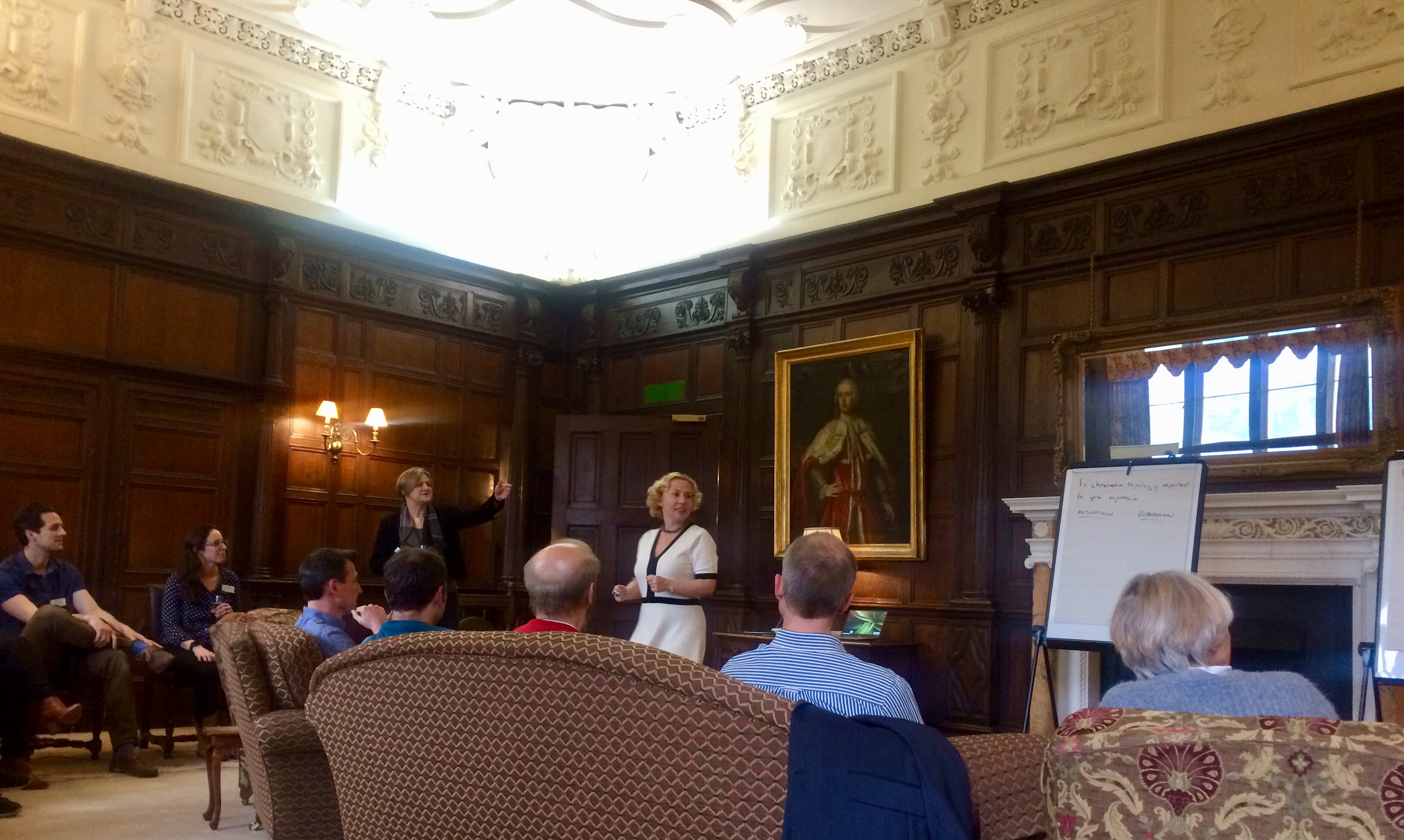
Mid-scientific debate in the library at Wiston House
At a good conference, you will forge connections with peers that may turn into friendships and collaborations in the future. You will interact with leaders in your field, gaining invaluable opportunity to pick their brains for career advice and to watch a well-honed scientific intuition at work. In doing all of this, you will have been doing the dreaded “networking”, too.
Taking a break for a hike “up the downs” (photo by @sudpinglay)
When it’s all over, you will return to your home lab armed with new ideas to test, with a new solution to a hurdle you have faced, or with new critiques from your peers to answer.
All of these features of the ideal conference were on display at this Company of Biologists workshop. The small size, intimate setting, and considerate pace of the meeting allowed for time to ruminate over exciting presentations and talk through ideas with other attendees. Early career researchers and established faculty were equal participants both in numbers and in the share of the conference program allotted to them. This was truly the most thoughtfully organized workshop I have ever attended.
Researchers from the Tokyo University of Science, Japan, have used CRISPR gene editing technology to make snails with shells that coil the ‘wrong’ way, providing insights into the fundamental basis of left-right asymmetry in animals. These findings were recently published in Development.
If you look at a snail’s shell, the chances are it will coil to the right. But, occasionally, you might find an unlucky one that twists in the opposite direction – as fans of Jeremy the lefty snail (https://en.wikipedia.org/wiki/Jeremy_(snail)) will remember, these snails struggle to mate with the more common rightward-coiling individuals.
This chirality (direction of coiling) of snail shells is an outward manifestation of left-right asymmetry: a phenomenon seen across animal evolution and extending to humans – your heart is (probably) on your left side, while your liver is to the right. But how does this asymmetry come about? Researchers from Japan, writing in the journal Development, think they now have a definitive answer – for one species of freshwater snail (Lymnaea stagnalis) at least.
Successfully applying CRISPR gene editing technology to molluscs for the first time, Masanori Abe and Reiko Kuroda (working at Tokyo University of Science, but recently relocated to Chubu University, Japan), have now made snails with mutations in a gene called Lsdia1, which had previously been suggested – but not conclusively proven – to be involved in snail shell coiling; snails without a functional copy of Lsdia1 produce offspring with shells that coil to the left, showing that this single gene is responsible for rightward- coiling. Surprisingly, the researchers could see signs of asymmetry at the earliest possible stage of development – when the snail embryo was just a single cell. Moreover, the mutant snails could be reared to adults, when they produced exclusively leftward-coiling offspring. According to Kuroda: “It is remarkable that these snails with reversed coiling are healthy and fertile, and that this coiling can be inherited generation after generation (we now have 5th-generation leftward-coiling snails). Further, these results may have an implication for snail evolution and speciation – given that left- and rightward-coiling snails probably wouldn’t interbreed.”
Knocking out one gene in the snail Lymnaea stagnalis reverses shell coiling. In contrast to the wild-type dextral snail (right), a CRISPR-created snail shows sinistral coiling (left). Credit: Dr Hiromi Takahashi of the Kuroda laboratory.
It’s still not clear how Lsdia1 might control left-right asymmetry: the gene encodes a formin, a protein that is involved in regulating the cell’s internal skeleton, but more work is needed to understand how this influences the cellular behaviours that control handedness – which is something Kuroda and her colleagues are actively working on. But given that genes like Lsdia1 are found throughout the animal kingdom, similar mechanisms for controlling left-right asymmetry could be at play in other species – including our own. As Kuroda says: “Although diverse mechanisms have been proposed for different animals, we think a unified mechanism, involving formins and cellular chirality, is probable”. So while it may seem a big leap from snail shell coiling to human left-right asymmetries, it’s possible that future studies on how Lsdia1 works in snails might eventually help us understand why some babies are born with their heart on the right (which is of course the wrong) side of their chest.
Our call for images to fill our 2019-20 calendar was met with an amazing response – 62 entries showcasing the diverse beauty of developmental biology. Now it’s time for you vote for the 12 that will make it into print.
Because we want a range of organisms and styles in the calendar, and because picking 12 favourites from 62 is not the easiest task, we’ve decided to split the voting up into categories. Turns out 12 into 62 doesn’t go so well, and some categories were better represented than others, so we ended up with the following:
Mammals (vote for 2 out of 10)
Zebrafish (vote for 2 out of 8)
Vertebrate variety show (vote for 2 out 10)
Drosophila (vote for 2 out of 11)
Invertebrate variety show (vote for 2 out 11)
Plants, Fungi and Choanoflagellates (vote for 1 out of 7)
Art and Illustration (vote for 1 out of 5)
The cut offs are a little arbitrary but it’s the best scheme we could come up with. Inevitably, many beautiful images are going to miss out, but we hope the selection stands alone in showcasing the aesthetic side of research.
The pictures are arranged in galleries – click to expand the image and see the caption (there’s also a link to see the image full size). Below the galleries are independent polls to pick your favourites from each section. Both the galleries and the polls are arranged alphabetically by creator, and the poll text is the same as the file name (e.g. ‘Blin. mESC micropatterns’) which you can see below the caption. Please just vote once (well, twelve times!) – polls are set up to stop repeat voters by cookie.
Voting closes on Sunday 19 May 23:59 GMT
You can also let us know what your overall favourite is in the comments. Happy voting.
Mammals (Mus and Homo)
Epiblast Stem Cells cultured on micropatterns of various geometries. The cells were stained for LaminB1 (Green), Tbra (Blue) and GM130 (Red). By Guillaume Blin (MRC Centre for Regenerative Medicine, The University of Edinburgh)
Mouse embryos at E12.0 nuclei stained in 1) control and 2) compound mutants for AER-FGFs with a graded loss of fore and hindlimbs. By Christian Bonatto (National Cancer Institute, NIH)
Super-resolution image acquired via structured illumination microscopy of a single developing human neuron ectopically expressing the autism risk gene neuroligin-4X with enlarged growth cones. F-actin is in green, HA-tagged neuroligin-4X is in magenta, doublecortin is in cyan confirming its neuronal identity, and the nuclear marker DAPI is in grey. By Nicholas Gatford (Institute of Psychiatry, Psychology and Neuroscience, King’s College London)
The image shows a confocal section of the organ of Corti (sensory epithelium of the cochlea) in the inner ear of a neonatal (P1) mouse. Actin is stained using phalloidin (cyan), nerve fibres using anti-β3-tubulin (yellow), and nuclei using DAPI (red). This is a developmental stage before the onset of air-borne hearing (which occurs around P11 in mice), and during a period of path-finding by afferent nerve fibres, and refinement and pruning of their terminals. Many fibres temporarily contact non-sensory supporting cells, before they establish permanent synaptic connections with the mechano-sensory hair cells. By Daniel Jagger (UCL Ear Institute)
Transverse section of a chimaeric E6.5 mouse embryo generated by aggregation of Id1-Venus reporter ES cells with a wild-type morula, stained for Id1-Venus (green), Nanog (red) and T (blue). By Mattias Malaguti (MRC Centre for Regenerative Medicine The University of Edinburgh)
8-cell mouse embryo which has just aquired apical-basal polarity but we can still appreciate individual cells. E-Cadherin is labelled in red and pERM is labelled in white. By Sergio Menchero (CNIC Madrid)
E14.5 mouse embryo labeled for cartilage (Sox9-GFP, in biop-SpringGreen) and vasculature (highlighter ink circulated by injection in a blood vessel, in mpl-magma). Vasculature “lights up” the embryo, including within the developing bones of the limbs. Image taken using a microscope kindly sponsored by Zeiss during the 2018 Embryology Course at the Marine Biological Laboratory in Woods Hole, MA. By Paul Gerald Layague Sanchez (EMBL Heidelberg)
Magnified view of the aortic and mitral valves in a developing mouse heart. By Matt Stroud (BHF Centre of Excellence, King’s College London)
Cross section through a 36 days human cerebral organoid stained with Ascl1 (red) and Arl13b (green) to reveal a subset of cortical progenitors and the primary cilium projecting into the lumen of the neuroepithelial rosette, respectively. Nuclei are counterstained with DAPI. By Thomas Theil (Centre for Discovery Brain Sciences, The University of Edinburgh)
Gastrulating mouse embryo. Nuclear envelope labelled with LaminB1 (grey). Primitive-streak and ingressing mesoderm labelled with T-Brachyury (magenta). By Darren Wisniewski (MRC Centre for Regenerative Medicine The University of Edinburgh)
Please pick your favourite 2 images
(please remember to pick 2)
Zebrafish
Regenerating zebrafish heart after cryoinjury. By Srinivas Allanki (Max Planck Institute for Heart and Lung Research)
Larval zebrafish at 6 days post fertilisation registered to a regional neuroanatomical zebrafish atlas. By Dominic Burrows (MRC Centre for Neurodevelopmental Disorders, King’s College London)
Developing gill vasculature in a 120h old zebrafish and also features the heart. This image was taken using lightsheet microscopy in two transgenic lines, one that marks the endothelial actin and the other marks the endothelial nuclei. After acquisition it was processed as a colour coded depth projection. By Philippa Carr (Bateson Centre, University of Sheffield )
Vasculature in a double-transgenic line (Tg(kdrl:HRAS-mCherry)s916, Tg(fli1a:CAAX-eGFP)), visualized using light-sheet fluorescence microscopy.The three individual images are part of a timelapse acquisition, showing 50 hours post fertilization (hpf), 60hpf and 70hpf. Essential processes such as anastomosis (eye and fin), joining of vascular beds (head and spinal cord), and remodelling (subintenstinal vein and duct of Cuvier) can be observed.By Elisabeth Kugler (Department of Infection, Immunity & Cardiovascular Disease,University of Sheffield)
48 hpf zebrafish embryo expressing GFP in sensory neurons and expressing mCherry where the MAP Kinase Kinase Kinase LZK (map3k13) is expressed. LZK is a vertebrate homologue of dlk-1, which is essential for neuronal regeneration in C. elegans and Drosophila. By Hannah Markovic (UCLA)
The image shows the Lateral Line primordium of a zebrafish embryo labeled with a green fluorescent membrane protein. This group of cells migrates together from the head of the animal to the tip of the tail in a journey in which it will form proto-neuromasts or rosettes, which will be later deposited as clusters of cells called ‘neuromasts’. By Joaquin Navajas Acedo (Stowers Institute for Medical Research)
Spinning disc confocal image of mitochondria in the developing zebrafish fin at ~96hpf. By Damian Dalle Nogare ( Eunice Kennedy Shriver National Institute of Child Health and Human Development, NIH)
Transgenic zebrafish (Danio rerio) larva expressing red fluorescent protein in the developing mouth and olfactory epithelium. A subset of cells also express a construct that labels actin filament with green fluorescent protein. DAPI (blue) is used to label DNA in the nuclei of all cells. By Oscar Ruiz (Department of Genetics MD Anderson Cancer Center)
Please pick your favourite 2 images
(please remember to pick 2)
Vertebrate variety show
Chicken embryo at stage HH9. Picture was taken with an iPhone through the ocular lens of a dissecting stereomicroscope, after injecting the space between the yolk and the embryo with blue pen ink. The ink enables us to see the transparent embryo and highlights the membranes surrounding it as well as some yolk granules. The picture was taken by me at the Embryology Course 2018, Marine Biological Laboratory, Woods Hole – MA.By Andrea Attardi (Max Planck Institute for Molecular Cell Biology and Genetics, Dresden)
13-day chicken embryo. Diaphanization protocol with Alcian blue and alizarin red. By Felipe Zanghelini Benevenutti (Federal University of Santa Catarina, Brazil)
Scanning electron micrograph of hatchling catshark (Scyliorhinus canicula) dermal denticle (scale). By Rory Cooper (Animal and Plant Sciences University of Sheffield)
Our lab works to understand the regenerative ability of the axolotl salamander, and a key step in understanding how a limb can regenerate is to understand how the limb was initially developed. This image depicts a developing larval axolotl limb bud which has been stained for 3 key developmental genes. The genes shown are: FGF8 (Cyan), SHH (yellow), and PRRX1 (magenta). The method used to stain the genes in this image was fluorescent in situ hybridization, a method for directly labeling the mRNA within a cell. The image was generated using a confocal microscope. By Alexander Lovely (Department of Biology, Northeastern University, Boston)
13-day chicken embryo with diaphanization protocol with Alcian blue and alizarin red. By Daniely Ramos Luz (Federal University of Santa Catarina, Brazil)
My entry for the photo contest is an unhatched sea turtle. I took a picture of this little guy when I was working with sea turtle monitors at the Yawkey Wildlife Center in South Carolina. I helped to monitor the nests and would often find unhatched turtles which failed to resorb their yolk and emerge from the nest. By Catherine May (Boston College)
Early developed chicken embryo (HH9) electroporated in the left side. The electroporated side is detected by a GFP reporter and displayed as green dots on the beautiful chicken embryo. By David Morales Vicente (University of São Paulo, Brazil)
This is Greg. Greg is a 1 month old Austrofundulus limnaeus annual killifish who is the nicest juvenile in my undergraduate research experiment. He likes brine shrimp, blood worms, and would like to have his picture featured on a magazine if possible. Greg is a part of my experiment where I am trying to see if this particular fish species sex ratio is affected by fluctuating temperature patterns. Greg wants you to give him a chance. Trust in Greg. By Motutama Sipelii (Portland State University)
Alligator mississipiensis embryo at stage 13-14 immunostained against Myosin heavy chain showing the developing muscles and (red) and neurofilament labeling axons of nerves. By Daniel Smith Paredes (Department of Geology and Geophysics, Yale University)
Stage 35 chicken embryo, cleared and immunostained for DAPI (orange) and Pax3 (cyan) demonstrating the developing neural crest and spinal cord. Image was taken on the Nikon AZ-C2 macro-confocal with image analysis performed in Imaris. Image was taken in collaboration with Andrea Attardi at the Max Planck Institute of Molecular Cell Biology and Genetics during the Woods Hole 2018 Embryology course. By Laurel Yohe (Department of Geology and Geophysics, Yale University)
Please pick your favourite 2 images
(please remember to pick 2)
Drosophila
Micrograph shows Drosophila larval brain attached to the leg and eye imaginal discs. There is a glial migration from brain to these discs as indicated by glial cells (shown in red) in the imaginal discs, along with corresponding repo-gal4 driving GFP (glial marker). Glial cells are stained by anti-repo (red), Neural stem cells are marked by anti-Dpn (blue) and glial membranes are marked by repo-gal4::GFP (green). By Asif Ahmad Bakshi (Centre for DNA Fingerprinting and Diagnostics, Hyderabad, India)
The development of the Drosophila lamina neuropil occurs under tight spatiotemporal control that involves a signal relay between photoreceptors (gray), glia (cyan) and lamina precursor cells (magenta). Visualised in my image is a bundle of (joy) photoreceptor axons innervating and signalling to neuroepithelia located at the surface of the optic lobe. With this signal, the neuroepithelia acquire lamina precursor cell identity and form beautifully organised columns. The wrapping glia, in the optic stalk, also receives signalling molecules from the innervating photoreceptors and helps to promote differentiation in lamina precursor cells. Zeiss LSM confocal microscope. By Matthew Bostock (Department of Cell & Developmental Biology, UCL)
Drosophila whole ovary stained for f-actin (Red), nuclei (Cyan) and actin (Green). By Yujun Chen (Kansas State University Division of Biology)
Reproductive organ of a young (1-3 days old) adult Drosophila male. The male reproductive tract consists of a pair of testes, where mature sperm is produced. The sperm is temporarily stored in the seminal vesicles (sv) before being released into the ejaculatory ducts (ED). Here, the sperm is mixed with the seminal fluid produced in the accessory glands (AGs). Ejaculatory bulb (EB) is also visible. The sample expresses nuclear RFP (cyan) driven by the Gal4 driver traffic jam (tj) and it was stained for Actin (magenta) and the germline stem cell marker vasa (green). Image was acquired at the Wolfson Bioimaging Facility (University of Bristol). Scale Bar: 200 um. By Giuliana Clemente (University of Bristol)
Drosophila notum as the sensory organ precursors undergo their first division, seemingly shooting across the field. Partner of Numb ( PoN) is shown in green and microtubules (as reported by Jupiter) in Magenta. The picture is color blind friendly! By Louise Couton (Department of Biochemistry, University of Geneva)
A neuroepithelium called the ‘outer proliferation centre’ (OPC) together with the eye disc epithelium (top right) are shown in Cyan. The OPC is a broad crescent shaped structure that generates neurons of the lamina and medulla neuropils. All glia are shown in magenta. They are present in the eye disc, through the optic stalk and in the optic lobe. By The Fernandes Lab (Department of Cell & Developmental Biology, UCL)
The image is a lightsheet micrograph of a Drosophila undergoing the final stages of development(~24hrs prior to ecclosion). The anterior half of the developing fly has been dissected out from the pupal case. The posterior pupal case can still be seen in the upper part of the image. The fluorescent marker is DE-cadherin- lining apical junctions of cells. There is a surreal resemblance to a developing human foetus during its late stages- when all the major morphological development is complete. Although, the individual seems suspended in time and space, there are major internal structures still developing. By Suhrid Ghosh (Max Planck Institute of Molecular Cell Biology and Genetics, Dresden)
Drosophila melanogaster ovary 24 hours after pupal formation stained for Lamin-C and Fasciclin III (in red), VASA (green), Traffic Jam (white), and DAPI (blue). By Lena Kogan (Biological Sciences Department, Columbia University NY)
Drosophila melanogaster pupa expressing a histone marker. It was imaged on our MultiView Light-Sheet Microscope (MuVi-SPIM) at 10x magnification. By Dimitri Kromm (EMBL Heidelberg)
Combined single molecule Fluorescent in situ hybridisation and immunofluorescence of developing Drosophila larval brain. Shown here are Syncrip protein (green), syncrip RNA (red) and DNA (blue). By Jeff Lee (Dept. of Biochemistry, University of Oxford)
Drosophila mutant showing a decreased eye size compared to wild type. This line is not able to generate descendants with wild type flies. By Marisa Merino (Department of Biochemistry, University of Geneva)
Please pick your favourite 2 images
(please remember to pick 2)
Invertebrate variety show
Live embryo of the beetle Tribolium castaneum. During early development, this egg was injected with mRNA encoding a photoconvertible fluorescent protein that is localised to nuclei. Several nuclei were then irradiated with short-wavelength light to induce a conformational change in the fluorescent protein, thereby causing it to fluoresce in a different wavelength (shown here in magenta rather than the unconverted cyan). This strategy allows the targeted labeling of one or more cells and the tracing of those cells and their descendents throughout development. In this case, I labeled a group of cells came to form part of an extraembryonic tissue that covers the embryo and supports its development. By Matt Benton (Department of Zoology, University of Cambridge)
Developing oocytes (red) in the marine cnidarian Hydractinia symbiolongicarpus enclosed in sporosacs – cell boundaries are in green and nuclear staining in blue. By Eleni Chrysostomou (Centre for Chromosome Biology & Regenerative Medicine Institute, National University of Ireland Galway
Embryonic development of the ascidian Phallusia mammillata. 3D rendering of membrane imaging with light-sheet microscopy covering the zygote, cleavage, gastrulation, neurulation, tailbud and larval stages. By Ulla-Maj Fiuza (EMBL Heidelberg)
Small worm of Platynereis dumerilii (annelid) with dividing cells, in the brain and the posterior part, labelled in green (EdU incorporation and chase). By Eve Gazave (Institut Jacques Monod, Paris)
My picture is of an interesting and uncommon animal: a larva of the colonial tunicate Symplegma rubra, after metamorphosis. The five projections are the ampullae, the first structures of the blood vessels. In the center, there is the oozooid, during organogensis. Some larval organs are seen, such as the ocellus (red circular structure). I observed this larva in the marine station Cebimar (Centro de Biologia Marinha da Universidade de São Paulo). By Stefania Gutiérrez (University of São Paulo, Brazil)
Tardigrade embryo (Hypsibius exemplaris) with membranes and mitochondria labeled. By Kira Heikes (UNC Chapel Hill.)
Live Hawaiian Bobtail Squid (Euprymna scolopes), stained with vital dyes (CellMask, LysoTracker and Hoechst) to understand its cellular and sub-cellular organisation during development. Blue is labelling cellular nuclei, green – cell plasma membranes and red – lysosomes that are important for cellular waste removal. This species is a candidate model organism that yet holds many answers to developmental biology questions, such as nervous system and eye development. The image was taken during the MBL 2018 Embryology Course with the confocal microscope provided by Zeiss. Animals were supplied by the cephalopod researcher Carrie Albertin. By Martyna Lukoseviciute (Weatherall Institute of Molecular Medicine, University of Oxford)
Squid embryo (Doryteuthis pealeii) with nuclei (pink), actin (cyan) and neurons (green). By Tessa Montague (Zuckerman Institute, Columbia University NY)
DIC and fluorescence image of Hydractinia male sexual (left) and feeding polyps (right) on a chitin bed. Chitin is shown in green. Noncycling cells probed with cyclin-dependent kinase inhibitor (CDKI) are shown in yellow, which are mainly in nematocytes, male gonophore, and gastrodermis. By Indu Patwal (Centre for Chromosome Biology, National University of Ireland Galway)
Ctenophore gastrula. Cyan: DAPI; Yellow: tubulin. By Miguel Salinas-Saavedra (Centre for Chromosome Biology, National University of Ireland Galway)
Surface view of an embryo of the brachiopod Novocrania anomala during early gastrulation. Image shows the cell membrane outlines in the ectodermal surface as stained by BODIPY-FL (F-actin staining). Animal pole is top and vegetal pole is bottom where the blastoporal opening is visible. Adults were collected in 60 meter depth waters near Storingavika in Bergen, Norway, spawned and fertilized in the laboratory. Image is a maximum intensity projection of the original stack taken on a Leica SP5 confocal microscope. By Bruno Vellutini (Max Planck Institute of Molecular Cell Biology and Genetics, dresden)
Please pick your favourite 2 images
(please remember to pick 2)
Plants, Fungi and Choanoflagellates
Transverse section of an Arabidopsis hypocotyl with disrupted vascular organisation. By Peter Etchells (Durham University)
3D TEM reconstruction of the colonial choanoflagellate Salpingoeca rosetta. Choanoflagellates are the closest unicellular relatives to the animal kingdom and some species such as S. rosetta are capable of developing into multicellular colonies. This makes S. rosetta a powerful model to investigate the development, origin and evolution of animal multicellularity. Shown are apical vesicles (pink), food vacuoles (green), endocytotic vacuoles (fuschia), ER (yellow), extracellular vesicles (grey), filopodia (external, purple), flagellar basal body (light blue), flagellum (dark green), glycogen storage (white), Golgi apparatus and vesicles (purple), intercellular bridges (external, yellow; septa, red), large vesicles (brown), microvillar collar (light orange), mitochondria (red), nonflagellar basal body (dark orange), and nuclei (dark blue). By Davis Laundon (The Marine Biological Association, Plymouth UK)
An Arabidopsis inflorescence expressing a fluorescent reporter for the APETALA3 gene, which promotes petal and stamen identity (green). Cell walls were stained with rpopidium iodide (magenta). By Nathanaël Prunet (Department of Molecular, Cell and Developmental Biology, UCLA)
Development of the three-cell asexual spore of the rice blast fungus. The spore starts as a swelling of the aerial hypha, changing from a sphere, to a symmetrical oval two cell stage and finally transforming into the spindle shaped spore seen at the end of the montage. By Hiral Shah (Bharat Chattoo Genome Research Centre The Maharaja Sayajirao University of Baroda Gujarat India)
Root of Medicago truncatula treated with auxin transport inhibitors, known to induce structures similar to symbiotic nodules. By Ioannis Tamvakis (Sainsbury Laboratory University of Cambridge)
The development of a lateral root in Arabidopsis thaliana. The sample has been cleared and stained with Calcofluor White to outline the cell walls and the green fluorescent nuclei represent a protein expressed specifically in the outer cell layer of developing lateral root. By Robertas Ursache (University of Lausanne, Switzerland)
Cross section through a developing fruit (carpel) of Austrobaileya scandens. The photograph was taken with a light microscope under 10X magnification and stained with safranin and alcian blue to have a better tissue contrast. The fruit of A. scadens is formed by multiple individual carpels, here we are looking at one of those that is going to grow fleshy in order to disperse the seed. By Cecilia Zumajo-Cardona (City University of New York, New York Botanical Garden)
Please pick your favouriteimage
Art and illustration
A single amino acid, a dot ( . ), becomes a chain of amino acids – a protein, a line (——), moves in space to become a plane, then a volume. Like in drawing, a dot progresses to a line, to meander in space. So it seems that drawing might be an appropriate way to experiment, to ‘be like’ the protein. Proteins pervade life and collectively they take an infinite variety of forms. Protein’s fold, most of the time this is what a protein is trying to do. The folded protein is the relaxed protein, in its ‘natural’ state, not wasting any energy. Folding is a process, it happens through time, in an ‘energy landscape’ often imagined to be conical (cone shaped) with the folded protein resting in the very bottom – the point of least energy in the landscape. As the protein moves from the top of this landscape to the bottom (it might not make it) it embarks on an explorative journey of the space; body and environment continuously co-creating each other. There are fast and slow tracks, uphills and downhills, dead-ends and if in trouble then a ‘chaperone’ will come to help find the way together. This dynamic process is hard to imagine and the images you find in scientific textbooks don’t exactly give the game away (generally a cone, a few uphills or downhills). How else can we imagine this complexity? What other images could we see? After making countless drawings with molecular biologist JJ Phillips, we take a day to look at them all together: drawings of cones, of simple figures moving in cones, colour coding energy levels, line drawings of movement series, planes in space. It struck me that the bottom of the cone is like the centre of a maze. We like the idea. We try drawing a maze. First we need an underlying grid architecture, not a generic pattern but one that is true to the dynamic pattern of the protein journeying. We change the granularity of the grid for different stages of the process. Colour brings dimensions and guides the pathways that are created. The environment and the body co-create one another as the maze undermines the distinction between figuration and abstraction. The drawing process has its own stochasticity and the image, following its own creative process, reveals something true of the dynamic pattern of protein folding. The idiosyncrasies of this living process have informed and given rise to a new artistic process. ‘Organic development in a work of art is at least analogous to, and probably identical with, organic development in nature; in an organic-artistic scheme the essence of art is in processes rather than its products; and such artistic ‘events’ as are thrown up are significant merely in that they reflect past, present and future aspects of the dominant process’. (Thistlewood, 1981). By Gemma Anderson (University of Exeter and Falmouth School of Art, http://www.gemma-anderson.co.uk/)
These are three hand-crafted coasters, made from perler beads. They represent pictures of three classic developmental biology model organisms, the fruitfly Drosophila melanogaster, a hatching chick Gallus gallus domesticus, and the flower of Arabidopsis thaliana. Pertinent to their natural habitat, the fruitfully is shown in a blue background representing its flight in the sky, the chick is shown in a green background representing the grass it forages and the flower is shown in a brown background representing the soil it grows in. These were created by me for use by any artsy developmental biologist who can use this during coffee breaks in lab. By Sumbul Jawed Khan (Sci-Illustrate and https://www.linkedin.com/in/sumbul-jawed-khan/)
This drawing illustrates the remarkable self-organization capacity of cerebral organoids that allows them to recapitulate human brain development in vitro. Each color represents a different type of cell, and the dorsal and ventral areas are separated by a defined boundary – like a yin and yang symbolizing the balance between distinct but complementary entities. By Beata Edyta Mierzwa (Ludwig Institute for Cancer Research and the University of California, San Diego, and www.beatascienceart.com)
The image depicts different views (cranial, ventral, dorsal, right, caudal and left) of a digital 3D model of the embryonic human heart at Carnegie stage 12. The model is based on work from Antoon Moorman’s group at the Academic Medical Centre in Amsterdam. My model is an example of scientific illustration, and is not volume-reconstructed from histological sections, confocal images or micro-CT data. By Kalin Narov (Embryo Safari, https://www.embryosafari.com/)
This drawing combines embryos and structures from embryos. Featuring: Bats, Drosophila, Xenopus, Parhyale, Ascidians, Chicken. By B. Duygu Özpolat (Eugene Bell Center for Regenerative Biology and Tissue Engineering, Marine Biological Laboratory Woods Hole)
Please pick your favouriteimage
Thanks for voting – if you made an error, just email aidan.maartens@biologists.com and we’ll correct the numbers!
The non-profit publishing groups can make a real difference
The very best part of being a scientist is cracking mysteries of the universe and what is in it. The second best thing about this profession, in my opinion, is being part of a scientific community. Conversations, conferences, workshops, collaborations, networking, support and constant inspiration from one another serve as a magic glue that keeps us together and motivates us to keep pushing the boundaries of the unknown. A perfect example of fostering a scientific community is the astounding job done by The Company of Biologists. As you may know, The Company of Biologists is a not-for-profit publishing organisation dedicated to supporting and inspiring the biological community. After attending the truly inspiring and excellent workshop on ‘Chromatin-Based Regulation of Development’ this April, I now genuinely understand the differences between profit-oriented and community-oriented publishing groups. The Company of Biologists is on a profound mission to give back to the community, for example, by organising workshops that aim to bring together scientists who would normally rarely meet, across the globe from the same or different fields. This holds real power and serves an important purpose for both scientists individually and science on the whole. I would like to thank The Company of Biologists for creating such great initiatives and also share my experiences of this fantastic workshop from an early career scientist’s view (I’m currently a final year PhD student in Tatjana Sauka-Spengler lab in the University of Oxford).
The highlights of the ‘Chromatin-Based Regulation of Development’ workshop
The workshop was organised by Benoit Bruneau (Gladstone Institutes, UCSF, USA) and Joanna Wysocka (Stanford University, USA) for 30 participants (17 invited speakers and 13 selected early-career researchers like me). The entire workshop was fully funded by the Company of Biologists: even the selected early-career researchers did not have to pay for attendance, which – let’s be honest – is somewhat unheard of! It was situated in a beautiful English manor house, Wiston House, in the South Downs National Park on the south coast of England. The venue, including my room, was so gorgeous that for four days I got to feel a little bit as if I belonged to the Royal family.
The purpose of the workshop was to bring together scientists from different places, expertise and career levels, yet it felt as if it was a non-hierarchical environment, where all scientists had an opportunity to contribute to all aspects of this great workshop equally. For instance, each one of us, no matter who, a PhD student or a professor, had an opportunity to officially present their research and answer questions for 30 minutes followed by an informal discussion and further questions during coffee breaks or at the cosy Wiston house bar. Also, dinners were organised superbly, where we were randomly allocated a different seat every night, getting a chance to speak with different scientists. This way, I got to sit and connect with scientists, such as Joanna Wysocka or Stefan Mundlos, that I would have been most likely too shy to sit next to in a ‘real-life’ setting. Overall, it was very nice to not only discuss scientific questions but also to get advice for future career steps and to talk about life-work-balance, women in science problems and other relatable questions.
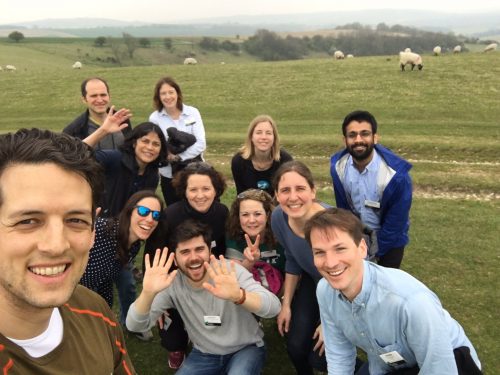
Hiking break around the Wiston house grounds with the participants before the evening session.
The workshop topics
The workshop consisted of nine sessions. The first two sessions focussed on 3D genome regulation during various developmental processes – from embryo patterning to limb development to synthetic Hox cluster building. During the workshop, we extensively scrutinised whether topologically associating domains (TADs) are actually central for gene regulation or rather if they only play genome-packaging/replication roles. Surprisingly, a lot of presented data challenged the importance of TADs for gene regulation (as nicely and critically reviewed in another participant’s – Roel Neijts’ – Node post (https://thenode.biologists.com/meeting-report-chromatin-based-regulation-of-development-an-excellent-workshop-by-the-company-of-biologists/events/)).
Other sessions delved into enhancers, their sequence variations and gene regulation during early development. We got to learn how histone variants at enhancer sites can play a predominant part in tissue-specific gene regulation and when misregulated can lead to severe cranial facial syndromes. Or how some enhancers are critical for sex determination and how knocking them out can lead to sex reversal despite the inherited X or Y chromosome combinations. Overall, the emerging trend indicated that active enhancers usually stay active despite TAD perturbation and that their close proximity to promoters is, most of the time, enough to drive correct gene expression; by contrast, inactive enhancers – even when they are ectopically brought to promoters – almost never get ‘turned-on’. Therefore, maybe chromatin marks rather than chromatin confirmation are more instructive when it comes to gene regulation?
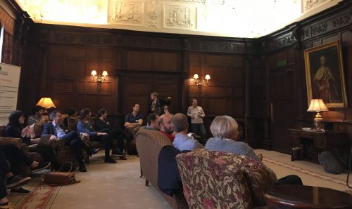
The organiser Joanna Wysocka is opening our debate evening on questions: ‘Is chromatin topology important for gene expression?’ and ‘Does phase-separation enable gene regulation within a crowded nucleus?’
We also looked into how transcription factors (TFs) regulate chromatin dynamics and nucleosome phasing. For instance, how do TFs work together with morphogens or in a combinatorial TF manner to create certain types of chromatin that correspond to tissue-specific gene expression? Or, how are they capable of forming liquid-like aggregates allowing a local increase in the concentration of scarcely available TFs in both plants and animals? Undeniably, the topic of phase-separation was extensively deliberated during both the talks and our evening debate. It seems that for us, biologists, this subject is still somewhat of a phenomenon when it comes to gene regulation. However, a steadily increasing number of experiments show that phase-separation might play very important roles, as illustrated by heterochromatin formation driven by HP1a phase-separation or polycomb body aggregation, which is important for gene repression and even formation of a repressive 3D chromatin.
Food for thought
Lastly, this brilliant workshop taught me a lot not only about the different shapes and shades of chromatin but also about its formation, dynamics and roles in developmental gene regulation. It was rather eye-opening to see how important it can be to learn from experts in a completely transparent and relaxed environment, where no one was afraid of sharing unpublished data, hypotheses and ideas. This was truly freeing and inspiring, and I could start shaping my own ideas and endorse a different way of thinking about my own research and future directions. Different emerging methods of studying chromatin were brought into the light of this workshop, e.g., not only looking at chromatin marks and accessibility using molecular biology methods , but also trying to visualize chromatin by employing different new imaging tools (after all – seeing is believing!) or turning to biophysical principles of embryo development and underlying chromatin-mediated gene expression dynamics. Taking even one step further, towards Feynman’s school of thought (“What I cannot create, I do not understand”), we got to hear about the importance of synthetic systems to generate in vitro chromatin, engineer chromatin contacts using optogenetics tools or produce synthetic embryo-like structures (e.g. gastruloids) in order to genuinely understand the basic principles and rules of many yet unanswered questions of chromatin-based regulation of development. As you might have already thought, usually there is no perfect single methodology to solve complex biological puzzles, and only collaborations between different fields can facilitate the quality and speed of putting all the pieces of such puzzles together. Importantly, workshops like this one that I had a privilege to attend bring experts from different fields together, which definitely accelerates the progress of science and inspires a lot of young scientists to tackle even the most complex problems of our universe in new ways.
By Ghislain Gillard, Maria J. Gomez Lamarca, Robert Krautz, Rosa Park, David Salvador-Garcia, Yara Sanchez-Corrales and Jelle van den Ameele
On the 28th of January, the Cambridge Fly Club held its very first Symposium in the beautiful environment of Wolfson College, Cambridge, UK. This meeting, titled “Past, Present and Future of Drosophila research” was supported by a small meeting grant from the Company of Biologists and brought together 130 scientists, mostly from Cambridge, UK, who work with the fruit fly Drosophila melanogaster.
The Cambridge Fly Club is a group of PhD students and postdocs who organise a couple of scientific meetings with pizza and drinks every other month to maintain a positive dynamic within the friendly and collaborative fly community in Cambridge. However, this year, we had a special anniversary to celebrate, because 25 years ago, Andrea Brand and Norbert Perrimon published their landmark paper in Development describing the Drosophila GAL4 system (Brand and Perrimon, 1993). The GAL4 system enables targeted gene expression in virtually any cell type and has facilitated many breakthrough discoveries in physiology and developmental biology.
A modern woman’s guide to fly design
We were very lucky that Andrea Brand (WT/CRUK Gurdon Institute) agreed to give a plenary lecture about how she came to develop the GAL4 system with Norbert Perrimon. For many of the attendees, this talk was the highlight of the symposium, and for some of them even “the best talk they had ever seen”. Andrea worked on yeast transcription as a PhD student and was then a postdoctoral Fellow in Mark Ptashne’s lab at Harvard University before joining the Perrimon lab for a second postdoc. The Ptashne lab were studying the yeast transcriptional activator GAL4 and found, surprisingly at the time, that GAL4 could activate transcription in organisms other than yeast. Inspired by this knowledge and by a seminar given by Walter Gehring on his lab’s unpublished work on lacZ enhancer trapping, Andrea realized that substituting GAL4 for lacZ would enable cell fates to be manipulated in vivo, rather than merely labelled. To put theory into practice, Andrea moved to the Perrimon lab in early 1988, bringing her knowledge about yeast genetics and molecular biology together with Norbert’s expertise in Drosophila genetics. The ease of hopping the GAL4 within the Drosophila genome, through P-element transposition allowed to capture the expression pattern of new uncharacterized enhancers and resulted in large numbers of GAL4 lines being generated quickly. Andrea got her first result with the GAL4 system in September 1989 – she even showed us the page from her lab notebook!
Development of the GAL4 system was the result of hard work, dedication and resilience and even a bit of luck: Andrea was introduced to Norbert by a chance meeting with a yeast colleague, Fred Winston. Andrea gave a great and positive message for all young scientists in the audience and inspired us with some of her creative poster designs – “a modern woman’s guide to fly design” did stick in our minds! Her message was to follow your dreams in spite of the inevitable hurdles that come your way.
Collaborative workshops
The plenary lecture was then followed by several high-quality workshops, delivered by leaders in their respective fields and mostly focused on transformative technologies from the past, present and future.
The different aspects of CRISPR/Cas9 in flies were nicely introduced in a very popular workshop by Simon Bullock from the MRC Laboratory of Molecular Biology. He talked about how to use CRISPR-Cas9 to generate mutant alleles, tag proteins and even perfom tissue-specific gene disruption. His talk was full of practical tips and advice, including potential pitfalls and how to recover the desired mutation. In a workshop on the art and history of genetic screens, Daniel St Johnston from the WT/CRUK Gurdon Institute presented an impressive overview of the various strategies to perform forward genetic screening, illustrating every approach with a historical landmark paper. Which mutagen to go for – EMS according to Daniel, or X-rays (without the protective plastic cover) according to Adelaide Carpenter – remains an unsolved question though.
Three other workshops were set up as collaborations between scientists from different institutes across Cambridge. Ben Sutcliffe (MRC Laboratory of Molecular Biology), Edward Allgeyer and George Sirinakis (WT/CRUK Gurdon Institute) gave an overview of imaging Drosophila tissues using three different advanced light microscopy techniques: single molecule localization microscopy, STED, and light-sheet microscopy. Considering the notorious difficulty of applying super-resolution methods to thick biological samples, Ed and George generated a lot of interest into how they custom-built 4Pi-SMS and STED microscopes for imaging different Drosophila tissues such as the ovary. Equally exciting was Ben’s imaging setup for capturing live tissue-dynamics of Drosophila embryos and larval salivary glands on a custom-built light-sheet microscope.
Alex Whitworth (MRC Mitochondrial Biology Unit), Cahir O’Kane (Department of Genetics) and Hansong Ma (WT/CRUK Gurdon Institute) convinced us that there is now a critical mass in Cambridge to study mitochondria in fruit flies. They introduced us to impressive and creative usage of powerful fly genetics to study mitochondrial inheritance and explained some exciting new discoveries with direct implications for neurodegenerative diseases. Finally, Greg Jefferis (MRC Laboratory of Molecular Biology), Matthias Landgraf and Marta Costa (Department of Zoology) teamed up to show that today’s high-level connectivity between Drosophila researchers had striking parallels in the Drosophila brain, whose neuronal networks they study with state-of-the-art techniques and powerful computational algorithms.
All the data, interpretations and scientific excitement that come from these technologies have to be shared of course. One platform that is widely used by the fly community is FlyBase, and Steven Marygold provided a very practical overview of how to perform large-scale queries in FlyBase. However, we all still like to publish our findings as a story, in the form of a journal article. Katherine Brown from Development, therefore, provided us with some very useful tips to make this actually happen – even the fly paper she published herself in Development 16 years ago turned out to offer room for improvement! She also generated new data for this symposium, summarising the success of Drosophila papers as opposed to other model organisms. These are more thoroughly discussed in her own recent blogpost.
A bright future for Drosophila research?!
To end this wonderful afternoon, Michael Akam, Andrea Brand, Cahir O’Kane, Katja Röper, Benedicte Sanson and Daniel St Johnston wholeheartedly joined in the game of discussing the future of Drosophila research. Young researchers clearly heard that it is not the model that makes great research, but the questions you ask. The powerful genetics, the possibility to study cell biology within a living organism, the emerging tools to study behaviour, or the relatively cheap maintenance of flies are only several of the many reasons why Drosophila will remain an important research model in the future. One reason to work with Drosophila was not discussed by the panel though: it makes you part of a wonderful, generous and vibrant community. Today’s symposium was a warm celebration of this great community.
This meeting has been organised thanks to the kind support of the Company of Biologists with several other sponsors which can be found together with the program of the event here.
Back in January, The Cambridge Fly Club held a symposium to mark 25 years since the publication of the famous Gal4/UAS paper (Brand & Perrimon, 1993 – published in Development); the organisers have posted a meeting report here. As part of this symposium, the organisers asked me to give a talk on ‘Publishing Fly Research’. Particularly given that I worked with Drosophila in Cambridge as a research assistant and PhD student and attended the odd Cambridge Fly Club talk back in the early 2000s, I was more than happy to oblige, though I wasn’t initially sure what I was going to talk about – surely publishing fly research is just like publishing any research?!
But I decided that I wouldn’t just trot out my ‘standard’ publishing talk, and that instead I’d try and take a look at the publishing landscape for the fly community, and at how it’s changed since the publication of that landmark paper. Some of the data I gathered surprised me, and given that the audience seemed to find my insights useful, I thought I’d (somewhat belatedly) share them here. I also hope that this post will be useful beyond just the fly community, though I’ve not crunched the numbers for other systems.
So, what I looked at was the number of fly papers published over recent decades, and where they were published, and compared this to the number of mouse papers (representing the most popular vertebrate model system) and total papers. I also looked at citation rates, as well as at submission rates to Development. An important caveat: my analyses are very crude, and are based on keyword searches in PubMed, Web of Science and Development’s own submission system. For certain there are both false positives and false negatives in the numbers, but hopefully the trends still hold.
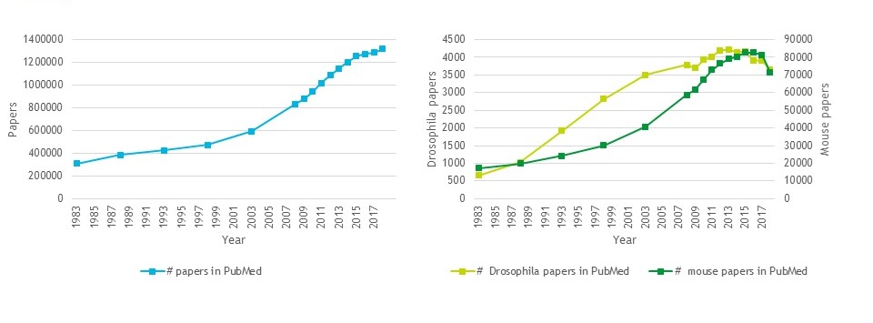
Looking over the past 35 years, it’s clear that publishing output has increased dramatically, and fly research has more than kept pace with this, ramping up massively in the 90s before plateauing in recent years.
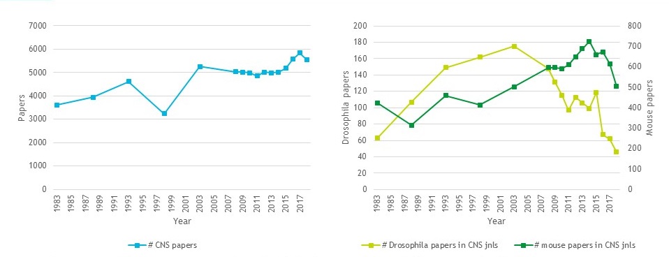
But what about the influence of the fly field? Using papers published in Cell, Science and Nature as a (very imperfect) proxy for how ‘hot’ a field is, you can see that we hit ‘peak fly’ in the early 2000s, and the numbers of fly papers published in those journals has declined since then.
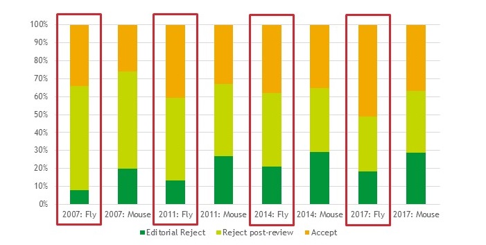
Incidentally, we see a not dissimilar trend in Development. So where are fly papers being published? Back in 1993, the top venues for Drosophila research were Development, PNAS and Genetics. In 2018, it was eLife, Scientific Reports and PLoS Genetics; the most popular journals in the early 90s still make the top 10, but have been pushed down the pecking order primarily by large, broad scope OA journals (PLoS One and Nature Communications completed the top 5 in 2018).
It’s also the case that fly papers aren’t as well cited as mouse papers. I didn’t have access to historic citation data, but for publications in 2015, the average fly paper in the CNS journals cited half as well as the average mouse paper. The differences aren’t this dramatic for Development papers, but they are still there. And I think this is largely to be expected – the mouse community is larger than the fly community, so it’s natural that those papers will get more citations. Plus, I suspect a fly paper is much more likely to cite mouse work than the other way around – given the current ecosystem that values (perhaps overly values) mammalian and translational relevance.
So is it getting harder to publish fly papers? Looking at submission and acceptance rates in Development over the past 10 years, it seems that what has declined is submission rate – fly papers submitted to us are just as likely to be published as they were a decade ago, and they’ve got an above-average acceptance rate.
We just don’t see as many papers submitted to us as we used to (2017 submission rate was 60% that of a decade earlier). Whether this reflects changing journal preferences of authors, or a decline in the volume of Drosophila developmental biology papers, I can’t say. But I think it’s fair to argue that fly work doesn’t have an overly hard time with us.
I was also asked to comment in my talk on how fly researchers can maximise their chances of getting their work published and seen by a broad audience. It’s important to state from the outset that – at Development at least – fly papers are treated just the same as any paper: what we’re looking for are studies that advance our understanding of developmental processes, or report techniques or resources of broad interest to our community. We don’t need translational relevance and we don’t need you to replicate or confirm your findings in another system. But we are looking for papers that we think will be of interest beyond a small group of researchers in a particular area, and it’s helpful if you as authors can make it clear where that interest lies. This is important not only to get your paper past the editor and referees, but also to make sure it gets read once it’s published. Remember that the vast majority of people who come across your paper will only read the title. A small proportion might click through to see the abstract, and even fewer will go on to read the full paper. So, while I’m never going to advocate overselling a paper, it will help to make clear the broader context up-front, and make the title and abstract appealing to non-fly people. Avoid fly-specific jargon and gene names (refer to E-cadherin, not shotgun!), and help the non-aficionados to realise the potential link to their own work.
Concrete examples are often more helpful than generic advice. So, and to avoid embarrassing anyone but myself, I went back and looked at my own very first paper – which I’m proud to say was published in Development. But looking at it now, the title and abstract fail to follow most of the advice given above. Just before my talk, I had a quick go at pulling the abstract apart and rewriting with my editor hat on. My new version is far from perfect, but hopefully it serves to exemplify the points I’ve made – scroll through the images below to see the first version, its flaws revealed, and a few little rewrites that at least start to make it more accessible. The title isn’t great either in terms of accessibility, though I remember Matthew and I agonised over the wording. If I had the chance again, I’d go with a simpler version – something like “Egfr signalling regulates ommatidial orientation in the Drosophila eye”. I fear the word ‘ommatidial’ would put most readers off immediately, but it’s hard to avoid – suggestions in the comments please!
The Company of Biologists (Development’s not-for-profit publisher) is currently seeking proposals for Workshops to be held during 2021.
The Workshops provide leading experts and early-career researchers from a diverse range of scientific backgrounds with a stimulating environment for the cross-fertilisation of interdisciplinary ideas. The programmes are carefully developed and are intended to champion the novel techniques and innovations that will underpin important scientific advances.
Workshop proposals should take into account the following points:
Proposals should focus on cutting-edge scientific research in topic areas that are new, novel and not covered by traditional conferences.
Proposals that concentrate on emerging or cross-disciplinary themes are particularly encouraged.
Organisers should be experts in their field, with sufficient standing to attract world-class speakers and attendees.
Proposals should indicate how the Workshop will contribute to establishing new collaborations or research directions.
Each Workshop will consist of 30 delegates to include 10 early-career researcher places to be applied for and proposers should ensure diversity in the proposed attendee list. You will need to propose 20 speakers (but they don’t have to be confirmed).
As the scientific organiser your involvement will be focused on the science. We will undertake all of the logistical arrangements, liaise with the venue, organise speaker travel, help with arranging the programme and fund the meeting.
The first human genome sequence cost billions of dollars. Today, we’ve broken through the thousand dollar barrier and are heading towards $100. In this episode of Genetics Unzipped we talk to pioneering geneticist George Church about his plans for the ‘Zero Dollar Genome’ and his thoughts on the future of genome reading, writing and editing.
Plus, we find out what happened when one scientist’s interest in personal genomics got a little too close to home, when Manuel Corpas invited his whole family to share their personal genomes with the world.
If you enjoy the show, please do rate and review and spread the word. And you can always send feedback and suggestions for future episodes and guests to podcast@geneticsunzipped.com
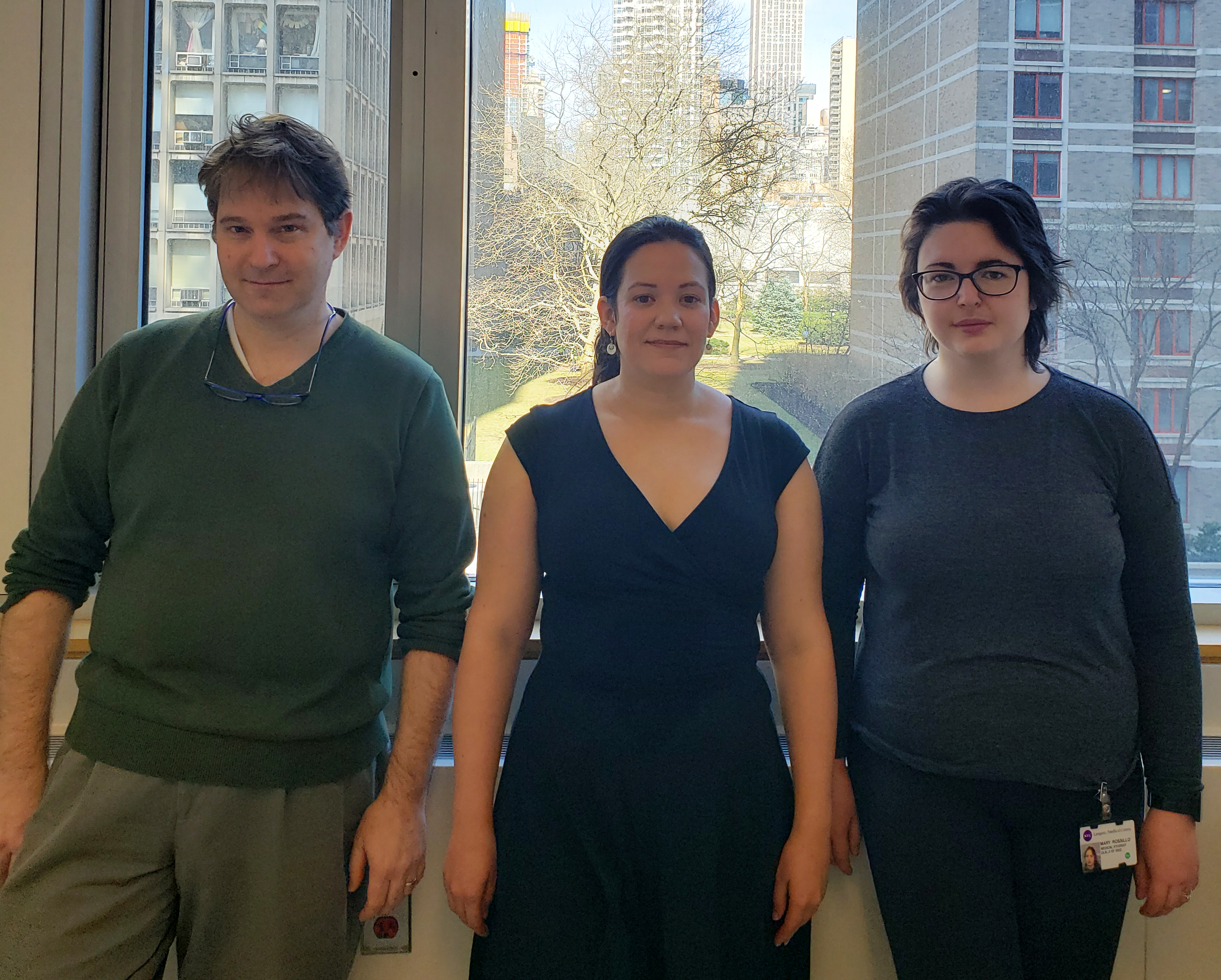
A fundamental aim in developmental biology is to understand how the various cell types of the body are specified by differential gene regulation. Caenorhabditiselegans nervous system development provides a powerful system for studying this, as exemplified by a new Development paper reporting on how the BAG neurons that help the worm sense oxygen and carbon dioxide are specified. We caught up with first authors Julia Brandt and Mary Rossillo and their supervisor Niels Ringstad (Associate Professor at the Skirball Institute of Biomolecular Medicine and Department of Cell Biology at New York University) to find out more about the story.
From left to right: Niels, Julia and Mary.
Niels, can you give us your scientific biography and the questions your lab is trying to answer?
NR I’ve had a longstanding interest in molecular neurobiology. This started during my graduate studies with Pietro De Camilli at Yale University, where I studied molecules required for the synaptic vesicle cycle. During my postdoctoral training with Bob Horvitz at the Massachusetts Institute of Technology I was introduced to behavioural genetics of C. elegans as a method to identify molecular mechanisms that are required for the function of specific neuron-types or for specific neurotransmitter signals. My own group continues to study the genetic basis of C. elegans behaviours to advance understanding of neuromodulation and sensation.
Julia and Mary: how did you each come to join the Ringstad lab, and what drives your research?
JB I’ve always been interested in how specific cells acquire the characteristics that distinguish them from other cells. The nervous system is where this question really stands out. There are hundreds of kinds of neuron, and each kind has a unique morphology and distinctive physiology.
MR I was drawn to the lab by the possibility of using C. elegans to understand how neurons sense respiratory gases. The worm gas-sensing neurons are specified by a transcription factor called ETS-5, which has a vertebrate homologue that is required to specify neurons in the mammalian brainstem that sense respiratory gases in arterial blood. I thought that this similarity offered the opportunity to leverage techniques in developmental genetics to interrogate mechanisms that might be involved in human pathology, specifically fatal respiratory disorders like Sudden Infant Death Syndrome. Because I am training to be a physician-scientist, I wanted my graduate research to impact clinical medicine.
Why are the BAG neurons an interesting developmental model, and what was known about their specification before your work?
NR We have colleagues studying chemosensation or neurodevelopment who previously identified and characterized mutants that are defective in BAG development. Elissa Hallem at the University of California, Los Angeles, had identified the ETS-5 transcription factor through an independent study. Roger Pocock at Monash University in Australia collaborated with us in our study of ETS-5, and his group independently identified two other transcription factors that are important for BAG development, EGL-13 and EGL-46. From this work, we had the sense that the genetics of BAG neuron development would be complicated and rich. Indeed, our own genetic screens yielded mutants that did not come up in Elissa’s or Roger’s studies. Our screens also yielded a mutation that causes ectopic expression of a BAG fate – a phenotype that is the mirror-image of the more common absence-of-BAG fate phenotype. This mutation was the starting point for the work that Julia and Mary did.
JB The BAG neurons exhibit some remarkable physiological characteristics, and they have an interesting and easy-to-measure function. BAGs are able to sense the respiratory gas carbon dioxide as if it were an odour. The carbon dioxide that they sense is coming from environmental microbes, which the worms eat. We think that worms prefer to eat dead microbes, or microbes that are growing slowly, and BAGs are used to sense whether microbes are metabolically active. I think that the development of these neurons is interesting because understanding how the BAGs are put together can help us understand how they do what they do.
MR I agree with Julia. The BAG neurons are an interesting developmental model because they have remarkable physiology that is imparted by a developmental programme. And that programme requires a transcription factor, which is conserved between worms and humans. I like the idea that, by studying the developmental mechanisms that make a BAG neuron a BAG neuron, we can identify molecules required for its chemosensory function.
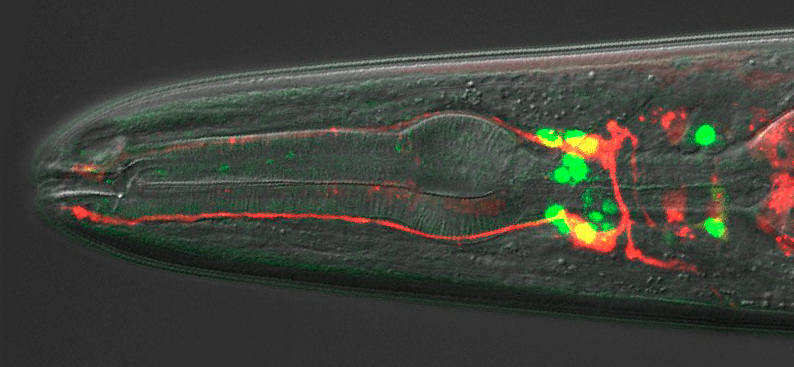
A wild-type adult hermaphrodite expressing an integrated ETS-5::GFP translational reporter and a Pflp-17::dsRed transcriptional reporter.
NR I agree with Julia and Mary. Understanding the developmental programme that generates a BAG neuron is going to teach us how these neurons execute a pretty cool function. And we now have the opportunity to study how that developmental programme is initiated and organized, which is new territory for us.
Can you give us the key results of the paper in a paragraph?
JB, MR & NR Chemosensory BAG neurons of C. elegansrequire the transcription factor ETS-5 to express a host of genes that are required for their specialized function. How expression of ETS-5 is regulated during development was unknown. Julia and Mary have found that ETS-5 expression is regulated by another transcription factor – VAB-3 – which is the nematode homologue of Pax6/Eyeless. Remarkably, the vab-3 locus encodes VAB-3 isoforms that have opposing functions with respect to regulating expression of a BAG fate. One isoform represses expression of ETS-5 in cells that are not supposed to become BAGs. Another isoform, however, is expressed in BAGs and is required for them to fully differentiate. Julia and Mary concluded that the decision to express one VAB-3 isoform or another is crucial for determining which cells become BAG neurons.
Have you got any ideas what controls which vab-3 isoform is expressed in a given cell?
JB, MR & NR That’s the key question! We had a short-lived feeling that we had learned something when we realized that ETS-5 expression was regulated by VAB-3. But now we feel like we have simply re-framed the question as one that concerns promoter-selection in the vab-3 locus. But we’re not completely in the dark, and we now have some ideas about what is going on at the vab-3 locus. Mary has been pushing hard to establish methods for purifying and sequencing chromatin associated with sparsely expressed transcription factors, and some of her studies are identifying factors that bind to vab-3 promoters. Stay tuned.
Why might the system of broad repression/selected activation of cell fates have evolved in animal development?
JB, MR & NR Our first thought is that biological systems aren’t designed or engineered, so we shouldn’t be surprised when we discover that they are rife with needless complexity and inefficiency. During evolution, a set of transcriptional repressors and activators might have been randomly permuted until they generated a pattern of ETS-5 expression that worked. We might be looking at the mess that was left behind in that workshop. But it is also possible that we are looking at a legacy of an ancestral and now-obsolete developmental programme. Cells with the characteristics of BAG cells might have been more widespread in the ur-nematode, and over time more efficient body plans that used those cells for other purposes became the norm. A final thought we have entertained is that there is some drive to carefully regulate expression of a BAG fate. A good control system will use both positive and negative regulators of its output, which allows rapid but controlled responses to inputs.
When doing the research, did you have any particular result or eureka moment that has stuck with you?
JB: Finding a mutant is always a eureka moment. I had found a large number of mutants that lacked expression of a BAG marker. It was pretty surprising to find a mutant with extra cells expressing that marker. And it was exciting to discover that the phenotype was caused by ectopic expression of ETS-5.
MR Julia had found that vab-3 mutated to cause ectopic expression of ETS-5, and she had tested a number of vab-3 alleles. We had the idea that one isoform was repressive and the other not, but we were confused by reports that vab-3 is highly expressed in BAG neurons. It was satisfying to design isoform-specific reporters and see that the long repressor isoform was not expressed by BAGs. This really crystallized our model of a promoter-selection event that patterns expression of a BAG fate.
Finding a mutant is always a eureka moment
And what about the flipside: any moments of frustration or despair?
JB & MR: Some of the mutants we used are difficult to work with because they are barely viable or the mutations cause lethality. ceh-32 was awful. It is an essential gene, but we also found that ceh-32 reporter transgenes could cause lethality. We had to figure out how much DNA to use to make the ceh-32::GFP trangenics that we needed for the project.
So, what is next for you two after this paper?
JB I’m wrapping up a post-doc and deciding on the next steps for my career.
MR I am halfway through my dissertation research. Now I’m focusing on using mRNAseq and ChIPseq to generate a genome-wide map of ETS-5 targets. I’m hoping this will lead to novel genes that function in carbon dioxide sensing.
Where will this work take the Ringstad lab?
NR We’re still trying to link the developmental programmes that specify a BAG cell to genes required for gas-sensing. Julia and Mary have put another transcription factor on the board – the isoform of VAB-3 that promotes a BAG fate. I think we want to know how that factor functions together with ETS-5 and what genes it regulates.
Finally, let’s move outside the lab – what do you like to do in your spare time in New York?
JB I enjoy life in Brooklyn, spending time with my family and reading.
MR I enjoy distance running, yoga and spending time with my cat, Ernie.
NR Family life, home repairs, and I’m trying to catch up on all the maths and physics I didn’t do at university because I was too preoccupied with biology.
 (No Ratings Yet)
 (7 votes)


")


 (20 votes)
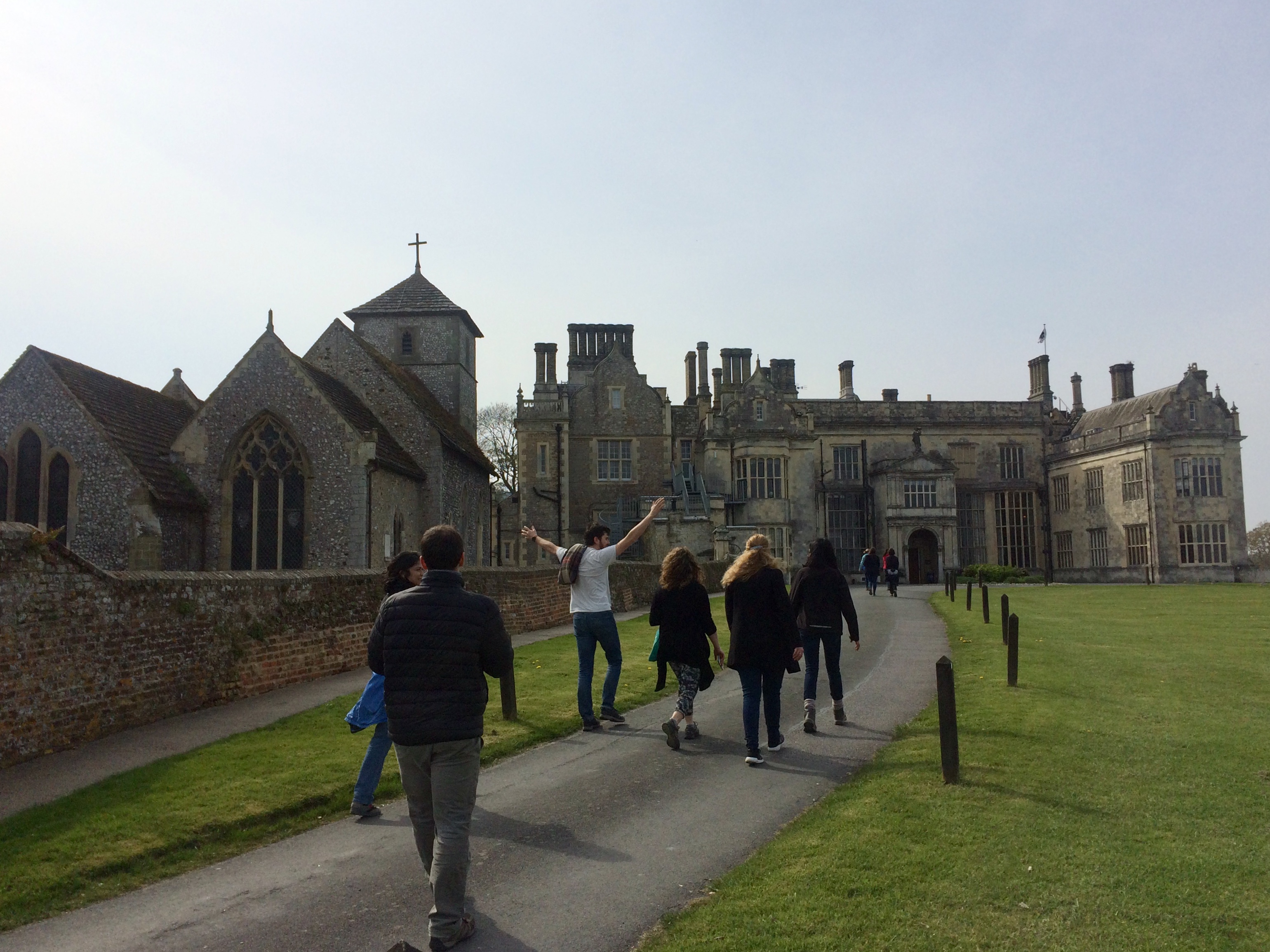


 The first human genome sequence cost billions of dollars. Today, we’ve broken through the thousand dollar barrier and are heading towards $100.