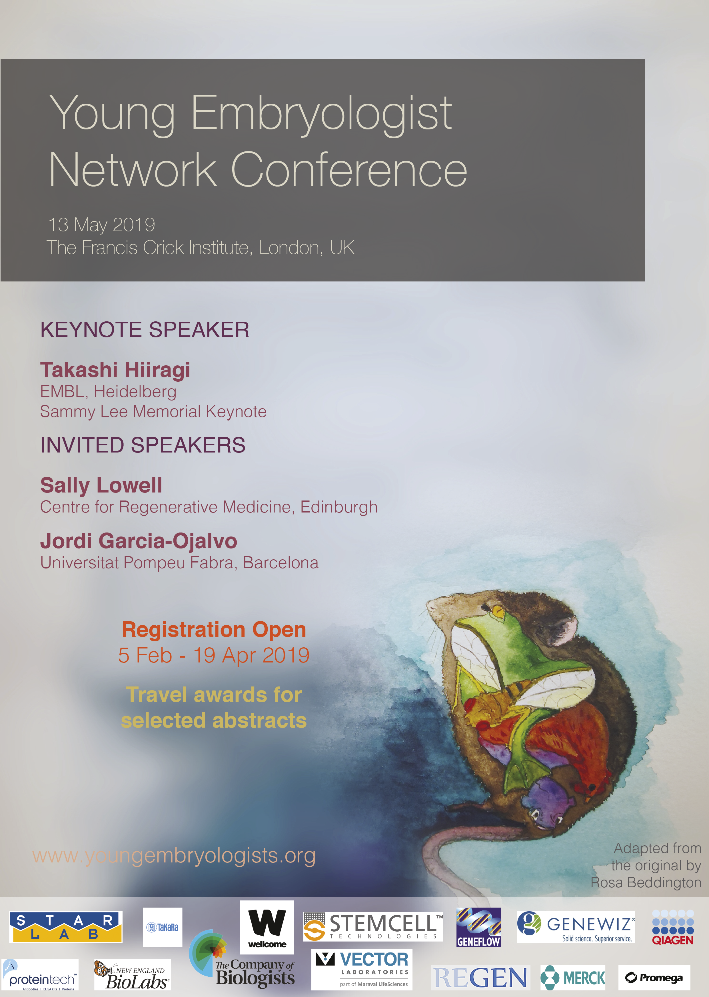
The Young Embryologist Network (YEN), is an academic body aiming to bring together early career scientists within the wide field of developmental biology, in order to provide opportunities to present talks and posters, network and collaborate, and gain research or career advice.
YEN was set up in 2008 by graduate students in the prestigious Department of Cell and Developmental Biology at University College London. Every year, the YEN hosts an annual conference at a UK research institution with great success. The conference is entirely organised by graduate students and junior post-doctoral scientists, and has remained free to attend since 2008, due to the generosity of sponsors and grants.
The 2019 conference is being held on the 13th of May 2019 at the Francis Crick Institute in London.
The deadline for Registration is the 19th April. Register here:
The Beddington Medal is the BSDB’s major commendation to promising young biologists, awarded for the best PhD thesis in Developmental Biology defended in the year previous to the award. Rosa Beddington was one of the greatest talents and inspirational leaders in the field of developmental biology. Rosa made an enormous contribution to the field in general and to the BSDB in particular, so it seemed entirely appropriate that the Society should establish a lasting memorial to her. The design of the medal, mice on a stylised DNA helix, is from artwork by Rosa herself. We would like to congratulate the 2019 winner of the Beddington Medal, David Munro, and would like to take this opportunity to give a brief overview of his career and the PhD project that was awarded the Beddington medal.
Jim Smith introduced the Beddington medal with heartfelt memories of Rosa Beddington and her time at the NIMR. Please read more of his thoughts here.
Some complicated selfies were taken as the medal was passed over before David went on to present the work that has deserved him this award.
In the words of his PhD supervisor:
“The really impressive thing about David’s work is that he did not come to my lab to fit in with an existing line of research but created one of his own”. Jamie Davies, University of Edinburgh.
David received his undergraduate degree in Sport and Exercise Science at the University of Stirling (2010-2014). With this, he achieved a first-class honours degree and the prize for the best overall performance throughout a physiology related degree (British Physiological Society Undergraduate Prize). His dissertation project investigated associations between ADRB2 mutations (an adrenaline receptor gene in humans) and athlete status/athletic ability measurements. Subsequently, he was awarded a University of Stirling Head of School Summer Bursary Award to remain in Stirling during the summer of 2014 and investigate the relationship between transcribed ultra-conserved regions of RNA (T-UCRs) and the development of diet-induced insulin resistance in humans (Summer 2014). He then moved to the University of Edinburgh for his MSc by Research in Biomedical Sciences (2014-2015). Again, he received a distinction and was awarded the Class Prize for best student. During this time, he studied the physiology of S-acylation the regulation of skeletal muscle energy expenditure by an obesity-associated phospholipase as part of two research placements.
David has been awarded the Beddington medal for his exceptional work performed during his 3-year MRC-funded PhD at the University of Edinburgh with Prof Jamie Davies and Dr Peter Hohenstein (2015-2018): The thesis is titled ‘Mechanisms of kidney vascularisation and the roles of macrophages in renal organogenesis’. During his PhD, he gave several oral and poster presentations at national and international conferences, supervised students (including a Gurdon Summer Studentship Awardee), established numerous international collaborations, was awarded travel grants (including a BSDB Conference Grant), and reviewed manuscripts for leading journals (including Cell Reports, Angiogenesis, and Scientific Reports). He is now a post-doctoral fellow at the UK Dementia Research Institute (University of Edinburgh; 2019- present), continuing research in macrophage biology under the supervision of Prof Josef Priller. His current focus in on brain macrophages (microglia) in development, neurodegeneration, and aging.
Thesis description
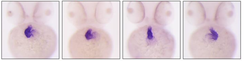
Kidneys are specialised organs that clean the blood, removing waste while retaining what is useful. This requires a complex vasculature, and its formation as a foetus develops is poorly understood. I started my PhD research by using advanced microscopy techniques to visualise how blood vessels form in three-dimensions in the mouse kidney. In doing so, I identified when and from where the first blood vessels enter the kidney, and how blood vessels pattern at the edge of the kidney throughout development.
Blood vessels can form through angiogenesis (branching of new vessels from pre-existing ones) and/or vasculogenesis (assembly of new vessels from the coalescence of endothelial precursor cells). It has long been thought that a combination of both processes occurs during kidney vascularisation; however, my thesis work indicates that this concept may not be correct. My data instead suggest that kidney vascularization relies on growth and remodelling of pre-existing vessels (angiogenesis) and does not depend on vasculogenesis at any point (Publications 1 and 5 in CV). When assessing the entire 3D vascular tree of the kidney, isolated endothelial cells were never observed at any developmental age. Instead, all vessels, including the newly forming ones, were connected to pre-existing vessels that could be traced to the major circulatory vessels.
I then focused on the blood vessels at the edge of the kidney, which I found to consistently and accurately pattern around a special collection of cells – the cap mesenchyme. The cap mesenchyme contains cells that eventually become the cleaning tubes of the kidney, the nephrons. This cell population undergoes rounds of splitting at the kidney’s periphery. As this happens, I demonstrated that blood vessels migrate through the newly opened regions between the separating cap mesenchymal populations (Publication 1 in CV). This occurs in cycles throughout development and is likely to be vital for the oxygenation of the kidney’s outer region, the site where important processes such as nephron formation take place.
I determined that a signalling molecule, semaphorin-3f, and its receptor, neuropilin-2, were expressed in a pattern consistent with them having roles in this cyclical patterning of blood vessels; however, using mouse models where the genes for these molecules were deleted, I established that they were not vital for this process (Publication 2 in CV).
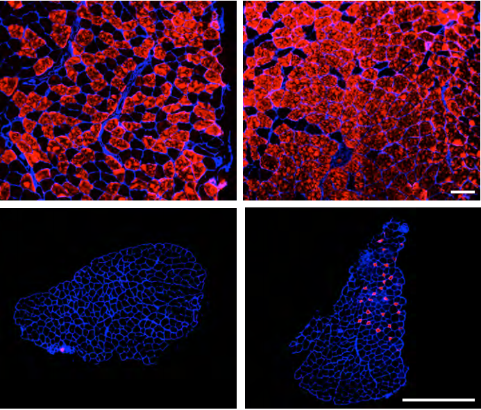
I next shifted my research focus towards a specialised cell type known as the macrophage (macro = big; phage = eater) in the developing kidney (Publication 3 in CV). Macrophages are immune cells best known for clearing foreign and damaged cells. These cells have vital roles during animal development, but little is known about their specific functions during kidney development.
Macrophages arrived in the mouse kidney early during its development, where they were required to clear away misplaced cells to ‘set-the-stage’ for early kidney development (Publication 6 in CV, under review). Throughout later development, most macrophages wrapped around blood vessels and I demonstrated their ability to eat endothelial cells (which usually line the blood vessels) and red blood cells (which are carried within them) within the kidney. I also established that kidney macrophages produced many molecules linked to blood vessel development, and so I examined the consequences of macrophage-loss on blood vessel formation. Blood vessels normally form continuous networks in the kidney; however, when macrophages were depleted (by blocking a macrophage-survival signalling pathway), connections between renal blood vessels were reduced (Publication 6 in CV).
Publications
Munro DAD, Hohenstein P, Davies JA. 2017. Cycles of vascular plexus formation within the nephrogenic zone of the developing kidney. Scientific Reports. 7: 3273.
Munro DAD, Hohenstein P, Coate TM, Davies JA. 2017. Refuting the hypothesis that semaphorin-3f/neuropilin-2 guide endothelial patterning around the cap mesenchyme in the developing kidney. Developmental Dynamics. 246:1047-1056.
Munro DAD, Hughes J. 2017. The Origins and Functions of Tissue-Resident Macrophages in Kidney Development. Frontiers in Physiology. 8:837. (Review)
Mills CG, Lawrence ML, Munro DAD, El-Hendawi M, Mullins JJ, Davies JA. 2017. Asymmetric BMP4 signalling improves the realism of kidney organoids. Scientific Reports. 7:14824.
Munro DAD, Davies JA. 2018. Vascularizing the kidney in the embryo and organoid: questioning assumptions about renal vasculogenesis. Journal of the American Society of Nephrology. (Perspectives article).
Munro DAD, et al. Macrophages restrict the nephrogenic field and promote endothelial connections during kidney development. eLife 2019;8:e43271 DOI: 10.7554/eLife.43271
A postdoctoral position is available in the laboratory of Dr. Jessica Mark Welch in the Bay Paul Center to study the spatial organization of microbial communities in the human mouth. The successful candidate will use fluorescence in situ hybridization, spectral imaging microscopy, and computational image analysis to investigate microbial community structure and will interact closely with collaborators at other institutions as well as with the vibrant and collegial MBL scientific community.
The position is for 2 years, and may be extended beyond this period contingent on securing additional funding.
Physical requirements: This position requires fine motor skills and willingness to work with potentially biohazardous materials and standard laboratory chemicals including fixatives and solvents.
Basic qualifications: A Ph.D. in biological sciences or a related field is required.
Preferred qualifications: Experience with confocal microscopy, computational image analysis, bioinformatics, and/or microbiology is desirable.
Instructions: To apply, please visit the MBL Employment Opportunities website: https://www.mbl.edu/hr/employment/. The following documents are required: (1) a cover letter describing your interests, skills, and prior research experience, including any specific experience with the job responsibilities listed above; (2) a curriculum vitae/resume; and (3) the names and contact numbers of three persons who can be contacted for letters of reference, at least one of whom must have acted as your supervisor in a previous research position.
The Rohner Lab at the Stowers Institute for Medical Research has an opening for a Postdoctoral Researcher to develop an independent project investigating the molecular, genetic, and developmental mechanisms of how cavefish maintain health under diabetes-like phenotypes. The lab has previously found that the cavefish Astyanax mexicanus develop high-blood sugar and insulin resistance as part of their natural strategy to survive in the caves but without the usually associated health problems (Riddle et al. Nature. 2018 Mar 29;555(7698):647-651). Visit http://research.stowers.org/rohnerlab/ for more information.
The selected candidate will investigate the molecular mechanism underlying these impressive adaptations. The candidate will closely work with the core facilities at the institute to perform single-cell RNA sequencing, proteomics, and functional validation in vitro and in vivo. The candidate will receive strong support from the core facilities that provide advice, training and service to enhance the Institute’s interdisciplinary and collaborative research programs. Current core facilities are staffed by over 100 scientists with expertise in bioinformatics, cytometry, histology, imaging, microarray, next generation sequencing, transgenic and ES cell technologies, proteomics and molecular biology. The Stowers Institute offers a highly competitive compensation and benefits package.
The position is funded for two years through a grant by the Juvenile Diabetes Research Foundation and can be renewed for up to five years in order to allow enough time to develop a research program/publication record that makes the postdoc a strong candidate for an independent position. The Rohner Lab has a strong commitment for mutual success and is dedicated to providing support for all lab members.
Minimum requirements include a doctoral degree in the life sciences, chemistry, or biomedical engineering. Experience in one or more of the following areas is desirable: molecular biology, developmental biology, genetics, genomics, evodevo, physiology.
In addition to excellent verbal and written communication skills, successful candidates must be dynamic and highly motivated, work independently and creatively, able to work in a team-oriented environment, and proficient at problem solving.
Application Instructions: To apply, please submit (1) a brief cover letter, (2) a current CV, and (3) contact information for two professional references to Dr. Nicolas Rohner at nro@stowers.org cc: careers@stowers.org.
About the Stowers Institute for Medical Research
The Stowers Institute for Medical Research is a world-class basic biomedical research organization focused on improving our understanding of fundamental mechanisms of biology and using this knowledge to guide the development of innovative treatments to improve human health.
Our dedicated scientists collaborate across a variety of disciplines, studying many different aspects of health and disease. A primary goal of our research is to understand the principles that guide the function and behavior of living organisms and individual cells. Discoveries resulting from this kind of research often prove to be major milestones along the path toward novel therapies and cures (visit www.stowers.org). Jim Stowers, founder of American Century Investments, and his wife, Virginia, opened the Institute in 2000. Since then, the Institute has spent over 900 million dollars in pursuit of its mission.
Currently, the Institute is home to almost 550 researchers and support personnel; over 20 independent research programs; and more than a dozen technology-development and core facilities. The Institute has been ranked 3rd place by the Scientist for best places to work in the world: https://www.the-scientist.com/features/best-places-to-work-academia-2012-40676
Kansas City is an emerging metropolitan city in the Midwest with a high quality of living and affordability. Visit https://www.visitkc.com for information about living and working in Kansas City.
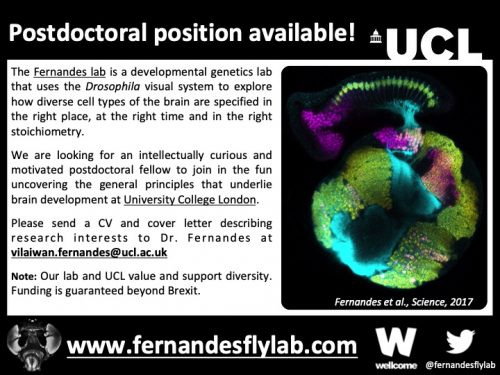
We are looking for an intellectually curious and motivated postdoctoral fellow to join in the fun exploring how glia regulate neurogenesis and neuronal fate-specification. Our approaches are interdisciplinary and involve genetics, live imaging, computational modelling, single cell RNA sequencing, etc. Available projects include: (1) Understanding how signals from glia impart unique neuronal identities (a follow up to Fernandes et al., Science, 2017). (2) Characterising glial diversity in the visual system. (3) Exploring glial involvement in neuroepithelial proliferation.
Applicants should have a PhD in a relevant subject area (or be close to completing their degree), excellent communication skills, a collaborative spirit and a kind heart. The ideal candidate will have a strong background in molecular biology, cell and/or developmental biology as well as experience with imaging. Knowledge of signal transduction, Drosophila genetics and bioinformatics are a plus but not essential.
Formal applications will be accepted online through UCL’s job portal till May 3rd, 2019. If you cannot meet this deadline but would like to apply, please contact Dr. Fernandes as soon as possible.
Proposed start date: August 1st, 2019 (Flexible).
For more details please contact Dr. Fernandes at vilaiwan.fernandes@ucl.ac.uk (along with a CV and cover letter describing research interests).
Note: Our lab and UCL value and support diversity. Funding is guaranteed beyond Brexit.
The Bressan Laboratory (www.bressanlab.com) at the University of North Carolina Chapel Hill is inviting applications for a postdoctoral fellow interested in developmental Cell Biology and Physiology research. The focus of the position will be to explore the genetic and molecular events that control cellular diversity during cardiovascular development. Specifically, candidates will conduct direct in vivo over expression, live imaging, cell sorting, primary culture, and next generation sequencing to explore how alterations in transcriptional activity and cellular mechanics influence physiological fate in the embryonic heart. The applicant is expected to manage an independent research project and to train students and other fellows in the laboratory.
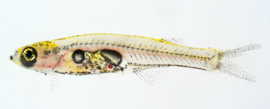
The correct patterning of embryonic tissues is essential for normal development. Aberrant patterning can lead to developmental abnormalities and pathogenic defects. Therefore, studying developmental patterning is important to better understand disease. The zebrafish embryo is a fantastic model for studying patterning during development owing to its optical clarity, small size and large clutch number. When coupled with dynamic transgenic lines, the picture of what occurs in the cell during these processes is starting to emerge.
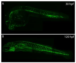
The vasculature of zebrafish expands throughout development, providing developing tissues with oxygen and nutrients.
Understanding how the ubiquitous second messenger, the calcium ion, regulates cellular physiology during development has become an important question in biology. This is where my story begins. I arrived in Sheffield after obtaining a PhD position, eager for what lay ahead. The project was exciting; using new transgenic lines and cutting-edge microscopy to study the function of a poorly understood gene, tmem33, which we hypothesised would regulate calcium signalling within the developing vasculature. The preliminary morpholino knockdown data generated in my host lab before I started my PhD hinted at a vast cache of riches waiting to be uncovered. This data suggested a role for tmem33 in both vascular and kidney development. I was given some new toys to play with, a light sheet microscope and an endothelial-specific calcium reporter line. My first experiment in the lab was to analyse the mutant zebrafish line generated by my supervisor around which my PhD was supposed to focus. It was, of course, completely normal. No vascular defects here. It looked like my project was dead in the water before it had begun. My next few experiments yielded additional dead ends – analysing 34 TRP channels by in situ hybridisation, looking for specific vascular enrichment (spoiler alert – there wasn’t any).
Thankfully, science moves very quickly. My original project went from dead, to very dead as several papers at the time heavily criticised morpholino-based approaches (Kok et al, 2014; Schulte-Merker and Stainier, 2014; Stainier 2017; Robu, 2007) to back alive within the space of about six months. A single paper (Rossi et al, 2015) brought hopes for a revived project. In a series of elegant experiments, the authors described a situation where knockdown of the egfl7 gene by morpholino induced a robust vascular phenotype, but when this gene was mutated using genome editing the phenotype was absent. Interestingly, the authors showed that the egfl7 mutant displayed nonsense mediated decay of egfl7 transcripts and that when egfl7 morpholinos were injected into egfl7 mutants, the mutants were protected against the effects of the morpholino. The synthesis of these data was that there existed genetic compensation in the mutants but not in the morphants.
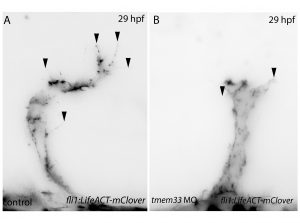
Finally, the stroke of luck I needed, our tmem33 mutants also showed nonsense mediated decay, so I set about injecting tmem33 morpholinos into our tmem33 mutants and I found strikingly similar results. This suggested that the reason the tmem33 mutants displayed no phenotype was because they displayed a kind of genetic protection, likely via a genetic compensation mechanism. I now had the beginnings of a successful project, a year in to my PhD. Rossi et. al used a new technology to address their issues, CRISPR interference (CRISPRi) – a modified version of the CRISPR/Cas9 system using an inactivated form of Cas9 (dCas9). The authors were able to reproduce the same phenotype they observed via morpholino knockdown using CRISPRi. I applied CRISPRi to knock down tmem33 and was able to reproduce our morpholino knockdown data of tmem33.
Filopodia are essential for cellular migration. Tmem33 morphants display reduced filopodia and delayed vascular migration.
Next, inspired by the conditional CRISPR approaches described in Ablain et al (2015), I sought to conditionally knock down tmem33 in endothelial cells by driving dCas9 expression under the control of the endothelial-specific fli1a promoter. Preliminary experiments using transient conditional knockdown were promising – only endothelial cells showed a phenotype! The next step was to generate a transgenic line which stably expressed dCas9 in endothelial cells.
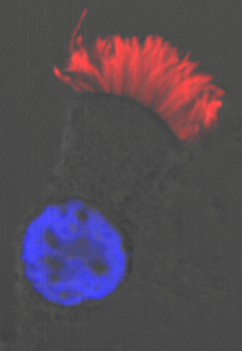
Throughout this process, I had been studying tmem33 function with regards to endothelial cell physiology during angiogenesis. Since it was known that tmem33 functioned within the endoplasmic reticulum, using a calcium reporter line I began to test whether tmem33 knockdown altered endothelial calcium signalling. I found that tmem33 knockdown reduced observable endothelial calcium oscillations, which coincided with reduced endothelial cell migration. This was validated by both morpholinos and CRISPRi. Furthermore, I began using both knockdown approaches to position the tmem33 within the hierarchy of developmental angiogenic signalling. I found that tmem33 functions downstream of VEGF signalling but upstream of Notch and ERK signalling, identifying an essential function for calcium oscillations in mediating the response to Vascular Endothelial Growth Factor (VEGF) and inducing downstream signalling pathways essential during angiogenesis.
Calcium signalling during endothelial development.
Throughout my PhD, I’ve learned how the zebrafish can be a powerful tool for understanding basic biology and how a PhD is not a linear endeavour. You have to follow the research (and the results of others) and see where it takes you. I’ve also found that phenotypes (or rather, the lack of them) can be hard to interpret. If you think about it, nothing would be alive today if there weren’t contingency plans built into the genome. Unexpected negative results (I’m looking at you again, non-phenotypic mutants) are not worthless. In fact, they’re probably more interesting.
References
Kok, Fatma O., et al. “Reverse genetic screening reveals poor correlation between morpholino-induced and mutant phenotypes in zebrafish.” Developmental cell 32.1 (2015): 97-108.
Robu, Mara E., et al. “p53 activation by knockdown technologies.” PLoS genetics 3.5 (2007): e78.
Rossi, Andrea, et al. “Genetic compensation induced by deleterious mutations but not gene knockdowns.” Nature 524.7564 (2015): 230.
Savage, Aaron M., et al. “tmem33 is essential for VEGF-mediated endothelial calcium oscillations and angiogenesis.” Nature communications 10.1 (2019): 732.
Schulte-Merker, Stefan, and Didier YR Stainier. “Out with the old, in with the new: reassessing morpholino knockdowns in light of genome editing technology.” Development 141.16 (2014): 3103-3104.
Stainier, Didier YR, et al. “Guidelines for morpholino use in zebrafish.” PLoS genetics 13.10 (2017): e1007000.
A postdoctoral position is available immediately for a highly-motivated individual interested in understanding the molecular mechanisms of brain development. The individual will join Dr. Martin Riccomagno’s laboratory at the University of California, Riverside (http://www.riccomagnolab.org). Our laboratory uses a combination of molecular biology, histology, tissue culture, mouse transgenesis, and cutting-edge imaging approaches to investigate the developmental mechanisms that regulate neural circuit formation.
The successful applicant will be involved in NIH-funded projects in one of the following research areas:
1) Regulation of neural migration by adhesive cues
2) Developmental profiling of caspase-dependent refinement
The salary for this position is commensurate with experience and determined by NIH guidelines.
Position requirements:
A PhD or MD-PhD degree in a biological science
A solid background in neuroscience or developmental biology
Previous experience working with genetic mouse models
Proficiency in cellular and molecular biology
Excellent communication skills
A demonstrated ability to work independently and learn new techniques
The scientific environment at UC Riverside is highly collaborative and multidisciplinary. One of the most significant resources available is the quantity and quality of valuable scientific interaction and support from fellow cell biologists and neuroscientists both within and beyond the Department of Molecular, Cell and Systems Biology (MCSB). UCR is also home to the Center for Glial-Neuronal Interactions (CGNI). CGNI provides a forum for scientific interactions among the members of the UCR neuroscience community, and organizes an annual research symposium that attracts neuroscientists from throughout southern California, and speakers from around the world.
Interested applicants should apply by sending a cover letter, CV, and contact information for three references to: martinmr@ucr.edu
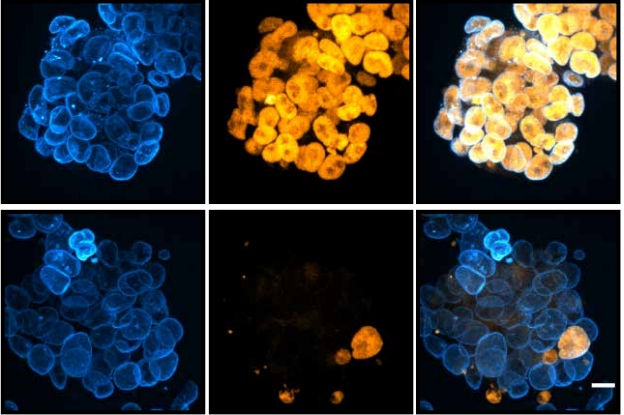
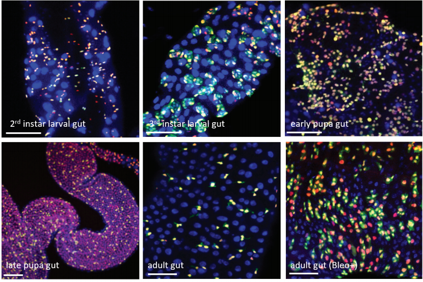
With air pollution on the rise, our respiratory system is continually abused by a barrage of harmful substances that we breathe in with each inhalation. Fortunately, we are equipped with highly specialised ciliated cells, the multiciliated cells (MCCs), which differentiate hundreds of motile cilia on their apical surface1,2. These cilia beat rhythmically to drive mucus that entraps air borne toxins and pathogens out of the airways – a process called mucociliary clearance in clinical circles. The importance of mucociliary clearance in respiratory physiology is perhaps best exemplified by patients afflicted with diseases, genetic as well as acquired, in which the cilia on MCCs do not function properly. In such instances, individuals suffer from recurrent lung infections that often lead to irreparable damage called bronchiectasis3. Given their importance, it is understandable why the MCCs have lately attracted a lot of attention. MCCs are also present in organs such as the central nervous system (within the brain ventricles and spinal canal), where they promote circulation of cerebrospinal fluid (CSF), and within the reproductive system where they are required for mixing of reproductive fluids and germ cell transportation. Pathological consequences of defective MCCs in these organs include hydrocephalus (distention of the brain ventricles due to improper CSF flow) and infertility, respectively1,2.
A particularly intriguing aspect of the MCC differentiation program is the ability of their precursors to support an explosive production of basal bodies (specialised centrioles), which are required to nucleate the assembly of multiple cilia. While most cells duplicate centrioles only once during the cell cycle, how post-mitotic MCC precursors can generate hundreds of these organelles has intrigued cell biologists for decades. However, it is only over the past few years that we have made substantial progress in identifying some of the key genetic determinants and important cell biological pathways that govern the differentiation of the MCCs. Notably, in a seminal piece of work published in 2012, Kinter and colleagues identified a small coiled-coil containing nuclear protein Multicilin (aka Mcidas), related to the celebrated DNA replication factor Geminin, as necessary and sufficient for MCC development4. In their study, which mainly utilized Xenopus embryos that differentiate MCCs on the epidermis, they could show that morpholino-mediated inhibition of Mci function completely abolished MCCs, while its overexpression led to the production of ectopic MCCs at the expense of other epidermal cell-types. Since Mci lacks a DNA binding domain, when a subsequent paper by the same group showed that the protein functions in association with the E2f family of cell-cycle transcription factors to activate MCC-specific gene expression (genes for production of multiple basal bodies and cilia)5, our understanding of MCC development achieved a certain degree of completeness. More importantly, mutations in MCI were concomitantly identified in patients with the respiratory disorder reduced generation of multiple motile cilia, underscoring the importance of the MCC developmental pathway in the etiology of severe airway disease6.
It was soon realised though, that there is more to MCCs than Mci. We and others found that another Mci-like protein, Gmnc (aka Gemc1), has identical effects on MCC development. Zebrafish, Xenopus and mice deficient in Gmnc function completely lack MCCs from various tissues in which these cells normally differentiate7,8,9. Moreover, overexpression of Gmnc can drive the production of supernumerary MCCs. In addition, these studies also showed that Gmnc functions upstream of Mci, since Mci is not induced in the absence of Gmnc, but Mci is incapable of inducing Gmnc expression. Despite all of this information, one outstanding question that has lingered in the field is how the activities of two highly related proteins can have near identical effects on the MCC developmental program –both proteins being necessary as well as sufficient for MCC development. We reasoned that evaluating the function of Mci with a stable genetic mutant in the mouse would be the best way forward, as the phenotypes that have been described for the Gmnc mutant mice will allow the appropriate comparisons to be made, in the context of a mammal. Surprisingly, in contrast to the current notion of Mci function, which posits its requirement in MCC specification as well as differentiation, we now report in Development that mice lacking Mci function can generate MCC precursor cells in normal numbers10. These precursors can express the full suite of genes that have been linked to the transcriptional regulation of ciliogenesis. However, they are completely unable to generate any basal bodies for multiciliation, and instead differentiate a single motile-like cilium.
A multiciliated cell from the human airway. Cilia are stained with antibodies to acetylated tubulin (red) and the nucleus is labelled with DAPI (blue).
Moreover, we could show that while Gmnc can activate genes that regulate motile ciliogenesis, Mci preferentially activates genes that promote the biogenesis of multiple basal bodies. Finally, we were also able to demonstrate that the difference in the transcriptional activities of the two proteins possibly lies in the differences in their ability to associate with E2f4 versus E2f5, and thereby target the regulation of different sets of MCC-specific differentiation genes. In sum, our study has clarified that Gmnc functions at the top of the transcriptional hierarchy for making MCCs: it induces MCC precursors and also activates the expression of Mci. Subsequently, Mci programs these cells to generate multiple basal bodies and complete the process of multiciliation10.
So what’s next for Mci? While we do need a much better understanding of the molecular basis of Mci-mediated transcriptional regulation, the future looks even brighter for the protein to enable us to tackle a more profound and unsolved issue in MCC development: What is the precise cellular pathway that allows the MCCs to generate multiple basal bodies? Current view posits that there are two ways by which this is achieved. First, in the mother centriole-dependant pathway, the mother centriole is thought to produce a small number of centrioles (this is the pathway utilised for centriole duplication during regular cell cycle). However, it is via a unique MCC-specific alternative pathway, the deuterosome-dependant pathway, that more than 90% of the basal bodies are produced. Although deuterosomes, electron dense ring-shaped structures that function in centriole biogenesis in MCCs, were originally believed to arise de novo, more recent evidence has suggested that they are produced in close association with the daughter centriole11,12,13. Research into the mechanism by which MCCs make multiple basal bodies is in such a state of flux, that accumulating new data are again poised to revise our present view of the contributions the two pathways highlighted above. Three papers, all in preprint, make the tantalising proposition that in fact the centrosomal centrioles are completely dispensable for basal body generation in the MCCs14,15,16!
Sudipto Roy is a Senior Principal Investigator with the Institute of Molecular and Cell Biology, A*STAR, Singapore.
In the absence of the mother and the daughter centrioles, MCC precursors can generate deuterosomes quite normally, likely from the pericentriolar material (PCM) associated with the microtubule organizing centre (MTOC), and these deuterosomes produce the normal complement of basal bodies. Given that Mci mutant MCCs are totally incapable of generating multiple basal bodies, we can look forward to obtaining useful insights into this enigmatic process through more detailed investigations into the basal body generation defects of the MCC precursors in these mice. In the end, all of this information will add significant value for appreciating the underlying mechanistic basis of the pathological consequences associated with ciliary disorders that affect the formation of the MCCs and the motility of their cilia.
References:
BROOKS, ERIC R. & WALLINGFORD, JOHN B. 2014. Multiciliated Cells. Current Biology 24, R973-R982.
ZHOU, F. & ROY, S. 2015. SnapShot: Motile Cilia. Cell 162, 224-224.e1.
BUSTAMANTE-MARIN, X. M. & OSTROWSKI, L. E. 2017. Cilia and Mucociliary Clearance. Cold Spring Harb. Perspect. Biol. 9, a028241.
STUBBS, J. L., VLADAR, E. K., AXELROD, J. D. & KINTNER, C. 2012. Multicilin promotes centriole assembly and ciliogenesis during multiciliate cell differentiation. Cell Biol. 14, 140-7.
MA, L., QUIGLEY, I., OMRAN, H. & KINTNER, C. 2014. Multicilin drives centriole biogenesis via E2f proteins. Genes Dev. 28, 1461-71.
BOON, M., WALLMEIER, J., MA, L., LOGES, N. T., JASPERS, M., OLBRICH, H., DOUGHERTY, G. W., RAIDT, J., WERNER, C., AMIRAV, I., HEVRONI, A., ABITBUL, R., AVITAL, A., SOFERMAN, R., WESSELS, M., O’CALLAGHAN, C., CHUNG, E. M., RUTMAN, A., HIRST, R. A., MOYA, E., MITCHISON, H. M., VAN DAELE, S., DE BOECK, K., JORISSEN, M., KINTNER, C., CUPPENS, H. & OMRAN, H. 2014. MCIDAS mutations result in a mucociliary clearance disorder with reduced generation of multiple motile cilia. Commun. 5, 4418.
ARBI, M., PEFANI, D.-E., KYROUSI, C., LALIOTI, M.-E., KALOGEROPOULOU, A., PAPANASTASIOU, A. D., TARAVIRAS, S. & LYGEROU, Z. 2016. GemC1 controls multiciliogenesis in the airway epithelium. EMBO Rep. 17, 400-13.
TERRÉ, B., PIERGIOVANNI, G., SEGURA-BAYONA, S., GIL-GÓMEZ, G., YOUSSEF, S. A., ATTOLINI, C. S.-O., WILSCH-BRÄUNINGER, M., JUNG, C., ROJAS, A. M., MARJANOVIĆ, M., KNOBEL, P. A., PALENZUELA, L., LÓPEZ-ROVIRA, T., FORROW, S., HUTTNER, W. B., VALVERDE, M. A., DE BRUIN, A., COSTANZO, V. & STRACKER, T. H. 2016. GEMC1 is a critical regulator of multiciliated cell differentiation. EMBO J. 35, 942-60.
ZHOU, F., NARASIMHAN, V., SHBOUL, M., CHONG, Y. L., REVERSADE, B. & ROY, S. 2015. Gmnc is a master regulator of the multiciliated cell differentiation program. Biol. 25, 3267-73.
LU, H., ANUJAN, P., ZHOU, F., ZHANG, Y., CHONG, Y. L., BINGLE, C. D., & ROY, S. 2019. Mcidas mutant mice reveal a two-step process for the specification and differentiation of multiciliated cells in mammals. Development 146: dev172643 doi: 10.1242/dev.172643
SOROKIN, S. P. 1968. Reconstructions of centriole formation and ciliogenesis in mammalian lungs. Cell Sci. 3, 207-30.
ZHAO, H., ZHU, L., ZHU, Y., CAO, J., LI, S., HUANG, Q., XU, T., HUANG, X., YAN, X. & ZHU, X. 2013. The Cep63 paralogue Deup1 enables massive de novo centriole biogenesis for vertebrate multiciliogenesis. Cell. Biol. 15, 1434-44.
AL JORD, A., LEMAITRE, A. I., DELGEHYR, N., FAUCOURT, M., SPASSKY, N. & MEUNIER, A. 2014. Centriole amplification by mother and daughter centrioles differs in multiciliated cells. Nature 516, 104-7.
MERCEY, O., AL JORD, A., ROSTAING, P., MAHUZIER, A., FORTOUL, A., BOUDJEMA, A.-R., FAUCOURT, M., SPASSKY, N. & MEUNIER, A. 2018. Dynamics of centriole amplification in centrosome-depleted brain multiciliated progenitors. bioRxiv, 503730.
NANJUNDAPPA, R., KONG, D., SHIM, K., STEARNS, T., BRODY, S., LONCAREK, J. & MAHJOUB, M. 2018. Regulation of cilia abundance in multiciliated cells. bioRxiv, 478297.
ZHAO, H., CHEN, Q., HUANG, Q., YAN, X. & ZHU, X. 2018. Mother centrioles are dispensable for deuterosome formation and function during basal body amplification. bioRxiv, 373662.
Welcome to our monthly trawl for developmental biology (and related) preprints.
This month we found three hydra preprints, lots of developmental mechanics, a typically hearty serving of single cell transcriptomic analyses and a survey of the life of PIs.
The preprints were hosted on bioRxiv, PeerJ, andarXiv. Let us know if we missed anything, and use these links to get to the section you want:
Microglia actively remodels adult hippocampal neurogenesis through the phagocytosis secretome
Irune Diaz-Aparicio, Iñaki Paris, Virginia Sierra-Torre, Ainhoa Plaza-Zabala, Noelia Rodríguez-Iglesias, Mar Márquez-Ropero, Sol Beccari, Oihane Abiega, Elena Alberdi, Carlos Matute, Irantzu Bernales, Angela Schulz, Lilla Otrokocsi, Beata Sperlagh, Kaisa E. Happonen, Greg Lemke, Mirjana Maletic-Savatic, Jorge Valero, Amanda Sierra
Neuronal programming by microbiota enables environmental regulation of intestinal motility
Yuuki Obata, Stefan Boeing, Álvaro Castaño, Ana Carina Bon-Frauches, Mercedes Gomez de Agüero, Werend Boesmans, Bahtiyar Yilmaz, Rita Lopes, Almaz Huseynova, Muralidhara Rao Maradana, Pieter Vanden Berghe, Andrew J. Murray, Brigitta Stockinger, Andrew J. Macpherson, Vassilis Pachnis
Human cortical organoids expose a differential function of GSK3 on direct and indirect neurogenesis
Alejandro López-Tobón, Carlo Emanuele Villa, Cristina Cheroni, Sebastiano Trattaro, Nicolò Caporale, Paola Conforti, Raffaele Iennaco, Maria Lachgar, Marco Tullio Rigoli, Berta Marcó de la Cruz, Pietro Lo Riso, Erika Tenderini, Flavia Troglio, Marco de Simone, Isabel Liste-Noya, Stefano Piccolo, Giuseppe Macino, Massimiliano Pagani, Elena Cattaneo, Giuseppe Testa
Pluripotency factors regulate the onset of Hox cluster activation in the early embryo
Elena Lopez-Jimenez, Julio Sainz de Aja, Claudio Badia-Careaga, Antonio Barral, Isabel Rollan, Raquel Rouco, Elisa Santos, María Tiana, Jesus Victorino, Hector Sanchez-Iranzo, Rafael D Acemel, Carlos Torroja, Javier Adan, Eduardo Andres-Leon, Jose Luis Gomez-Skarmeta, Giovanna Giovinazzo, Fatima Sanchez-Cabo, Miguel Manzanares
Embryonic stem cells from Rhodes, et al.’s preprint
Cohesin disrupts polycomb-dependent chromosome interactions
JDP Rhodes, A Feldmann, B Hernández-Rodríguez, N Díaz, JM Brown, NA Fursova, NP Blackledge, P Prathapan, P Dobrinic, M Huseyin, A Szczurek, K Kruse, KA Nasmyth, VJ Buckle, JM Vaquerizas, RJ Klose
Whole-genome and RNA sequencing reveal variation and transcriptomic coordination in the developing human prefrontal cortex
Donna M. Werling, Sirisha Pochareddy, Jinmyung Choi, Joon-Yong An, Brooke Sheppard, Minshi Peng, Zhen Li, Claudia Dastmalchi, Gabriel Santpere, Andre M. M. Sousa, Andrew T. N. Tebbenkamp, Navjot Kaur, Forrest O. Gulden, Michael S. Breen, Lindsay Liang, Michael C. Gilson, Xuefang Zhao, Shan Dong, Lambertus Klei, A. Ercument Cicek, Joseph D. Buxbaum, Homa Adle-Biassette, Jean-Leon Thomas, Kimberly A. Aldinger, Diana R. O’Day, Ian A. Glass, Noah A. Zaitlen, Michael E. Talkowski, Kathryn Roeder, Matthew W. State, Bernie Devlin, Stephan J. Sanders, Nenad Sestan
Human cortical neural stem cells generate regional organizer states in vitro before committing to excitatory neuronal fates
Nicola Micali, Suel-Kee Kim, Marcelo Diaz-Bustamante, Genevieve Stein-O’Brien, Seungmae Seo, Joo-Heon Shin, Brian G. Rash, Shaojie Ma, Nicolas A. Olivares, Jon Arellano, Kristen R. Maynard, Elana J. Fertig, Alan J. Cross, Roland Burli, Nicholas J. Brandon, Daniel R. Weinberger, Joshua G. Chenoweth, Daniel J. Hoeppner, Nenad Sestan, Pasko Rakic, Carlo Colantuoni, Ronald D. McKay
Identification of a human adult cardiac stem cell population with neural crest origin
Anna Höving, Madlen Merten, Kazuko Elena Schmidt, Isabel Faust, Lucia Mercedes Ruiz-Perera, Henning Hachmeister, Sebastian-Patrick Sommer, Buntaro Fujita, Thomas Pühler, Thomas Huser, Johannes Greiner, Barbara Kaltschmidt, Jan Gummert, Cornelius Knabbe, Christian Kaltschmidt
The Gag Protein PEG10 Binds to RNA and Regulates Trophoblast Stem Cell Lineage Specification
Mona Abed, Erik Verschueren, Hanna Budayeva, Peter Liu, Donald S. Kirkpatrick, Rohit Reja, Sarah K. Kummerfeld, Joshua D. Webster, Sarah Gierke, Mike Reichelt, Keith R. Anderson, Robert J Newman, Merone Roose-Girma, Zora Modrusan, Hazal Pektas, Emin Maltepe, Kim Newton, Vishva M. Dixit
A smooth muscle-like niche facilitates lung epithelial regeneration
Alena Moiseenko, Ana Ivonne Vazquez-Armendariz, Xuran Chu, Stefan Guenther, Kevin Lebrigand, Vahid Kheirollahi, Susanne Herold, Thomas Braun, Bernard Mari, Stijn De Langhe, Chengshui Chen, Xiaokun Li, Werner Seeger, Jin-San Zhang, Saverio Bellusci, Elie El Agha
Human liver organoids; a patient-derived primary model for HBV Infection and Related Hepatocellular Carcinoma
Elisa De Crignis, Fabrizia Carofiglio, Panagiotis Moulos, Monique M.A. Verstegen, Shahla Romal, Mir Mubashir Khalid, Farzin Pourfarzad, Christina Koutsothanassis, Helmuth Gehart, Tsung Wai Kan, Robert-Jan Palstra, Charles Boucher, Jan M.N. Ijzermans, Meritxell Huch, Sylvia F. Boj, Robert Vries, Hans Clevers, Luc van der Laan, Pantelis Hatzis, Tokameh Mahmoudi
Glioblastomas derived from genetically modified pluripotent stem cells recapitulate pathobiology
Tomoyuki Koga, Jorge A Benitez, Isaac A Chaim, Sebastian Markmiller, Alison D Parisian, Kristen M Turner, Florian M Hessenauer, Matteo D’Antonio, Nam-phuong D Nguyen, Shahram Saberi, Jianhui Ma, Shunichiro Miki, Antonia D Boyer, John Ravits, Kelly A Frazer, Vineet Bafna, Clark C Chen, Paul S Mischel, Gene W Yeo, Frank B Furnari
Molecular mechanisms of Evening Complex activity in Arabidopsis
Catarina S. Silva, Aditya Nayak, Xuelei Lai, Veronique Hugouvieux, Jae-Hoon Jung, Agnès Jourdain, Irene López-Vidriero, Jose Manuel Franco-Zorrilla, François Parcy, Kishore Panigrahi, Philip A. Wigge, Max Nanao, Chloe Zubieta
The quail as an avian model system: its genome provides insights into social behaviour, seasonal biology and infectious disease response
Katrina Morris, Matthew M Hindle, Simon Boitard, David W Burt, Angela F Danner, Lel Eory, Heather L Forrest, David Gourichon, Jerome Gros, LaDeana Hillier, Thierry Jaffredo, Hanane Khoury, Rusty Lansford, Christine Leterrier, Andrew Loudon, Andrew S Mason, Simone L Meddle, Francis Minvielle, Patrick Minx, Frederique Pitel, J Patrick Seiler, Tsuyoshi Shimmura, Chad Tomlinson, Alain Vignal, Robert G Webster, Takashi Yoshimura, Wesley C Warren, Jacqueline Smith
A draft genome sequence of the miniature parasitoid wasp, Megaphragma amalphitanum
Artem V. Nedoluzhko, Fedor S. Sharko, Brandon M. Lê, Svetlana V. Tsygankova, Eugenia S. Boulygina, Sergey M. Rastorguev, Alexey S. Sokolov, Fernando Rodriguez, Alexander M. Mazur, Alexey A. Polilov, Richard Benton, Michael B. Evgen’ev, Irina R. Arkhipova, Egor B. Prokhortchouk, Konstantin G. Skryabin
A robotic platform for fluidically-linked human body-on-chips experimentation
Richard Novak, Miles Ingram, Susan Clauson, Debarun Das, Aaron Delahanty, Anna Herland, Ben M. Maoz, Sauveur S. F. Jeanty, Mahadevabharath R. Somayaji, Morgan Burt, Elizabeth Calamari, Angeliki Chalkiadaki, Alexander Cho, Youngjae Choe, David Benson Chou, Michael Cronce, Stephanie Dauth, Toni Divic, Jose Fernandez-Alcon, Thomas Ferrante, John Ferrier, Edward A. FitzGerald, Rachel Fleming, Sasan Jalili-Firoozinezhad, Thomas Grevesse, Josue A. Goss, Tiama Hamkins-Indik, Olivier Henry, Chris Hinojosa, Tessa Huffstater, Kyung-Jin Jang, Ville Kujala, Lian Leng, Robert Mannix, Yuka Milton, Janna Nawroth, Bret A. Nestor, Carlos F. Ng, Blakely O’Connor, Tae-Eun Park, Henry Sanchez, Josiah Sliz, Alexandra Sontheimer-Phelps, Ben Swenor, Guy Thompson II, George J. Touloumes, Zachary Tranchemontagne, Norman Wen, Moran Yadid, Anthony Bahinski, Geraldine A. Hamilton, Daniel Levner, Oren Levy, Andrzej Przekwas, Rachelle Prantil-Baun, Kevin K. Parker, Donald E. Ingber
High-density spatial transcriptomics arrays for in situ tissue profiling
Sanja Vickovic, Goekcen Eraslan, Fredrik Salmen, Johanna Klughammer, Linnea Stenbeck, Tarmo Aijo, Richard Bonneau, Jose Fernandez Navarro, Ludvig Bergenstraahle, Joshua Gould, Mostafa Ronaghi, Jonas Frisen, Joakim Lundeberg, Aviv Regev, Patrik L Staahl
The mesoSPIM initiative: open-source light-sheet mesoscopes for imaging in cleared tissue
Fabian F. Voigt, Daniel Kirschenbaum, Evgenia Platonova, Stéphane Pagès, Robert A. A. Campbell, Rahel Kästli, Martina Schaettin, Ladan Egolf, Alexander van der Bourg, Philipp Bethge, Karen Haenraets, Noémie Frézel, Thomas Topilko, Paola Perin, Daniel Hillier, Sven Hildebrand, Anna Schueth, Alard Roebroeck, Botond Roska, Esther Stoeckli, Roberto Pizzala, Nicolas Renier, Hanns Ulrich Zeilhofer, Theofanis Karayannis, Urs Ziegler, Laura Batti, Anthony Holtmaat, Christian Lüscher, Adriano Aguzzi, Fritjof Helmchen
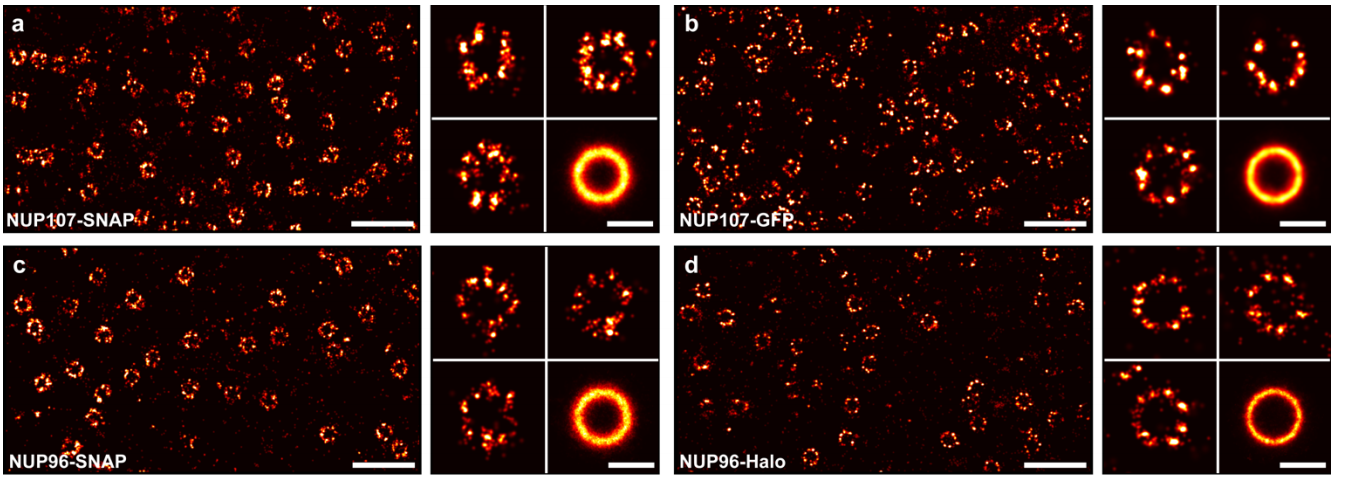
Nuclear pores as versatile reference standards for quantitative superresolution microscopy
Jervis Vermal Thevathasan, Maurice Kahnwald, Konstanty Cieśliński, Philipp Hoess, Sudheer Kumar Peneti, Manuel Reitberger, Daniel Heid, Krishna Chaitanya Kasuba, Sarah Janice Hoerner, Yiming Li, Yu-Le Wu, Markus Mund, Ulf Matti, Pedro Matos Pereira, Ricardo Henriques, Bianca Nijmeijer, Moritz Kueblbeck, Vilma Jimenez Sabinina, Jan Ellenberg, Jonas Ries
The Fruit Fly Brain Observatory: From Structure to Function
Nikul H. Ukani, Chung-Heng Yeh, Adam Tomkins, Yiyin Zhou, Dorian Florescu, Carlos Luna Ortiz, Yu-Chi Huang, Cheng-Te Wang, Mehmet K. Turkcan, Tingkai Liu, Paul Richmond, Chung-Chuan Lo, Daniel Coca, Ann-Shyn Chiang, Aurel A. Lazar
Comparing quality of reporting between preprints and peer-reviewed articles in the biomedical literature
Clarissa F. D. Carneiro, Victor G. S. Queiroz, Thiago C. Moulin, Carlos A. M. Carvalho, Clarissa B. Haas, Danielle Rayêe, David E. Henshall, Evandro A. De-Souza, Felippe Espinelli, Flávia Z. Boos, Gerson D. Guercio, Igor R. Costa, Karina L. Hajdu, Martin Modrák, Pedro B. Tan, Steven J. Burgess, Sylvia F. S. Guerra, Vanessa T. Bortoluzzi, Olavo B. Amaral
Ten myths around open scholarly publishing
Jonathan P Tennant, Harry Crane, Tom Crick, Jacinto Davila, Asura Enkhbayar, Johanna Havemann, Bianca Kramer, Ryan Martin, Paola Masuzzo, Andy Nobes, Curt Rice, Bárbara S Rivera-López, Tony Ross-Hellauer, Susanne Sattler, Paul Thacker, Marc Vanholsbeeck
 (1 votes)
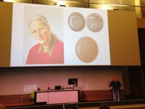 Jim Smith introduced the Beddington medal with heartfelt memories of Rosa Beddington and her time at the NIMR. Please read more of his thoughts
 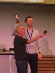 “The really impressive thing about David’s work is that he did not come to my lab to fit in with an existing line of research but created one of his own”. Jamie Davies, University of Edinburgh.
David received his undergraduate degree in Sport and Exercise Science at the University of Stirling (2010-2014). With this, he achieved a first-class honours degree and the prize for the best overall performance throughout a physiology related degree (British Physiological Society Undergraduate Prize). His dissertation project investigated associations between ADRB2 mutations (an adrenaline receptor gene in humans) and athlete status/athletic ability measurements. Subsequently, he was awarded a University of Stirling Head of School Summer Bursary Award to remain in Stirling during the summer of 2014 and investigate the relationship between transcribed ultra-conserved regions of RNA (T-UCRs) and the development of diet-induced insulin resistance in humans (Summer 2014). He then moved to the University of Edinburgh for his MSc by Research in Biomedical Sciences (2014-2015). Again, he received a distinction and was awarded the Class Prize for best student. During this time, he studied the physiology of S-acylation the regulation of skeletal muscle energy expenditure by an obesity-associated phospholipase as part of two research placements.
David has been awarded the Beddington medal for his exceptional work performed during his 3-year MRC-funded PhD at the University of Edinburgh with Prof Jamie Davies and Dr Peter Hohenstein (2015-2018): The thesis is titled ‘Mechanisms of kidney vascularisation and the roles of macrophages in renal organogenesis’. During his PhD, he gave several oral and poster presentations at national and international conferences, supervised students (including a Gurdon Summer Studentship Awardee), established numerous international collaborations, was awarded travel grants (including a BSDB Conference Grant), and reviewed manuscripts for leading journals (including Cell Reports, Angiogenesis, and Scientific Reports). He is now a post-doctoral fellow at the UK Dementia Research Institute (University of Edinburgh; 2019- present), continuing research in macrophage biology under the supervision of Prof Josef Priller. His current focus in on brain macrophages (microglia) in development, neurodegeneration, and aging.
Thesis description
Kidneys are specialised organs that clean the blood, removing waste while retaining what is useful. This requires a complex vasculature, and its formation as a foetus develops is poorly understood. I started my PhD research by using advanced microscopy techniques to visualise how blood vessels form in three-dimensions in the mouse kidney. In doing so, I identified when and from where the first blood vessels enter the kidney, and how blood vessels pattern at the edge of the kidney throughout development.
Blood vessels can form through angiogenesis (branching of new vessels from pre-existing ones) and/or vasculogenesis (assembly of new vessels from the coalescence of endothelial precursor cells). It has long been thought that a combination of both processes occurs during kidney vascularisation; however, my thesis work indicates that this concept may not be correct. My data instead suggest that kidney vascularization relies on growth and remodelling of pre-existing vessels (angiogenesis) and does not depend on vasculogenesis at any point (Publications 1 and 5 in CV). When assessing the entire 3D vascular tree of the kidney, isolated endothelial cells were never observed at any developmental age. Instead, all vessels, including the newly forming ones, were connected to pre-existing vessels that could be traced to the major circulatory vessels.
I then focused on the blood vessels at the edge of the kidney, which I found to consistently and accurately pattern around a special collection of cells – the cap mesenchyme. The cap mesenchyme contains cells that eventually become the cleaning tubes of the kidney, the nephrons. This cell population undergoes rounds of splitting at the kidney’s periphery. As this happens, I demonstrated that blood vessels migrate through the newly opened regions between the separating cap mesenchymal populations (Publication 1 in CV). This occurs in cycles throughout development and is likely to be vital for the oxygenation of the kidney’s outer region, the site where important processes such as nephron formation take place.
I determined that a signalling molecule, semaphorin-3f, and its receptor, neuropilin-2, were expressed in a pattern consistent with them having roles in this cyclical patterning of blood vessels; however, using mouse models where the genes for these molecules were deleted, I established that they were not vital for this process (Publication 2 in CV).
I next shifted my research focus towards a specialised cell type known as the macrophage (macro = big; phage = eater) in the developing kidney (Publication 3 in CV). Macrophages are immune cells best known for clearing foreign and damaged cells. These cells have vital roles during animal development, but little is known about their specific functions during kidney development.
Macrophages arrived in the mouse kidney early during its development, where they were required to clear away misplaced cells to ‘set-the-stage’ for early kidney development (Publication 6 in CV, under review). Throughout later development, most macrophages wrapped around blood vessels and I demonstrated their ability to eat endothelial cells (which usually line the blood vessels) and red blood cells (which are carried within them) within the kidney. I also established that kidney macrophages produced many molecules linked to blood vessel development, and so I examined the consequences of macrophage-loss on blood vessel formation. Blood vessels normally form continuous networks in the kidney; however, when macrophages were depleted (by blocking a macrophage-survival signalling pathway), connections between renal blood vessels were reduced (Publication 6 in CV).
Publications
“The really impressive thing about David’s work is that he did not come to my lab to fit in with an existing line of research but created one of his own”. Jamie Davies, University of Edinburgh.
David received his undergraduate degree in Sport and Exercise Science at the University of Stirling (2010-2014). With this, he achieved a first-class honours degree and the prize for the best overall performance throughout a physiology related degree (British Physiological Society Undergraduate Prize). His dissertation project investigated associations between ADRB2 mutations (an adrenaline receptor gene in humans) and athlete status/athletic ability measurements. Subsequently, he was awarded a University of Stirling Head of School Summer Bursary Award to remain in Stirling during the summer of 2014 and investigate the relationship between transcribed ultra-conserved regions of RNA (T-UCRs) and the development of diet-induced insulin resistance in humans (Summer 2014). He then moved to the University of Edinburgh for his MSc by Research in Biomedical Sciences (2014-2015). Again, he received a distinction and was awarded the Class Prize for best student. During this time, he studied the physiology of S-acylation the regulation of skeletal muscle energy expenditure by an obesity-associated phospholipase as part of two research placements.
David has been awarded the Beddington medal for his exceptional work performed during his 3-year MRC-funded PhD at the University of Edinburgh with Prof Jamie Davies and Dr Peter Hohenstein (2015-2018): The thesis is titled ‘Mechanisms of kidney vascularisation and the roles of macrophages in renal organogenesis’. During his PhD, he gave several oral and poster presentations at national and international conferences, supervised students (including a Gurdon Summer Studentship Awardee), established numerous international collaborations, was awarded travel grants (including a BSDB Conference Grant), and reviewed manuscripts for leading journals (including Cell Reports, Angiogenesis, and Scientific Reports). He is now a post-doctoral fellow at the UK Dementia Research Institute (University of Edinburgh; 2019- present), continuing research in macrophage biology under the supervision of Prof Josef Priller. His current focus in on brain macrophages (microglia) in development, neurodegeneration, and aging.
Thesis description
Kidneys are specialised organs that clean the blood, removing waste while retaining what is useful. This requires a complex vasculature, and its formation as a foetus develops is poorly understood. I started my PhD research by using advanced microscopy techniques to visualise how blood vessels form in three-dimensions in the mouse kidney. In doing so, I identified when and from where the first blood vessels enter the kidney, and how blood vessels pattern at the edge of the kidney throughout development.
Blood vessels can form through angiogenesis (branching of new vessels from pre-existing ones) and/or vasculogenesis (assembly of new vessels from the coalescence of endothelial precursor cells). It has long been thought that a combination of both processes occurs during kidney vascularisation; however, my thesis work indicates that this concept may not be correct. My data instead suggest that kidney vascularization relies on growth and remodelling of pre-existing vessels (angiogenesis) and does not depend on vasculogenesis at any point (Publications 1 and 5 in CV). When assessing the entire 3D vascular tree of the kidney, isolated endothelial cells were never observed at any developmental age. Instead, all vessels, including the newly forming ones, were connected to pre-existing vessels that could be traced to the major circulatory vessels.
I then focused on the blood vessels at the edge of the kidney, which I found to consistently and accurately pattern around a special collection of cells – the cap mesenchyme. The cap mesenchyme contains cells that eventually become the cleaning tubes of the kidney, the nephrons. This cell population undergoes rounds of splitting at the kidney’s periphery. As this happens, I demonstrated that blood vessels migrate through the newly opened regions between the separating cap mesenchymal populations (Publication 1 in CV). This occurs in cycles throughout development and is likely to be vital for the oxygenation of the kidney’s outer region, the site where important processes such as nephron formation take place.
I determined that a signalling molecule, semaphorin-3f, and its receptor, neuropilin-2, were expressed in a pattern consistent with them having roles in this cyclical patterning of blood vessels; however, using mouse models where the genes for these molecules were deleted, I established that they were not vital for this process (Publication 2 in CV).
I next shifted my research focus towards a specialised cell type known as the macrophage (macro = big; phage = eater) in the developing kidney (Publication 3 in CV). Macrophages are immune cells best known for clearing foreign and damaged cells. These cells have vital roles during animal development, but little is known about their specific functions during kidney development.
Macrophages arrived in the mouse kidney early during its development, where they were required to clear away misplaced cells to ‘set-the-stage’ for early kidney development (Publication 6 in CV, under review). Throughout later development, most macrophages wrapped around blood vessels and I demonstrated their ability to eat endothelial cells (which usually line the blood vessels) and red blood cells (which are carried within them) within the kidney. I also established that kidney macrophages produced many molecules linked to blood vessel development, and so I examined the consequences of macrophage-loss on blood vessel formation. Blood vessels normally form continuous networks in the kidney; however, when macrophages were depleted (by blocking a macrophage-survival signalling pathway), connections between renal blood vessels were reduced (Publication 6 in CV).
Publications
 (No Ratings Yet)