This editorial and interview appeared in Development, Vol 145 Issue 6
A new Editor-in-Chief for Development
Sarah Bray, Kate Storey, Katherine Brown
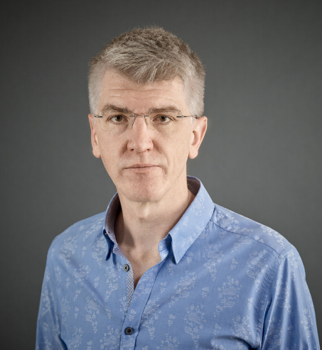
As many of you will be aware, The Company of Biologists initiated a search last year for a new Editor-in-Chief for Development, after Olivier Pourquié announced his intention to step down in September 2018. Following community consultation and a shortlisting and interview process, we are delighted to announce that James Briscoe will be the journal’s new Editor-in-Chief.
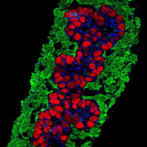
Many of you will already be familiar with James and his research. As a developmental neurobiologist working on the vertebrate spinal cord, he has a particular interest in using quantitative approaches to understanding how signalling pathways (particularly sonic hedgehog) regulate gene expression networks to control patterning, cell fate specification and growth of this tissue. His lab uses a range of in vivo, in vitro and in silico models to gain insight into this question at the molecular, cellular and tissue scales. For those interested in finding out more about James, we invite you to read our Spotlight interview with him elsewhere in this issue.
The position of Editor-in-Chief of Development is an important one in the developmental biology field – given Development’s position as a key community journal. The Editor-in-Chief is responsible for (among other things) appointing the editorial team, overseeing the handling of papers submitted to us, and setting new priorities for the journal. It was therefore important to us to gather input from the community as we set about appointing a successor to Olivier. We are hugely grateful to those of you – editorial board members, referees, authors and readers – who took the time to respond to our community consultation and provide feedback, not only on who they would like to see running Development, but also more broadly on how we are doing as a journal. Many of you are aware that, as a member of The Company of Biologists’ Board of Directors, James was part of the advisory group who initiated the consultation. He stepped away from any involvement in the process as soon as his name began to appear in nominations. The responses were collated by the three of us, and here we would like to share with you some of that feedback.
Your responses were, in general, consistent with our own assessment of the journal’s standing. We heard that Development is seen as a high-quality, rigorous venue for publication, with excellent academic editors, a ‘tough but fair’ review process and good production values. We were delighted that many respondents picked up on some of our more recent innovations – cross-referee commenting during peer review, openness to preprints, and our strong online presence, particularly through our community blog the Node. Given that the Node was launched at least in part in response to the consultation we conducted when looking for a new Editor-in-Chief to replace Jim Smith, it was fantastic to see how much traction the Node has now gained in the community. A recurring theme in the feedback on the journal’s strengths was that Development is the ‘journal of reference’ for the field, and that Development papers ‘stand the test of time’. If this is how we are seen in the community, we are clearly doing something right!
You also told us that Development can be seen as ‘hard to get into considering its impact factor’ and that competition from newer journals means that Development is sometimes seen as a less attractive choice, especially for early career researchers. These are of course issues of which we are all too aware. As signatories of the San Francisco Declaration on Research Assessment (DORA), we would argue that impact factor is a poor proxy for journal quality, and an even worse one for individual papers. That this measure still holds so much sway, particularly in certain geographic regions, is disappointing, and is something that The Company of Biologists – as a supporter of the newly revamped DORA project – is trying to change. Nevertheless, Development recognises that you as authors have a wide choice of journals to which you can submit, and the team will continue to work to make the journal an attractive option.
Looking to the future, we were delighted to hear that many respondents feel it is important for Development to maintain and strengthen the focus on stem cells, regeneration and human development. However, this is clearly not the only area in which developmental biology is growing: there was strong support for increased visibility in the genomics, biophysics, quantitative and systems biology, and evo-devo fields. These are all fields that Development has highlighted as future priorities for the journal, and the team looks forward to working with many of you to realise the potential of these areas.
Away from consideration of specific research areas, we heard that Development should do more to support and advocate for the field (this is actually something the Development team is actively working on – look out for news in an Editorial in the near future!) and that we should continue to support our communities through our charitable activities – and, if possible, grow these. Development and The Company of Biologists see these activities – meeting grants, travelling fellowships, workshops and so on – as a key part of our ‘raison d’être’, and you can rest assured that The Company will continue to give back to the community as much as we can.
On the specifics of the choice of new Editor-in-Chief, your feedback helped us to draw up a ‘wish-list’ for the kind of person we wanted to lead the journal. We were looking for an individual whose research is at the cutting edge of developmental biology, with interdisciplinary skills, broad interests in the field and a strong vision for the future of the journal – to build on the developments that Olivier initiated, bring new ideas to further strengthen the journal, and be an active advocate for Development. James fulfils all these criteria and more, as evidenced by the fact that his name came up over and over again in the feedback we received; in fact, he was suggested by almost half of those who provided specific names – around four times as often as any other single individual. The unanimous view was that James is an outstanding candidate to succeed Olivier, and we are delighted that he has agreed to take on this role.
Over the next few months, James will be working alongside Olivier to ensure a smooth handover when Olivier steps down in September. You’ll be hearing more from him later in the year when he officially takes over the editorship. As James gets ready to take the reins, he (and the journal more broadly) welcomes any feedback or suggestions that you might have for how you would like to see the journal develop. In the meantime, we hope you will join us in congratulating James on his appointment, and in wishing him luck as he steps up to this challenging and important position!
An interview with James Briscoe
Katherine Brown
James Briscoe is a group leader at The Francis Crick Institute in London. His lab’s research focusses on the developing vertebrate spinal cord, with a particular interest in how sonic hedgehog gradients, and the downstream signal transduction and transcriptional networks, regulate the development of this tissue. In September 2018, James will become the new Editor-in-Chief of Development. We met with James to discuss his career and research interests, the importance of interdisciplinary thinking in developmental biology, and his views on the current state and future opportunities in scientific publishing.
Let’s start at the beginning: how did you first become interested in biology?
There was never a moment of epiphany and I didn’t have a well thought out plan. At school I enjoyed science and did well at it, but I was not aware that a career in academic research could be a possibility. Around the time I was thinking about going to university, I watched the film Life Story, based on Jim Watson’s book The Double Helix, and read Richard Dawkins’ The Blind Watchmaker. I think both of these tipped me towards biology. I also remember reading Microbes and Man by John Postgate, which is less well known but a really superb book, and it’s probably at least partly a consequence of this that I ended up at Warwick University studying microbiology and virology.
During your PhD, you worked on interferon signalling in cell culture. What attracted you to that field, and what then prompted you to move into developmental neurobiology for your postdoc?
During my undergraduate degree, I became fascinated by how the study of microbiology and virology had taught us fundamental aspects of eukaryote molecular biology. This led to an interest in interferons – the secreted cytokines that are the major cellular response to viral infection – and I joined Ian Kerr’s lab at the Imperial Cancer Research Fund (ICRF; which later became Cancer Research UK and then merged to form part of The Francis Crick Institute) to study the mechanism of interferon signalling. By chance my timing was perfect, as we were identifying what became known as the Jak-STAT signalling pathway. Our neighbouring labs were also investigating signal transduction and elucidating pathways such as MAP kinase, RAS, PKA and more. I became very interested in understanding how cells perceive and respond to extracellular signals. Most of the groups at ICRF used cell lines but there were a few developmental biology labs, including those of David Ish-Horowitz, Julian Lewis and Phil Ingham. I went to their seminars and this introduced me to the beauty of embryos and to hedgehog signalling. I decided I wanted to continue studying signal transduction but to learn developmental biology so that I could investigate signalling in its natural environment rather than just in cell culture.
You joined Tom Jessell’s lab at what must have been an exciting time: sonic hedgehog (Shh) had only recently been cloned and it was known to be involved in spinal cord patterning, but little was understood about how the system worked. What were the questions you set out to answer during your time there?
Again, I was lucky with my timing. As you say, it was a really exciting time to be in the Jessell lab and at Columbia University. I wanted to understand Shh signalling and how it could function in a graded manner to control cellular responses. Very little was known about the molecular mechanism of signal transduction – I think even today we still have an embarrassingly poor understanding of this pathway – and I thought we could figure this out using the neural tube. Being in the Jessell lab, however, I soon became fascinated with and distracted by broader aspects of developmental neurobiology and how the spinal cord forms and functions. It became apparent that understanding the role of Shh in the spinal cord required studying the gene regulatory network that it controls and this has become the passion I’ve pursued ever since.
So what are the problems you’re working on now?
Broadly, they’re still the same problems that I was interested in when I was in Tom’s lab. For me, the fundamental question in developmental biology is how the right cells are produced in the right place, at the right time and in the right amounts in a developing tissue. The spinal cord turns out to be a fantastic system to address this and it’s revealing general principles that are applicable to many, if not most, developing tissues. Addressing these issues covers some of the most basic questions in biology. How is gene activity controlled? How is cell function determined? How are tissues shaped and organised from cells? Over the last couple of decades, developmental genetics has identified at least some of the key players in these processes. Now, new approaches and technology, from imaging to genomics to genome engineering, are providing unprecedented insight and resolution. I’ve been keen to move beyond purely qualitative explanations to a more dynamic and quantitative understanding of how the neural tube is formed and patterned. We’re trying to bridge scales from molecules to cells to tissues, to explain how the cells make the key decisions and how this guides the assembly of a functional, well-organised neural tube. I think this is of fundamental interest, but it also has practical implications for understanding disease and for progress in regenerative medicine and tissue engineering.
You’ve embraced mathematical modelling of developmental systems: why do you think this is important, and what are the challenges involved in trying to model something as complex as the developing spinal cord?
This was a decision I made about 10-12 years ago and, in part, it is because of the complexity of the problems we were investigating that I thought mathematical modelling was important. We were analysing increasingly complicated gene regulatory networks and we were trying to get away from simplistic reductionist descriptions to find explanations of how the system functioned as a whole. It’s often difficult to form an intuitive understanding of even relatively small systems if there are multiple interactions and feedback – it’s very easy to fool or confuse yourself. Mathematical modelling provides a rigorous way to describe and investigate a system. It tests whether your assumptions and interpretations are compatible. And different kinds of modelling can be useful at different levels of organisation, from the molecular to the tissue level, allowing us to look at the problem at different scales. I’ve been fortunate to have had great collaborators over the years that have taught me a lot of maths and physics. One thing I’ve learnt from this is that developing productive interdisciplinary collaborations takes a long time and requires a lot of trust and patience, but the investment can be very rewarding. We now think of mathematical modelling as just one of the available techniques that we use to tackle a problem, alongside more conventional molecular and cellular experiments.
More broadly, how important do you think it is for today’s young scientists to think interdisciplinarily?
Perhaps because it operates across multiple scales (both spatially and temporally), I think developmental biology has always been interdisciplinary. The molecular genetics revolution that transformed our field over the last 30-40 years is just one example of this. The importance of genetics is now taken for granted but it was once seen as pioneering and innovative. Today, it’s increasingly recognised that biologists need to be more quantitative and computationally literate and many of the applicants we see at The Francis Crick Institute have these skills. Nevertheless, it is something that needs to be strengthened through training at undergraduate and graduate levels. Having said that, one of the pleasures of academic research is that you’re continually learning and challenging yourself, so there are always opportunities to fill any gaps in your knowledge and learn something new. I often participate in advanced study courses run by organisations such as the Marine Biological Laboratory at Woods Hole and the Kavli Institute for Theoretical Physics at UC Santa Barbara. Even though I’m nominally ‘faculty’ on these courses, I usually end up learning at least as much as the students.
Developing productive interdisciplinary collaborations takes a long time and requires a lot of trust and patience, but the investment can be very rewarding
You recently moved into The Francis Crick Institute, which formed through the merger of NIMR at Mill Hill (where your lab was based), Cancer Research UK Lincoln’s Inn Fields and a number of other partners. How has the move been?
The planning took a long time, but the move itself went much more smoothly than I had anticipated. It’s been less than 18 months since we moved but it feels much longer; I guess that means we’ve settled in well. While I miss the charm and familiarity of our old institute, the new building more than makes up for it. The merger of the two institutes and our new partners mean I have new colleagues, which is stimulating and invigorating. I also love being in the centre of London – both because of the proximity to other academic institutions, and because we’re in a very vibrant part of town just minutes from the West End.
You’re active on Twitter: where do you see the value in social media for science and scientists?
I firmly believe that communication is a central part of science and academia: knowledge that isn’t passed on is wasted. Although not everything that appears on Twitter necessarily contributes to human progress, I enjoy being involved and I find it very useful. I get a lot of scientific information from Twitter. Particularly in fast-moving fields; for example, when CRISPR/Cas9 was coming on to the scene, I found Twitter was a great way to stay up to date and hear the latest developments. Also, like many scientists, I have friends and colleagues all over the world and Twitter is an easy way to stay in touch as well as to share and discuss (albeit briefly) papers and ideas.
You’ll be taking over as Development’s Editor-in-Chief in September, but you’ve been associated with our publisher, The Company of Biologists, for many years. Can you tell us a bit about your role as Director on The Company’s board and why you chose to get involved?
Yes, I’ve been involved a long time – since 2004. The Company of Biologists is a not-for-profit scientific publisher that publishes five journals: Development, Journal of Cell Science, Journal of Experimental Biology, Disease Models & Mechanisms and Biology Open. But unlike commercial publishers such as Elsevier and Springer Nature, we put the money that we make back into the scientific community. We give away about £1 million per year across our range of charitable activities: funding scientific societies such as the British Society for Developmental Biology (BSDB), sponsoring conferences, running workshops, and promoting research collaboration through travelling fellowships. We also support non-profitable, community-focussed activities such as the Node and preLights. There are 17 Directors of The Company and we’re almost all active scientists. We aren’t directly involved in the day-to-day business but we are responsible for the major strategic decisions, such as deciding to launch a new journal or start a big new project. In my role as Director, I was very involved in setting up the Workshops programme, and I’ve chaired the Grants Committee for the last few years, which decides how we spend our charitable funds. I’ve really enjoyed being on the board: it’s been a great opportunity to support the scientific community and I hope we’ve done at least a little bit of good.
Remember, by submitting your paper to Development or reviewing a paper for us, you’re supporting not only the journal but also all our other charitable activities
Scientific publishing is going through some significant changes at the moment, with the open access movement, developments in online publishing technology, the rise of preprints and so on. What do you think the future holds for small not-for-profit publishers like The Company of Biologists?
It’s definitely an exciting time in the publishing world. While all the changes create a lot of uncertainty both for people working in publishing and for scientists, I think there are lots of opportunities for small publishers such as The Company of Biologists. One of our advantages is that we’re run by scientists and we can respond in ways that help scientists and science and we’re not driven by profit margins. We’re also small enough and bold enough to experiment occasionally. Eight or nine years ago, we saw the increasing use of social media in our labs and heard calls from the community for an online discussion forum, so we launched the Node as a way to encourage informal discussion and communication about science within the developmental biology community. This has continued to grow in popularity. Just last month, we launched another experiment – preLights, the preprint highlighting and commenting service. This was in response to the growing numbers of preprints and it will be interesting to see how this develops in the coming months and years. More broadly, The Company’s goal is to support and inspire our fields and the people working in them and these aims will continue to drive our thinking and any future innovations.
Fundamentally, though, the journals and the research papers we publish remain at the heart of The Company of Biologists and it’s crucial that we all continue to read, write and referee for Development, if we are to continue to benefit from all the other initiatives and charitable support that The Company offers. Remember, by submitting your paper to Development or reviewing a paper for us, you’re supporting not only the journal but also all our other charitable activities.
Given the huge volume of published research, it’s increasingly difficult to keep up with the scientific literature. How much do you read and how do you choose what you read?
I agree it can be daunting keeping up with literature. I have a routine where I read a paper first thing in the morning when I get in to work (often I’ll choose it the night before). So I read at least one paper every day, although usually I’ll also read or review an article during the day. I try to read things outside my own research area, as this can provide ideas for my own work. I often pick up paper or preprint suggestions from people I follow on Twitter and I expect I will increasingly use preLights to get recommendations. I also think this is where community journals such as Development play a crucial role. Every issue of Development has papers handled by academic editors who are leaders in the field, so it offers a curated collection of the latest developmental biology research selected by experts. I always browse through the list of newly published papers and I often find myself picking one or two to read – it might be on neural development but as often as not it’s a Drosophila paper, an evo-devo study or even sometimes a plant paper.
And how would you like to see the journal (Development) evolve under your editorship?
I’m not taking over until late September 2018, so although I have lots of ideas I’m still refining my plans and taking suggestions. Olivier has done a great job during his time as Editor-in-Chief. He’s strengthened the journal, incorporating and encouraging new areas of research, and I certainly want to keep building on his accomplishments as developmental biology continues to change and grow over the next few years. I’m also very keen that Development continues to innovate and support our community. I’d welcome suggestions from readers, authors and referees about what we should do. What areas of science should we be encouraging in Development? What more can Development do to help our field? I’d urge anyone with thoughts or ideas to contact me.
Finally, is there anything that Development readers would be surprised to find out about you?
Some people find it surprising that I’m really a country boy at heart. Despite having lived most of my adult life in big global cities – London and New York – I was brought up in a 300-year-old thatched cottage on the Sussex Downs. I spent most of my teens working in stables and riding horses. We always kept lots of animals. As well as cats, dogs and rabbits we also had hens, ducks and goats; as a consequence, I can milk a goat.
 (1 votes)
 Loading...
 (No Ratings Yet)
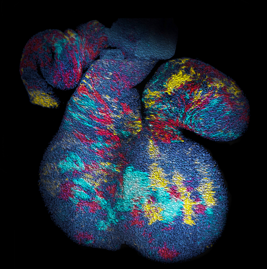
 (16 votes)