There are around 10 funded places available for early career scientists to attend the workshop, along with the 20 speakers. Deadline for applications = 16 February
Wellcome Trust Cancer Research UK Gurdon Institute and Wellcome Trust/MRC Stem Cell Institute
Salary – £25,728 – £29,799 or £31,604 – £38,833
Closing date: 16th February, 2018
Applications are invited for a postdoctoral research assistant/associate to work on functional validation of targets for lung regeneration using human organoids. This project will be jointly supervised by Dr Joo-Hyeon Lee (Stem Cell Institute; https://www.stemcells.cam.ac.uk/research/pis/lee) and Dr Emma Rawlins (Gurdon Institute; http://www.gurdon.cam.ac.uk/research/rawlins) and is funded by Astra Zeneca. Recent research in the two labs has focused on the development of organoid systems for studying lung development and maintenance (Lee et al., Cell, 2017; Nikolic et al., Elife, 2017). We now aim to recruit an outstanding individual who is interested in lung developmental mechanisms, and their potential therapeutic applications, in order to extend our current organoid work into lung regeneration.
Applicants should have a PhD in a relevant subject, or be close to completion of their degree. Expertise in general areas of developmental/stem cell biology including live cell imaging, image analysis, CRISPR genome editing, flow cytometry analysis, and cell signalling mechanisms would be suitable for this position. Experience of in vitro models would be an advantage.
The successful applicant will learn human lung organoid systems and have a strong publication record and an excellent aptitude for research and career development. We are looking for applicants who are collaborative with effective communication skills and enjoy working in a team. Proven capacity to design, execute, and interpret your own experiments is essential.
Limited funding: The funds for this post are available for 2 years in the first instance with year 2 of funding being contingent on progress made in year 1.
To apply online for this vacancy, please click on the ‘Apply’ button below. This will route you to the University’s Web Recruitment System, where you will need to register an account (if you have not already) and log in before completing the online application form.
Applications should include a CV and a brief statement outlining key areas of expertise and reasons why you would like to join the project. Informal enquiries can be addressed to Dr Joo-Hyeon Lee (jhl62@cam.ac.uk), or Dr Emma Rawlins (e.rawlins@gurdon.cam.ac.uk). Please quote reference PR14553 on your application and in any correspondence about this vacancy.
The University values diversity and is committed to equality of opportunity. The University has a responsibility to ensure that all employees are eligible to live and work in the UK. Benefits include generous maternity/paternity leave, flexible working and funds for returning carers and other family-friendly schemes.
CENTURI seeks to attract outstanding physicists, mathematicians or computer scientists with a theoretical and/or computational biology project.
The selected candidates, who are expected to bridge biology and other disciplines will be affiliated to two institutes and will be offered competitive funding.
Applicants should send a curriculum vitae with a complete list of publications, a 2- page summary of research achievements and projects in English, and the names and addresses of three references to Thomas LECUIT (info@centuri-livingsystems.org), before February 26th, 2018. Short listed candidates will be invited to submit a full proposal by 22nd April, and for an interview in May.
Over the past fews years I’ve done a lot of SciComm and really enjoyed everything I learned and all the experiences I got from running PhD websites and the Pint of Science Ireland events.
As many researchers are looking to get experience in scientific writing we’ve put together a “Digital Scientific Writer Traineeship” program.
If you are interested you can find our more here, would be great to hear from interested candidates:
Plenary talk given at the School of Biological Sciences symposium on Friday, 12 January 2018
Matthew Cobb is an inspiring advocate and communicator of science, in particular of biology. This is clearly reflected in his books and articles about the history of biology (and beyond), and his various radio programmes reflecting on past and contemporary science topics. Recent highlights are his book “Life’s Greatest Secret: The Race to Crack the Genetic Code” (shortlisted for the 2015 Royal Society Winton Prize for Science Books) as well as his BBC Radio programmes “Editing Life” (a well-balanced analysis of the opportunities and risks of the newly emerging CRISPR gene editing techniques) and ”Sydney Brenner: A Revolutionary Biologist“ (about the life of this outstanding scientist).
As a result of working on two BBC radio programmes and a project on scientific collaboration (supported by a Sydney Brenner Research Scholarship from the Cold Spring Harbor Laboratory), Matthew has been reflecting on what are the magic ingredients that produce great biology. The talk he presented at Manchester’s School of Biological Sciences Symposium on 12 January 2018 (see re-recording of this entire talkhere), was an inspiring and enlightening summary of the key ideas extracted from this endeavour.
In his talk, Matthew first gave a definition of “great biology” which he proposes as “influencing our thinking about life, from nucleic acids to ecosystems, in ways that are long-lasting and opening up new perspectives”; he pointed out that these traits are “not necessarily associated with large grant funding, publication in ‘high-ranking’ journals or practical or therapeutic implications”. He then moved on to explain what he thinks the five magic ingredients are that promote great biology, illustrating each point with enlightening examples of scientists and anecdotes from the biology’s past (one of them about amateur scientists!) – always with a view to current practice, and providing inspirational advice for the future. Certainly, I do not want to spoil the watching of Matthew’s talk by giving away the concrete examples, but shall briefly discuss the fundamental ingredients that Matthew highlighted, mixed in with some of my own thoughts.
The first advice is to be early in the game. To be at the forefront of new developments or even being a trend setter requires vision, meaning the ability to recognise today what will be important tomorrow, but also living with the right mind at the right time. Certainly, the chances of being in the early game is helped by keeping an open mind for the bigger picture and unanswered questions, rather than getting lost in detail. And there are measures that can be taken to this end. For example “The encyclopaedia of ignorance – life sciences”, edited by R. Duncan and M. Weston-Smith in 1977/78, was an inspiring attempt to make renowned scientists of the time think aloud about the future of the various life science disciplines; and I could imagine that this was a great catalyser for curious readers to awaken their pioneer spirits. Another important means to foster a culture that honours forward-looking thinking, is to free time for, and take an interest in, true dialogue and discussion. For this, we also need to generate the right environment and opportunities, such as to ensure that all new buildings have informal social spaces where people can meet and discuss. But also, when reviewing manuscripts, we need to be open to new ideas so that they can develop – rather than brush them away with a tunnel view. In a recent case of a fellowship application I experienced, ten lay reviewers gave maximum marks; in contrast, three specialist reviewers rated the application as being mere average, mostly based on technical detail and blind to the concepts and ideas behind that application and its potential to develop great science. It is the latter that we all need to recognise and foster.
The second advice is to think and plan long-term. Key to this strategy is the art of long-term objective setting guided by fundamental questions and their break-down into medium and short-term goals that can be dealt with at a time. Again, this is not made easy in the current science and science funding landscape, but all arguments are in favour of long-term developments. The “skimming-the-cream-and-moving-on” mentality might be good for the careers of individual scientists, but perhaps not so good for the actual science. Similar conclusions are drawn by the article “How should novelty be valued in science?” published by B. A. Cohen in Elife last year. Cohen’s analysis of arguments from the philosophy of science (how reliable knowledge is generated) and from the sociology of science (how science communities work most efficiently) clearly dismantle the excessive demand for novel mechanisms (as opposed to gradual development of concepts) that governs contemporary science funding and publishing policies. This trend has become a significant inhibitor of high quality science, and the alarming increase in reports about non-reproducibility of published results is a logic consequence of these developments, and poses a risk to the future of science.
The third advice is to choose and/or develop the right tools. Apart from the examples Matthew mentioned in his talk, I can mainly speak for the biomedical sciences; my personal experiences lie with Developmental Biology and Drosophila research within, both of which are under threat by the growing emphasis on translational (i.e. clinical) research. Many politicians and funders seem to focus increasingly on the final steps of the discovery pipeline, fixing their gaze on expected short-term returns. They overlook, and often don’t understand, that longer-term basic research (which is typically not performed on humans but on animal model organisms as the more versatile “tools”) generates the creative pool of ideas that will feed the bench-to-bedside pipeline in the future. But even within fundamental research, “tool choice” has shifted away from the use of genetic invertebrate model organisms, such as C. elegans or Drosophila, to the use of mouse models. This development fails to recognise that research concerning evolutionary conserved processes and mechanisms is done far more productively and economically in the smaller model organisms; as Hugo Bellen once stated [LINK]: “You get 10 times more biology for a dollar invested in flies than you get in mice”. I therefore believe that we would see greater biology if grant panels looked more critically at the justifications for the model organisms that are being proposed for projects – and that such a practice would free up funds for a greater variety of science.
The fourth advice is to build good teams. Matthew highlighted three factors. Ideally have at least one person with great vision in your team that has the genius to drive the conceptual ideas behind the work. Be complementary in your expertises. But, most importantly, foster a productive culture of discussion: be prepared to play around with ideas even if they appear mad at first sight; and enjoy and capitalise on friction coming from different opinions and opposing thoughts, so that the best ideas can emerge and be developed into scientific experiments.
The fifth ingredient, least accessible to advice, is luck! We all know this component as a potential maker and breaker of careers. This said, bad luck certainly breaks those who either lack the vision or disregard the above advice. However, the good scientists will be prepared: either by taking the long view that provides flexibility for alternative strategies and approaches, or by using failure as an opportunity to rethink and take new directions – perhaps providing a new chance to be early in the game! As Napoleon supposedly said: “I believe in luck, and the wise man neglects nothing which contributes to his destiny”.
But even if we are eager to follow this advice, the circumstances of the current science landscape might not be in support of great biology. Harmful metrics, counterproductive expectations, bureaucracy, a shift in public opinion, and harmful publishing policies “bully us into bad science” and promote self-focussed communities and mechanisms that inhibit true progress and passion for science. Some examples were given above and many more can be found in other publications (e.g. Lawrence; Cohen; Young; Smaldino; Martínez-Arias; Nerlich). But, as I argued in arecent blog, we must organise ourselves and engage as a community in dialogue with each other, clinicians, policy makers, funders and publishers – all with a view to improving the biology we do. For such dialogue, we need arguments and elevator pitches that are engaging and convincing. To this end, Matthew’s talk is a rich resource of ideas and well thought-out arguments that we all should listen to and take on board.
In our recently published paper, we discovered that the Hippo pathway transcription factors have an unexpected role in creating the conditions for the zebrafish body to extend posteriorly during embryogenesis, as well to form the precursors of the dorsal and ventral fins. Here is the backstory of the twists and turns that lead to these results, and why we think it is important.
It began with an email…
One day almost two years ago, I got an email from Jason Lai, a grad student in the lab of my friend and colleague Didier Stainier, asking if I would be interested in studying a mutant they made using CRISPR. Didier’s lab does great work studying heart development, but this mutant also has an amazing effect on the formation of the embryonic body, which I study, and so they asked if I would be interested in pursuing it. The email contained a movie, which shows that the body starts to form normally, but then as the tail starts to grow out it suddenly collapses. I was so excited by this movie that I immediately wrote back to start the collaboration, without doing any background reading first.
Comparison of sibling (left) and yap1;wwtr1 double mutant (right) embryos. The movie begins at the 3-somite stage.
Jason and Didier’s mutant eliminates the function of two transcription factors in the Hippo pathway called Yap1 and Wwtr1 (a.k.a. Taz). Single mutants in either Yap1 or Wwtr1 have no effect on forming the body, but when both are mutated the posterior body fails to form normally as shown in the movie. When I started reading the literature, I sadly discovered that in the fish species medaka a very interesting Yap1 mutant called hirame had already been described a year earlier in a Nature paper. In this paper, the authors proposed a model in which an apparently ubiquitous Yap1, acting through the Rho GTPase Activating proteins (called Arhgaps), provides tissue tension that allows the embryo to oppose the forces of gravity since the hirame embryos look like they have partially collapsed along the dorsal-ventral axis1. The authors hadn’t examined the formation of the posterior end of the body, but in a figure showing a hirame embryo it clearly looked to me like the posterior body was defective, much as we were seeing in zebrafish. The evidence for the role of the Arhgaps was actually based on a human cell culture system and not on any strong evidence from studying hirame embryos. However, since my lab had been starting to investigate the role of Arhgaps in zebrafish, I thought we could at least add a tiny bit to the literature by figuring out exactly which Arhgap was involved downstream of Yap1 and Wwtr1 in the embryo.
Never discount the value of serendipity
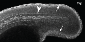
As luck would have it, my department had just hired a terrific new Assistant Professor named Young Kwon, who is an expert in Yap1 in Drosophila. When I told Young about this mutant that I was starting to work on, he offered me an aliquot of a commercial Yap1 antibody he had. I didn’t think it would be very valuable since the hirame paper implied Yap1 was everywhere, but never being one to turn down a free reagent I gave it a try. The results really shocked me, since not only was the Yap1 localized, but it was localized to the skin and notochord (Fig. 1)! The reason this was so surprising to me was that the mesoderm is the engine that drives the elongation of the body as people like Ray Keller have shown for a long time, with the notochord only providing a minor component, whereas the skin is something that most of us just ignore. Jason then followed up by looking at Wwtr1 with an antibody, and found exactly the same localization. We also looked at medaka Yap1, and this was also in the same places, so this was not just some zebrafish weirdness.
Figure 1 Yap 1 expression in the posterior end of a zebrafish embryo. Arrows show expression in the skin and arrowhead shows expression in the notochord.
Not knowing what to do we turned to RNA-seq, since I hoped that levels of Yap1 and Wwtr1 that did not show up with the immunostaining would still be working to activate mesodermal genes. Jason did the RNA-seq experiment in Didier’s lab, and while the embryonic localization of most of our top hits (none of which were arhgaps) were not known, when I did the in situs I found that they were all expressed in the skin and notochord. Thus, there was no easy solution to this conundrum.
I get by with a little help from my friends
I was constantly puzzling about what the skin and notochord had to do with the formation of the body when I suddenly remembered some recent beautiful papers from another friend and colleague, Scott Holley2-4. Scott also has been interested in how the vertebrate embryo forms, and particularly the formation of the somites. Scott and his lab have shown not only that Fibronectin and Integrins are important in the morphogenesis of the embryonic body, but the very surprising result that while Fibronectin is secreted by all cells, it only assembles into extracellular matrix around the borders of each somite, much the way a pillowcase covers a pillow. Part of this Fibronectin matrix occurs where the somites touch each other (the intersomitic boundary), whereas the rest of the matrix is where the somites touch the skin and notochord. What really grabbed my attention was a line in one of the papers that the Fibronectin “matrix pattern gives rise to the tissue mechanics of the mesoderm required for body elongation”3. In other words, Fibronectin is providing an adhesive surface that allows the mesodermal engine to grab onto in order to be able to drive elongation of the body.
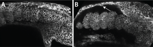
Scott kindly gave me the very detailed protocol for Fibronectin staining his lab had carefully worked out, and with this I could see that the Fibronectin matrix was present in yap1;wwtr1 double mutants, but clearly defective. Natalie, my lab manager, and I then examined normal embryos where we expressed a dominant-negative Fibronectin, and produced embryos that looked very much like the yap1;wwtr1 mutants. So if the issue was a failure of the mesoderm to attach to the skin and notochord we might expect to see adhesive defects between these tissues, and this is exactly what we saw. For example, imaging in live embryos we could see the skin rip away from the mesodermal tissue as the mutant phenotype got progressively worse, something we didn’t see in normal embryos (Fig. 2).
Figure 2 Loss of adhesion in yap1;wwtr1 embryos. (A) In wildtype embryos that skin adheres tightly to the mesoderm whereas in the mutant (B) the skin pulls away from the mesoderm (B).
So what does it mean?
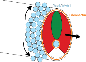
Our results show that the Hippo pathway transcription factors play a critical role in forming the posterior body of the early fish embryo by regulating a set of genes in the skin and notochord that allow the Fibronectin matrix to assemble normally at the border with the mesoderm. Thus, when the mesoderm starts elongating, it has something strong to grab onto and push against (imagine the difference between walking on a normal floor versus walking on one covered in oil). In addition, this Fibronectin matrix provides a surface for the skin cells themselves to undergo very interesting morphogenetic movements that Natalie elucidated, which allow the dorsal and ventral fins of the embryo to form (you will have to read the paper to learn more about that but it is summarized in Figure 3).
Figure 3 Model. Yap1 and Wwtr1, expressed in the skin and notochord (blue), activate genes necessary for the formation of a Fibronectin matrix (orange), which is required for the body to elongate as well as for the epidermal cells to migrate dorsally and ventrally to form the fins.
How all the Yap1 and Wwtr1 targets affect Fibronectin assembly is a big unanswered question. While the arhgaps are essentially unchanged in the yap1;wwtr1 mutants, of the many genes that do change, none are obviously known to affect Integrin and Fibronectin, so clearly there is much more to learn about this story.
CENTURI aims at recruiting postdocs willing to work in an interdisciplinary life-science environment.
This year, we will recruit postdocs on the interdisciplinary projects that are listed below.
Applications are invited from highly motivated individuals who are interested in fundamental mechanisms of neuronal migration and axon guidance. The main focus of our research is to understand the molecular and cellular mechanisms underlying the development of neural circuits using the embryonic spinal cord as a model system (http://www.ucmm.umu.se/english/research/sara-wilson/). The fellowship is funded for two years and is available immediately upon approvals. The laboratory is located at IMB, Umeå University, Sweden. IMB is an interdisciplinary department, which focuses on questions in basic and medical sciences and provides an interactive modern environment with easy access to good core facilities. The working ‘day to day’ language in the laboratory is English.
Requirements: Individuals with a background in developmental biology, neuroscience, molecular and or cell biology or related discipline and with a keen interest in developmental neuroscience are encouraged to apply. The successful candidate will have or about to receive a Ph.D. in a relevant discipline and be proficient in written and spoken English. Technical experience with imaging, vertebrate embryonic model systems – especially mouse handling and genetics, chick or mouse embryo electroporation and /or neurite outgrowth assays, is a big advantage although full training will be given. Experience with molecular/cellular biology will be positively evaluated. The most successful candidatewill have ahigh level of motivation, be creative, organized, rigorous and have the ability to work both independently and within a team.
Please submit your application (reference 2018SW200) by 23rd February 2018 to anita.dreyer-perkiomaki@umu.seby sending the following documents as pdf files:
1) A short cover letter (not more than 1 page) to include a description of your research experience and suitability for the position.
2) Curriculum Vitae including: publication list, technical expertise, names and contact information for three referees.
Informal enquiries may be directed to Dr. Sara Wilson (sara.wilson@umu.se).
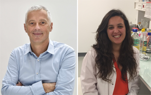
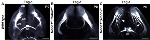
Vertebrate brain development is characterised by cell migration, as neurons are often born far from where they need to end up. Migration is regulated by guidance cues and their receptors, but, problematically, many of these molecules are expressed throughout the brain, complicating efforts to spatially and temporally pin down their function. A paper in the latest issue of Development addresses this problem using cell-specific knockouts for Slit and Robo, crucial migration regulators. We caught up with first author Chloé Dominici and her supervisor Alain Chédotal of the Institut de la Vision in Paris to find out more.
Alain and Chloé
Alain, can you give us your scientific biography and the questions your lab is trying to answer?
AC Our lab studies the cellular and molecular mechanisms controlling the development of brain connections, mostly focusing on so-called commissural neurons which interconnect the right and left side of the nervous system. We mainly use genetics and mice but have also started to study other organisms (birds, amphibians, marsupials, humans etc) to address the question of the evolution of midline guidance processes.
I started to study the development of commissural circuits during my PhD in Paris, using various neuroanatomical methods including electron microscopy. For my post-doc, I wanted to learn other techniques such as genetics, molecular biology and biochemistry and joined the lab of Corey Goodman at UC Berkeley who had just discovered the first vertebrate Semaphorin. It was an exciting time as little was known about midline crossing in vertebrates. Within a few years all the main guidance cues, Slits, semaphorins, netrins, ephrins, and their receptors were characterized. After starting my own group, I continued to work on axon guidance, but also neuronal migration, angiogenesis and myelination. In parallel, we have been developing new 3D imaging techniques combining whole-mount immunostaining, tissue clearing and light-sheet microscopy, to facilitate the phenotypic analysis of our mutant mice.
And Chloé, how did you come to be involved with this project?
CD When I first joined Alain’s lab, my project was aiming to understand the role of Slit2 in the development of motorneurons. We initially attempted to do so by using conditional lines and wanted to show, as a control, that we could also remove Slits in the floor plate. After several attempts, I wasn’t able to remove Slits in the motorneurons but the conditional line for the floor plate worked very well. So, we began to analyse these mutants by focusing mainly on the hindbrain as it had been previously reported that Slits were involved in the migration of precerebellar neurons, a model system in the lab.
Lightsheet microscopy of the forebrain in different genetic conditions, from Fig 1, Dominici, et al. 2018
What were the limitations of previous genetic studies on the role of Slit and Robo in precerebellar neuron migration, and how did you set out to overcome these?
AC & CD The current model proposes that Slits act as repulsive factors for precerebellar neurons (PCNs), acting through Robo receptors. Accordingly, Slits are found at the floor plate, that most PCNs do not cross, and in the facial nucleus that one class of PCNs, the pontine neurons, avoid during migration. This conclusion was primarily supported by the phenotypic analysis of Slits and Robo knockouts in which many PCNs fail to stop at the midline or migrate over the facial nucleus. However, the existing lines were full knockouts. As Slits and Robos are expressed in many cell types and control the development of many organs, it was possible that some of the PCN migration defects were secondary to anomalies in other systems. To address this question, we have used conditional knockouts to selectively delete Slits and Robos from a limited number of cells. This was particularly challenging for Slits as there are three mouse orthologues with high redundancy. For instance, they are all co-expressed by floor plate.
Wild type mouse brain showing Barhl1 in green and Robo3 in red. Movie 1 from Dominici, et al., 2018
Can you give us the key results of the paper in a paragraph?
AC & CD We show that the floor plate-specific deletion of all Slits perturbs midline crossing of spinal cord commissural axons (many accumulate at the midline as expected from lower repulsion). But surprisingly, it does not have any noticeable consequence on the development of precerebellar neurons, which still stop at the midline. Likewise, the simultaneous deletion of Slits from the floor plate and the facial nucleus does not alter pontine neuron migration. Moreover, precerebellar neurons lacking Robo1and Robo2 receptors (the two main mediators of Slit repulsion) migrate normally. Together these results suggest that the migration of precerebellar neurons is independent of Slit/Robo signalling and that other guidance cues control midline crossing in this system. They also show that the mechanisms controlling midline crossing differ between spinal cord and hindbrain commissural neurons.
Do you have any clues as to which other signalling cues might be involved in stopping precerebellar neurons at the midline?
AC & DC Semaphorins are obvious candidates as they are repulsive and control midline crossing in the spinal cord as well as in the cortex. However, this is a large protein family (around 30 members), and in most cases conditional knockouts are not available. Semaphorins are also redundant and single knockout often display minor axon guidance defects. Moreover, none of the semaphorin knockouts that were analysed display PCN migration. Alternatively, Ephrins/Eph receptors are also good candidates and a previous study showed that some PCN neurons cross the midline in EphA 4mutants. However, I would rather support the existence of additional guidance cues, outside the usual suspects. In addition, it is likely that different guidance mechanisms control the migration of the different types of PCNs.
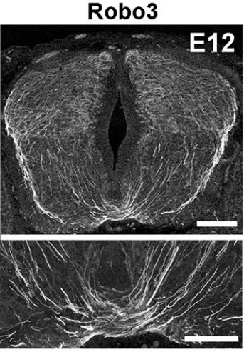
Robo3 in E12 spinal cord sections, from Fig 4, Dominici, et al., 2018
AC & CD Most midline guidance cues (Slits, Netrins, Semaphorins) are expressed in other cell types, including commissural neurons themselves. Therefore, even if the corresponding knockouts have clear axon crossing phenotypes, the direct demonstration of the floor plate as a main source of these factors was lacking. This pushed us to generate conditional knockout lines (Slit2 and Netrin 1) to selectively ablate them from specific cells. In addition, we wanted to use the mice to study the function of these molecules in the postnatal and adult nervous system, which is impossible with the classic knockouts which all die shortly after birth.
The increase in CNS size during vertebrate evolution has put commissural neurons at a larger distance from the floor plate, in particular in the hindbrain, which might explain why they might rely less on floor plate cues, at least initially. This might have been accompanied by a relocation of the main guidance cues to other cell types, closer to commissural neurons.
The imaging in your paper is stunning: can you give us an introduction to technique and how has it changed your view of developing nervous system?
AC & CD The 3DISCO-tissue clearing method was initially developed by Ali Ertürk and Frank Bradke who published it in 2011 in Nature Medicine. The basic principle is to use a series of solvents to solubilize lipids and homogenize the refractive index of the sample (such as a whole embryo, or an adult mouse brain). The method is fast, cheap and works with a wide array of tissues and species. Our addition to this field was to adapt whole-mount immunostaining on top of tissue clearing. Fluorescent dyes are not altered during clearing and samples can be quickly imaged in 3D using a light-sheet microscope. This has significantly facilitated and accelerated the phenotyping of our axon guidance mutants and also diminished the number of samples needed to understand the defects (as they can be oriented in all directions using a 3D Imaging software). We can also be much more precise in the evaluation of the phenotypes of compound mutants. Dissecting the embryo is not needed with this method, therefore you can also easily extend your analysis to organs that are outside the CNS, something that would be too time-consuming using classic histology.
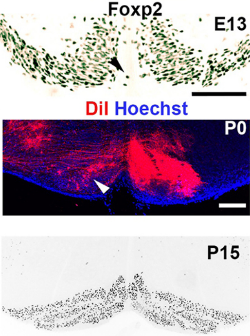
Tracking inferior olive neurons, from Fig 8, Dominici, et al. 2018
When doing the research, did you have any particular result or eureka moment that has stuck with you?
CD When I began to study motorneuron development, I wanted to visualize motor pools in 3 dimensions. This coincided with the emergence of the first tissue clearing techniques and commercial light sheet microscopes. We first used the 3DISCO clearing protocol, but GFP, the reporter used to label axons in our lines, quickly bleached after clearing. I wondered whether we could still visualise GFP using immuno-labelling. I decided to test an in toto immunostaining on a small portion of spinal cord and then cleared it, but I wasn’t really expecting to see anything. To my surprise, it worked! The images were incredible! We then optimised the technique and I could later apply the same technique to visualise migrating precerebellar neurons in 3D.
Not only were we able to visualise the migration of this neuronal population like never before, but we also were able to show that conditional Slits removal had no impact on precerebellar neuron migration.
And what about the flipside: any moments of frustration or despair?
CD Whilst it was a eureka moment, it was also a moment of despair when we realised that conditional removal of Slits in motor neurons showed no clear phenotype. Moreover, the generation of the mouse lines (more than 30) for my project was very difficult. In some mutants we had a one in sixteen chance of getting the right combination of genes. Indeed, the revision of this paper took almost a year due to the difficulty of obtaining additional mutants.
What next for you Chloé?
CD My PhD was quite intense. So I decided to take a year off after defending my PhD in 2016. I travelled and looked for multiple other opportunities. After taking a step back from research, I have realised that it is still something I am incredibly passionate about and would like to pursue. I am currently applying for postdocs.
Where will this work take the Chédotal lab?
AC Our work shows that the midline puzzle is not solved. Commissural neurons are highly diverse: some migrate to the midline, some cross it, others don’t, some project to both sides of the CNS and so on. How could a single unifying model account for this diversity? It has been considered for a long time that midline crossing mechanisms were similar in all bilaterians but this is not the case. We are now trying to identify new regulators of midline crossing using genetic screens, proteomics and transcriptomics. We are also comparing guidance mechanisms in multiple vertebrate species to determine if changes in receptors and ligands played a role in brain evolution, by modifying neuronal connectivity and migration. Some of our preliminary results seem to support this hypothesis.
Finally, let’s move outside the lab – what do you like to do in your spare time?
AC Cooking is one of my favorite hobbies and is the best method I know to rake my mind off science. This said, running an experiment and cooking a dish have a lot in common.
CD Unfortunately during my PhD, I had little time to do much extracurricular activities. But when I managed to squeeze some experiments, I took some time out of Paris to visit my family and friends. This really allowed me to take a step back from the stress of the PhD.
We are looking to appoint a talented post-doctoral researcher to work on an exciting new regenerative medicine project. In this project we will evaluate the effectiveness of using a physiological matrix scaffold created by organ decellularisation to support the differentiation of stem cells into insulin-producing beta cells. This collaborative 3 year project is funded by Diabetes UK and is based in the research groups of Dr Natasha Hill at Kingston University and Dr Aileen King at Kings College London (Guys Campus).
The Person
The project will involve stem cell culture and differentiation, pancreas decellularisation, fluorescence microscopy, flow cytometry, methods to evaluate dynamic insulin release and calcium flux, and in vivo evaluation of beta cell function. Candidates with a relevant PhD degree, ideally with experience in stem cell differentiation or organ decellularisation, and who are interested in developing new treatments for people with diabetes are particularly encouraged to apply.
Further information
Candidates must be able to demonstrate their eligibility to work in the UK in accordance with the Immigration, Asylum and Nationality Act 2006. Where required this may include entry clearance or continued leave to remain under the Points Based Immigration Scheme.
Please also submit a CV and covering letter outlining your interest and suitability for the role.
This is a fixed term contract for 3 years.
To apply click here to go to the Kingston University website.
If, for accessibility reasons, you need to apply in an alternative format, please email jobs@kingston.ac.uk for an application pack or call the HR Shared Services team on 020 8417 3118.

 (1 votes)
 (No Ratings Yet)
 Matthew Cobb is an inspiring advocate and communicator of science, in particular of biology. This is clearly reflected in his